. 2010 Apr;1(1):55-86.
doi: 10.1210/TEAM.9781879225718.ch2.

Pathogenesis of Osteoporosis

Affiliations

Pathogenesis of Osteoporosis

Sundeep Khosla. Transl Endocrinol Metab. 2010 Apr.

Abstract

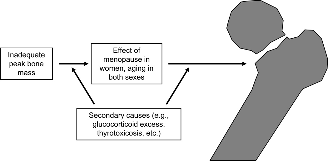
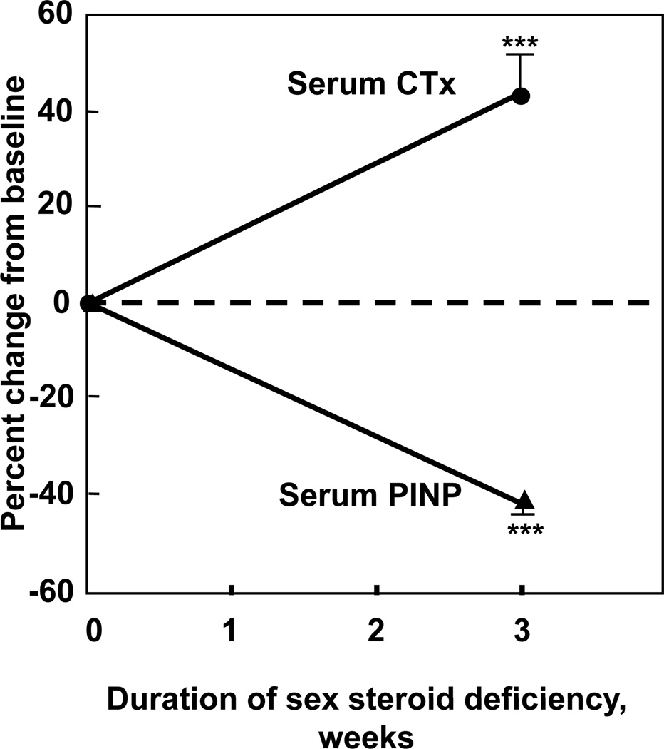
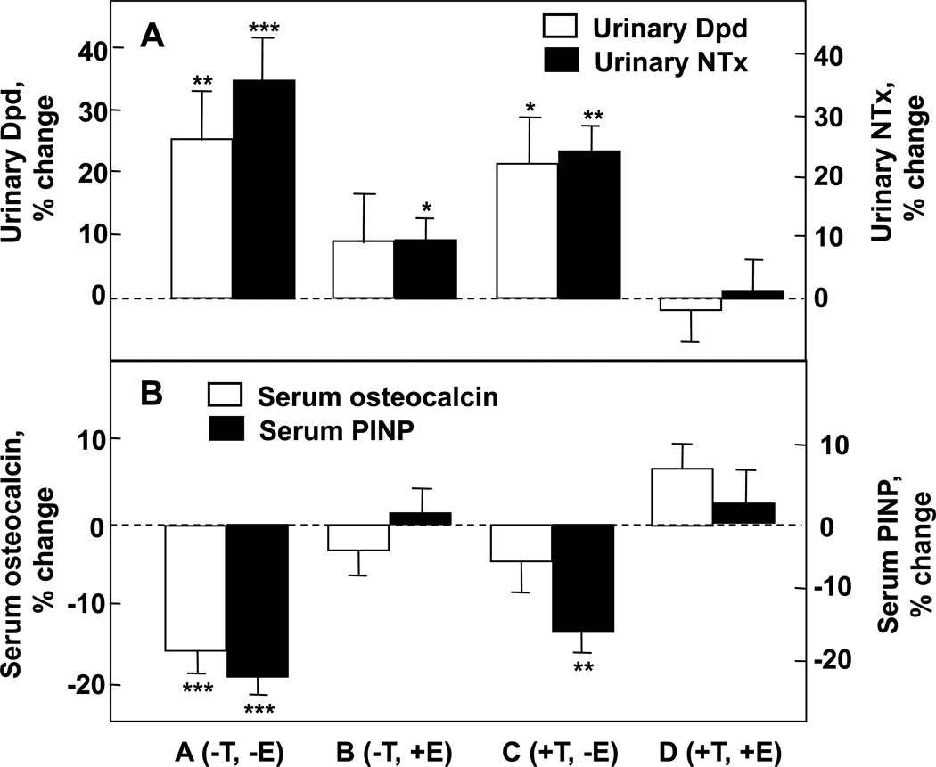
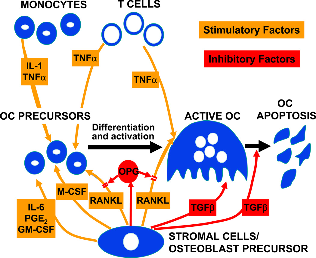
As for most multifactorial disorders, the pathogenesis of osteoporosis is complex, and a different set of mechanisms may be operative in any given individual. However, there are certain common causes of bone loss and increased fracture risk with aging in most people. These include genetic factors contributing to the acquisition of peak bone mass, illnesses affecting skeletal growth and development, sex steroid deficiency following the menopause in women and with aging in men, and intrinsic, age-related changes in bone metabolism. Superimposed on these factors are specific secondary causes of bone loss, such as corticosteroid use or other illnesses affecting bone metabolism that may contribute to fracture risk in individuals exposed to these factors. The past decade has witnessed tremendous advances in our understanding of each of these various causes of bone loss, leading to the development of novel, mechanism-based therapeutic approaches to prevent and treat this important public health disorder.

Keywords: bone loss; fractures; osteoporosis.


Figures

Figure 1
Figure 1
Overall schematic for the pathogenesis of osteoporosis. Please see text for details.
Figure 2
Figure 2
Bone mineral content (BMC) in the total body (TB) accrual in boys (open circles) and girls (closed circles). Reproduced from Bailey et al. (6), with permission.
Figure 3
Figure 3
(A) Values for vBMD (mg/cm3) of the total vertebral body in a population sample of Rochester, Minnesota women and men between the ages of 20 and 97 years. Individual values and smoother lines are given for premenopausal women in red, for postmenopausal women in blue, and for men in black. (B) Values for cortical vBMD at the distal radius in the same cohort. Color code is as in Panel A. All changes with age were significant (P < 0.05). Reproduced from Riggs et al. (16), with permission.
Figure 4
Figure 4
Bone formation (serum osteocalcin [OCN], bone alkaline phosphatase [BAP] and C-terminal propeptide of type I collagen [PICP]) and bone resorption (urinary C-telopeptide of type I collagen [CTx] and N-telopeptide of type I collagen [NTx]) markers in perimenopausal (peri MP), early postmenopausal (Early pMP), and late postmenopausal (Late pMP) women. Reproduced from Garnero et al. (35), with permission.
Figure 5
Figure 5
(A) Short term increase in the bone resorption marker, serum C-telopeptide of type I collagen (CTx) and decrease in the bone formation marker, serum N-terminal propeptide of type I collagen (PINP) following the acute induction of estrogen deficiency in postmenopausal women (adapted from (36)). ***P < 0.001.
Figure 6
Figure 6
Percent changes in (A) bone resorption markers (urinary deoxypyridinoline [Dpd] and N-telopeptide of type I collagen [NTx]) and (B) bone formation markers (serum osteocalcin and N-terminal extension peptide of type I collagen [PINP]) in a group of elderly men (mean age 68 yrs) made acutely hypogonadal and treated with an aromatase inhibitor (Group A), treated with estrogen alone (Group B), testosterone alone (Group C), or both (Group D). See text for details. Asterisks indicate significance for change from baseline: *, P < 0.05; **, P < 0.01; ***, P < 0.001. Adapted from Falahati-Nini et al. (52), with permission.
Figure 7
Figure 7
Summary of stimulatory and inhibitory factors involved in osteoclast (OC) development and apoptosis. Please see text for details. Reproduced from (125), with permission.

References

    1. Riggs BL, Khosla S, Melton LJ. Sex steroids and the construction and conservation of the adult skeleton. Endocr Rev. 2002;23:279–302. - PubMed
    1. Khosla S, Melton LJ, Riggs BL. Estrogen and the male skeleton. J Clin Endocrinol Metab. 2002;87:1443–1450. - PubMed
    1. Cooper C, Eriksson JG, Forsen T, Osmond C, Tuomilehto J, Barker DJP. Maternal height, childhood growth and risk of hip fracture in later life: a longitudinal study. Osteoporos Int. 2001;12:623–629. - PubMed
    1. Barker DJ. The Wellcome Foundation Lecture, 1994. The fetal origins of adult disease. Proc Biol Sci. 1995;262:37–43. - PubMed
    1. Dennison EM, Syddall HE, Sayer AA, Gilbody HJ, Cooper C. Birth weight and weight at 1 year are independent determinants of bone mass in the seventh decade: the Hertfordshire cohort study. Pediatr Res. 2005;57:582–586. - PubMed

LinkOut - more resources