

Antimicrobial peptides: their role as infection-selective tracers for molecular imaging
- PMID: 25243191
- PMCID: PMC4163393
- DOI: 10.1155/2014/867381
Antimicrobial peptides: their role as infection-selective tracers for molecular imaging
Abstract
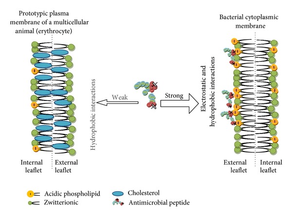
Antimicrobial peptides (AMPs) are a heterogeneous class of compounds found in a variety of organisms including humans and, so far, hundreds of these structures have been isolated and characterised. They can be described as natural microbicide, selectively cytotoxic to bacteria, whilst showing minimal cytotoxicity towards the mammalian cells of the host organism. They act by their relatively strong electrostatic attraction to the negatively charged bacterial cells and a relatively weak interaction to the eukaryote host cells. The ability of these peptides to accumulate at sites of infection combined with the minimal host's cytotoxicity motivated for this review to highlight the role and the usefulness of AMPs for PET with emphasis on their mechanism of action and the different interactions with the bacterial cell. These details are key information for their selective properties. We also describe the strategy, design, and utilization of these peptides as potential radiopharmaceuticals as their combination with nuclear medicine modalities such as SPECT or PET would allow noninvasive whole-body examination for detection of occult infection causing, for example, fever of unknown origin.
Figures
Similar articles
-
The use of technetium-99m radiolabeled human antimicrobial peptides for infection specific imaging.Mini Rev Med Chem. 2008 Sep;8(10):1039-52. doi: 10.2174/138955708785740670. Mini Rev Med Chem. 2008. PMID: 18782056 Review.
-
Antimicrobial peptides as an opportunity against bacterial diseases.Curr Med Chem. 2015;22(14):1665-77. doi: 10.2174/0929867322666150311145632. Curr Med Chem. 2015. PMID: 25760092 Review.
-
Application of Antimicrobial Peptides of the Innate Immune System in Combination With Conventional Antibiotics-A Novel Way to Combat Antibiotic Resistance?Front Cell Infect Microbiol. 2019 Apr 30;9:128. doi: 10.3389/fcimb.2019.00128. eCollection 2019. Front Cell Infect Microbiol. 2019. PMID: 31114762 Free PMC article.
-
Selective phenylalanine to proline substitution for improved antimicrobial and anticancer activities of peptides designed on phenylalanine heptad repeat.Acta Biomater. 2017 Jul 15;57:170-186. doi: 10.1016/j.actbio.2017.05.007. Epub 2017 May 5. Acta Biomater. 2017. PMID: 28483698
-
Mammalian Antimicrobial Peptides: Promising Therapeutic Targets Against Infection and Chronic Inflammation.Curr Top Med Chem. 2016;16(1):99-129. doi: 10.2174/1568026615666150703121819. Curr Top Med Chem. 2016. PMID: 26139111 Review.
Cited by
-
Antimicrobial Peptides as Anticancer Agents: Functional Properties and Biological Activities.Molecules. 2020 Jun 19;25(12):2850. doi: 10.3390/molecules25122850. Molecules. 2020. PMID: 32575664 Free PMC article. Review.
-
Multi-label classification and features investigation of antimicrobial peptides with various functional classes.iScience. 2023 Oct 18;26(12):108250. doi: 10.1016/j.isci.2023.108250. eCollection 2023 Dec 15. iScience. 2023. PMID: 38025779 Free PMC article.
-
An Overview of the Potentialities of Antimicrobial Peptides Derived from Natural Sources.Antibiotics (Basel). 2022 Oct 26;11(11):1483. doi: 10.3390/antibiotics11111483. Antibiotics (Basel). 2022. PMID: 36358138 Free PMC article. Review.
-
Nanoantibiotics containing membrane-active human cathelicidin LL-37 or synthetic ceragenins attached to the surface of magnetic nanoparticles as novel and innovative therapeutic tools: current status and potential future applications.J Nanobiotechnology. 2020 Jan 2;18(1):3. doi: 10.1186/s12951-019-0566-z. J Nanobiotechnology. 2020. PMID: 31898542 Free PMC article. Review.
-
The Tat protein transport system: intriguing questions and conundrums.FEMS Microbiol Lett. 2018 Jun 1;365(12):fny123. doi: 10.1093/femsle/fny123. FEMS Microbiol Lett. 2018. PMID: 29897510 Free PMC article. Review.
References
-
- Basu S, Chryssikos T, Moghadam-Kia S, Zhuang H, Torigian DA, Alavi A. Positron emission tomography as a diagnostic tool in infection: present role and future possibilities. Seminars in Nuclear Medicine. 2009;39(1):36–51. - PubMed
-
- Palestro CJ. Radionuclide imaging of infection: In search of the grail. Journal of Nuclear Medicine. 2009;50(5):671–673. - PubMed
-
- Rennen HJJM, Boerman OC, Oyen WJG, Corstens FHM. Imaging infection/inflammation in the new millennium. European Journal of Nuclear Medicine. 2001;28(2):241–252. - PubMed
-
- Boerman OC, Dams ETM, Oyen WJG, Corstens FHM, Storm G. Radiopharmaceuticals for scintigraphic imaging of infection and inflammation. Inflammation Research. 2001;50(2):55–64. - PubMed
Publication types
MeSH terms
Substances
LinkOut - more resources
Full Text Sources
Other Literature Sources
