. 2014 Nov;94(4):771-93.
doi: 10.1111/mmi.12802. Epub 2014 Oct 12.

Diguanylate cyclase NicD-based signalling mechanism of nutrient-induced dispersion by Pseudomonas aeruginosa

Affiliations

Diguanylate cyclase NicD-based signalling mechanism of nutrient-induced dispersion by Pseudomonas aeruginosa

Ankita Basu Roy et al. Mol Microbiol. 2014 Nov.

Abstract

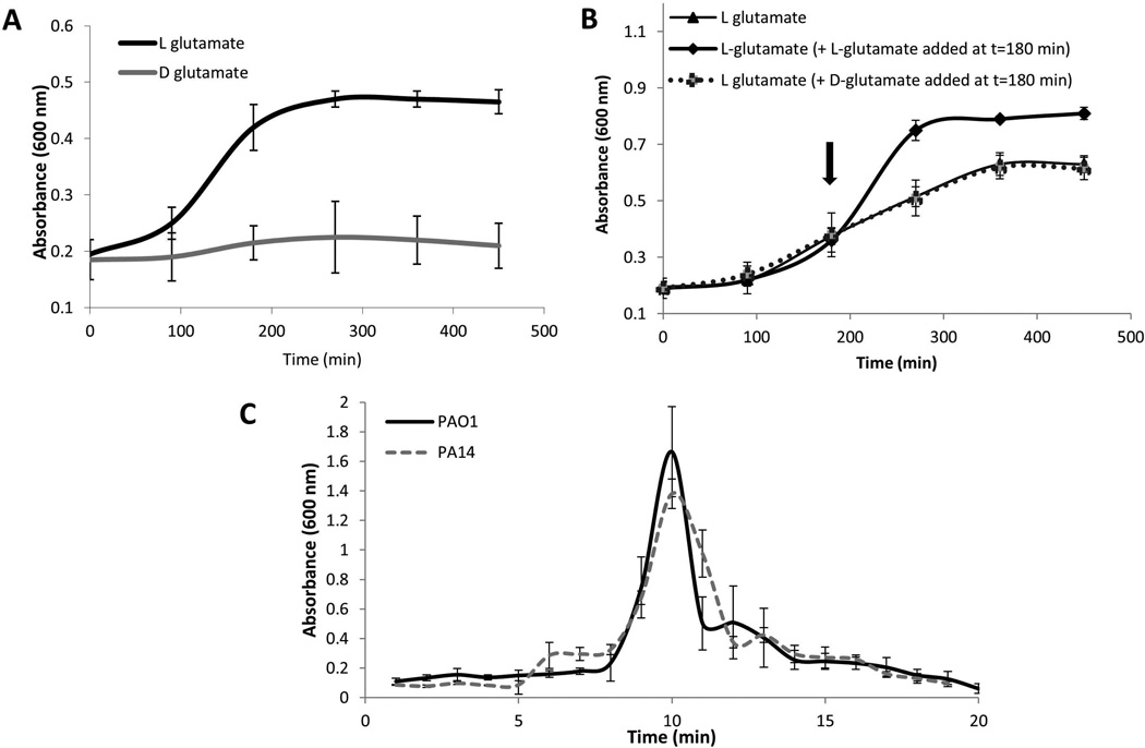
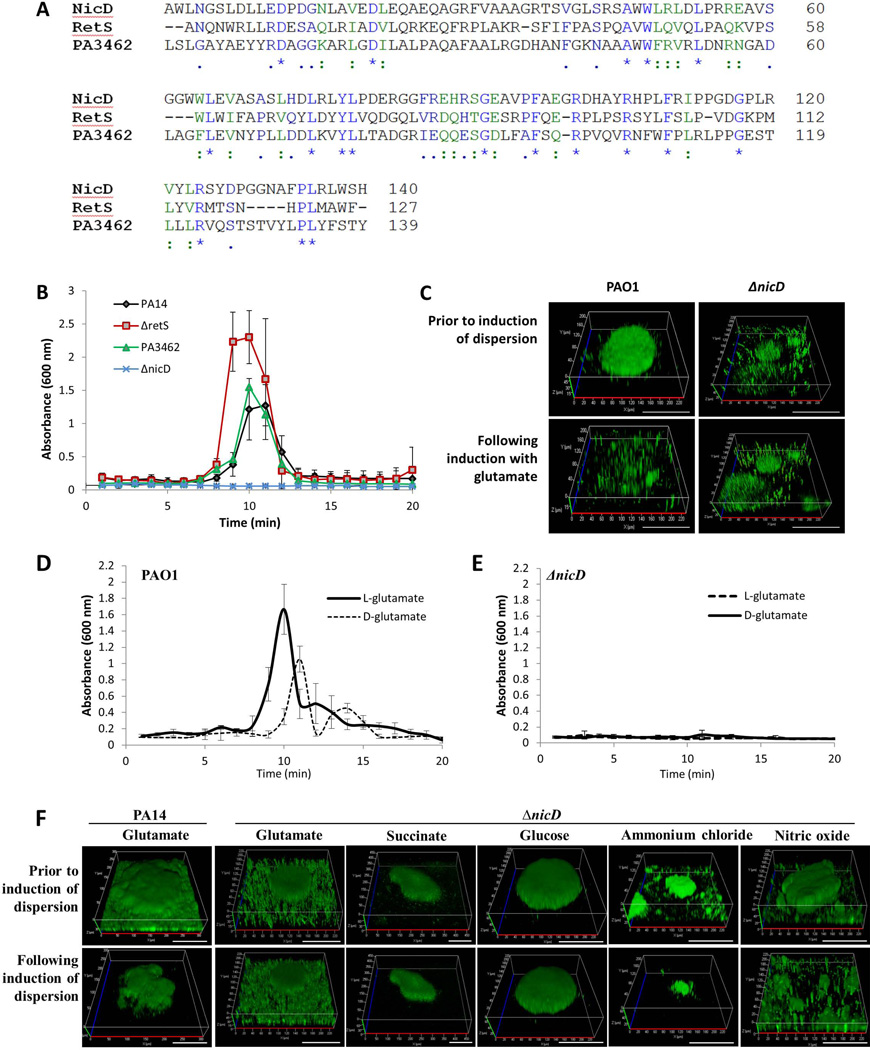
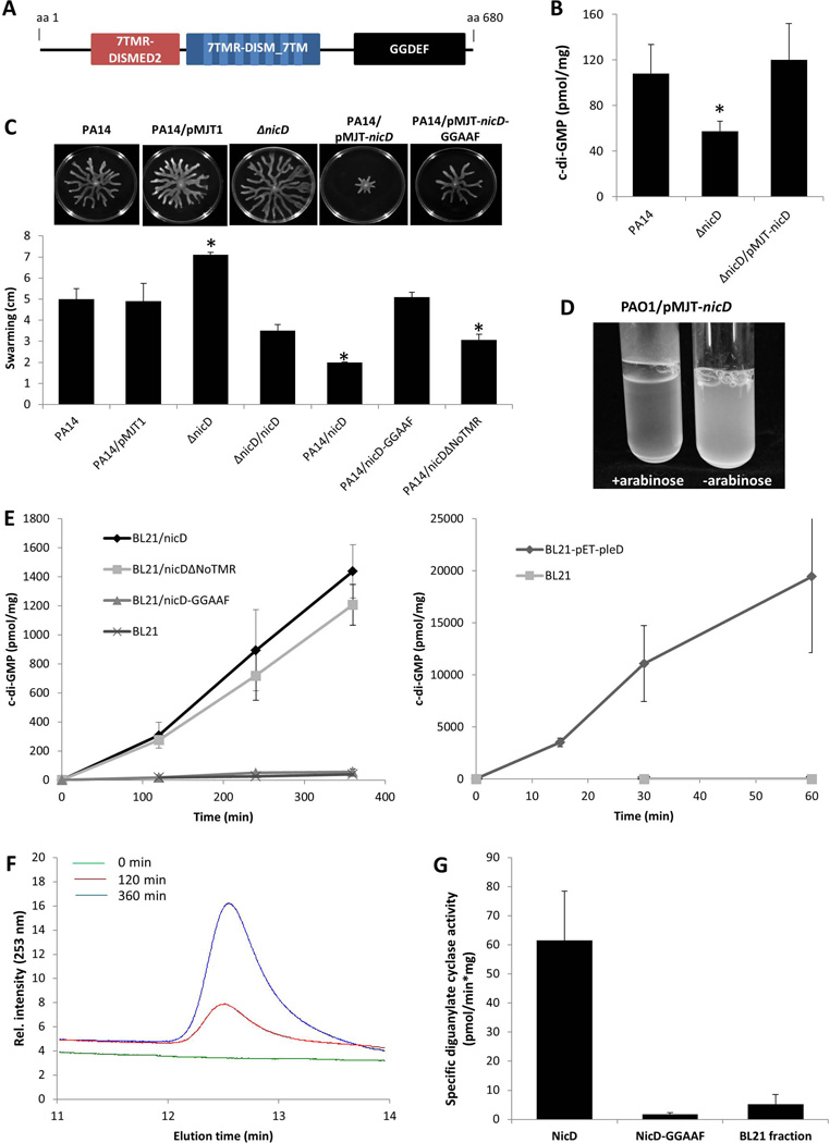
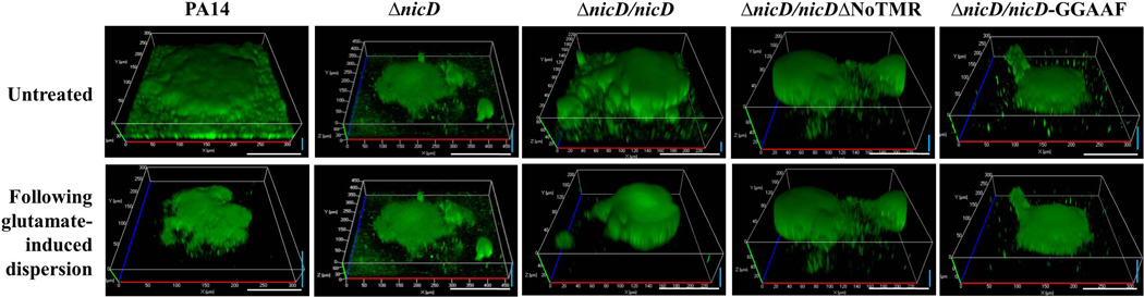
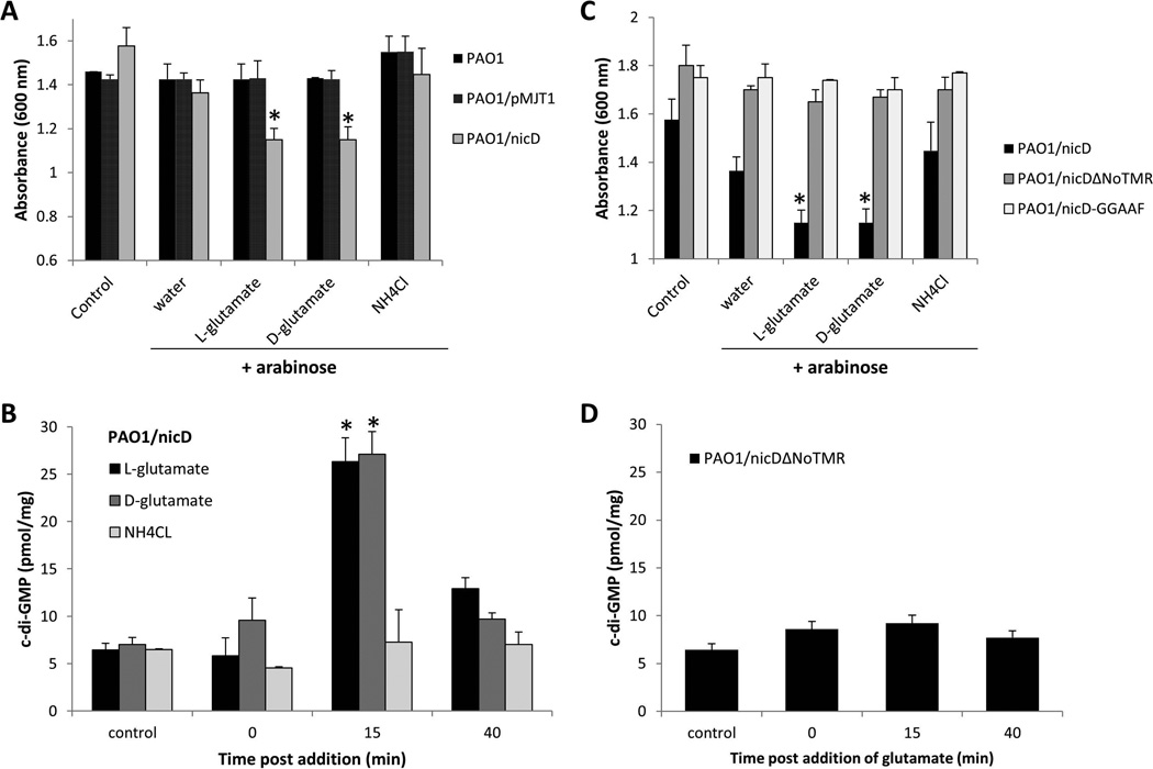
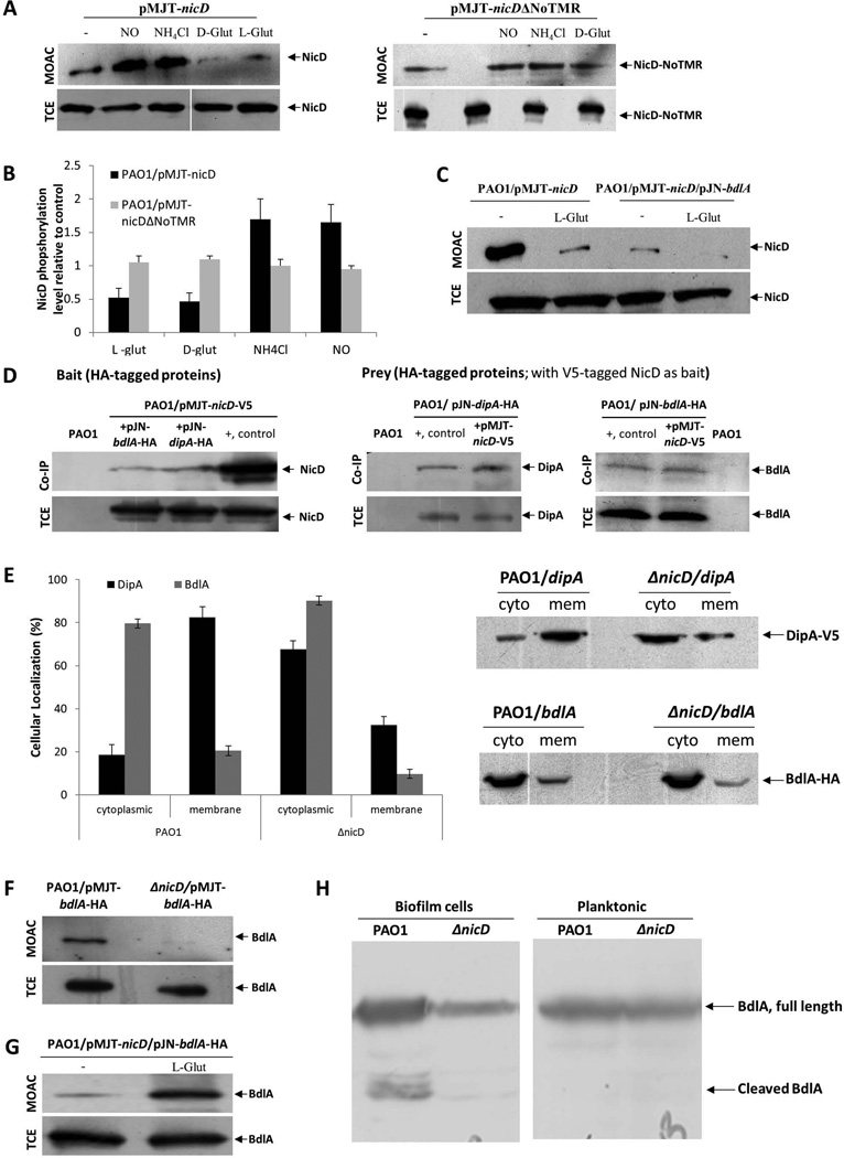
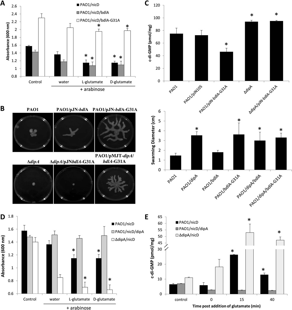
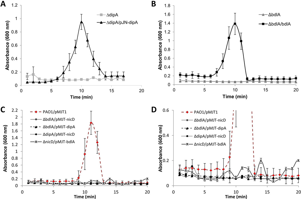
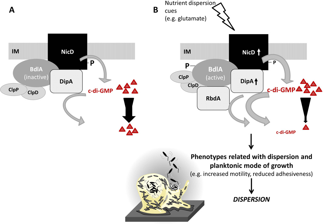
Dispersion enables the transition from the biofilm to the planktonic growth state in response to various cues. While several Pseudomonas aeruginosa proteins, including BdlA and the c-di-GMP phosphodiesterases DipA, RbdA, and NbdA, have been shown to be required for dispersion to occur, little is known about dispersion cue sensing and the signalling translating these cues into the modulation c-di-GMP levels to enable dispersion. Using glutamate-induced dispersion as a model, we report that dispersion-inducing nutrient cues are sensed via an outside-in signalling mechanism by the diguanylate cyclase NicD belonging to a family of seven transmembrane (7TM) receptors. NicD directly interacts with BdlA and the phosphodiesterase DipA, with NicD, BdlA, and DipA being part of the same pathway required for dispersion. Glutamate sensing by NicD results in NicD dephosphorylation and increased cyclase activity. Active NicD contributes to the non-processive proteolysis and activation of BdlA via phosphorylation and temporarily elevated c-di-GMP levels. BdlA, in turn, activates DipA, resulting in the overall reduction of c-di-GMP levels. Our results provide a basis for understanding the signalling mechanism based on NicD to induce biofilm dispersion that may be applicable to various biofilm-forming species and may have implications for the control of biofilm-related infections.


Figures

Figure 1
Figure 1. Dispersion occurs in response to D-glutamate despite D-glutamate being a poor growth substrate for P. aeruginosa
Growth of P. aeruginosa PAO1 in the presence of D-glutamate (130 mg/L) or L-glutamate (130 mg/L) as the sole carbon source (A) and (B) upon addition of extra D- or L-glutamate (130mg/L) at the 3 hr time point. P. aeruginosa was grown in minimal medium at 37°C with shaking at 220 rpm and absorbance was recorded at 600 nm every 90 min over a period of 8 hr. Experiments were repeated at least three times Error bars indicate standard deviation. (C) Dispersion by P. aeruginosa PA14 and PAO1 biofilms in response to L-glutamate, as indicated by measurements of effluent absorbance. P. aeruginosa strain PA14 harbors a mutation in ladS that renders the protein non-functional (Mikkelsen et al., 2011). Dispersion assays were carried out using biofilm tube reactors. Experiments were repeated at least three times Error bars indicate standard deviation.
Figure 2
Figure 2. Dispersion in response to nutrient cues requires NicD
(A) Alignment of the NicD 7TMR-DISMED2 with the 7TMR-DISMED2 domains of RetS and PA3462. ":" denotes residues with strong conservation (score >0.5 in PAM250 matrix) and "." denotes residues with weak conservation (score ≤0.5); "*", denotes identical amino acids. Identical amino acid residues are shown in blue while similar amino acids are highlighted in green. (B) Inactivation of nicD but not retS or PA3462 renders P. aeruginosa dispersion-deficient in response to glutamate as indicated by measurements of effluent absorbance. Biofilms were grown for 5 days under flowing conditions in tube reactors prior to induction of dispersion inducing conditions. Experiments were repeated at least three times. Error bars indicate standard deviation. (C) Inactivation of nicD impairs dispersion of P. aeruginosa PAO1 biofilms to L-glutamate as indicated by confocal microscopy. Biofilms were grown for 5 days in flow cells under flowing conditions. Confocal images were acquired at the same position, prior to and post-induction of dispersion. Representative images are shown. Size bar, 100 µm. Dispersion by P. aeruginosa PAO1 biofilms (D) and ΔnicD biofilms (E) in response to D- and L-glutamate as indicated by measurements of effluent absorbance. Biofilms were grown for 5 days in tube reactors under flowing conditions prior to induction of dispersion inducing conditions. Experiments were repeated at least five times. Error bars indicate standard deviation. (F) Confocal images of P. aeruginosa PA14 ΔnicD biofilms prior to and following the addition of glutamate, succinate, glucose, ammonium chloride and nitric oxide to induce biofilm dispersion. Inactivation of nicD renders P. aeruginosa dispersion-deficient in response to glutamate, glucose and succinate but not ammonium chloride or nitric oxide. Shown confocal images were acquired at the same position, prior to and post-induction of dispersion. Size bar, 100 µm.
Figure 3
Figure 3. nicD harbors in addition to 7TMR-DISMED2 and 7TMR-DISM_7TM domains a GGDEF domain, and encodes an active diguanylate cyclase
(A) NicD (PA4929) is predicted to be a membrane-bound protein harboring three domains including a periplasmic 7TMR-DISMED2 sensory domain, a membrane-spanning 7TMR-DISM_7TM domain and a cytoplasmic GGDEF domain. Model of NicD domain organization and putative subcellular localization of NicD domains. NicD comprises of three domains: an extracellular 7TMR-DISMED2 (7 Trans-membrane Receptor with Diverse Intracellular Signaling Modules extracellular) domain, a membrane-spanning 7TMR-DISM_7TM (7TMR-DISM, 7 Trans-membranes) domain, and a cytoplasmic diguanylate cyclase (GGDEF) domain. Domain localization is based on TMHMM prediction. (B) C-di-GMP levels in P. aeruginosa PA14, ΔnicD and ΔnicD/pMJT-nicD biofilms. C-di-GMP was quantitated using an HLC-based method with commercially available c-di-GMP as standard. c-di-GMP (pmol/mg) refers to c-di-GMP levels (pmol) per total cell pellet protein (in mg). Experiments were repeated at least three times. Error bars indicate standard deviation. *, significantly different from wild type biofilms; p < 0.05 as determined by ANOVA and SigmaStat (C) Swarming motility of P. aeruginosa PA14, ΔnicD, and strains overexpressing nicD, nicD-GGAAF, and nicDΔNoTMR. The swarming diameter was recorded following 48 hr of growth on NA agar. Representative images of swarming motility P. aeruginosa PA14, ΔnicD, and strains overexpressing nicD, nicD-GGAAF, and nicDΔNoTMR are shown above the graph. P. aeruginosa PA14 harboring the empty vector pMJT1 was used as vector control. Experiments were repeated at least three times. Error bars indicate standard deviation. *, significantly different from wild type biofilms; p < 0.05 as determined by ANOVA and SigmaStat. (D) Image of P. aeruginosa PAO1/pMJT-nicD planktonic culture prior to and following addition of arabinose to induce nicD expression. –arabinose, no arabinose was added; +arabinose, 1% arabinose was added to the growth medium. (E) Formation of c-di-GMP by total cell extracts of E. coli BL21 overexpressing nicD, nicD variants and pleD. DGC assays were carried out using a total of 200 µg of E. coli BL21 cell extracts overexpressing nicD and nicDΔNoTMR lacking the N-terminal 7TMR-DISMED2 domain (NicDΔNoTMR) or nicD-GGAAF in which the GGDEF motif was substituted with GGAAF under the control of the IPTG-inducible promoter of the pET101D vector or a total of 20 µg of E. coli BL21 cell extracts overexpressing pleD cloned into pET11b (Merritt et al., 2007, Paul et al., 2004) were used. Cell extracts of E. coli BL21 not harboring any vector (200 and 20 µg) were used as controls while E. coli BL21 overexpressing pleD were used as positive control. Experiments were carried out in triplicate. Error bars indicate standard deviation. (F) Elution profiles of c-di-GMP produced by purified NicD. The reaction mixtures were analyzed by HPLC for the presence of c-di-GMP 0, 120, and 360 min post initiation of DGC assays. Representative peaks corresponding to c-di-GMP are shown. (G). Specific diguanylate cyclase activity of purified NicD and NicD-GGAAF. Diguanylate cyclase assays were performed using 25 µM GTP and 70 µg of Ni-affinity chromatography purified protein. BL21 control, protein purified from E. coli cell extracts not harboring any vector was used as negative control. Experiments were carried out in triplicate. Error bars indicate standard deviation.
Figure 4
Figure 4. Dispersion in response to glutamate requires NicD DGC activity and the presence of the periplasmic 7TMR-DISMED2 domain
Biofilms formed by P. aeruginosa PA14, ΔnicD, and ΔnicD mutant strains overexpressing nicD, nicD-GGAAF and nicDΔNoTMR under the control of the arabinose-inducible promoter of the pMJT1 vector were grown for 5 days in flow cells under flowing conditions. Multicopy expression of nicD but not nicD-GGAAF or nicDΔNoTMR restores biofilm dispersion upon glutamate exposure to wild type levels. Confocal images were acquired at the same position, prior to and following induction of dispersion by L-glutamate. Representative images are shown. White size bar, 100 µm. Blue vertical size bar (indicating height), 40 µm.
Figure 5
Figure 5. NicD DGC activity is induced upon exposure to glutamate, resulting in increased cellular c-di-GMP levels
(A) Absorbance of culture medium of exponential phase P. aeruginosa PAO1/pMJT-nicD, prior to addition of arabinose (control) and following addition of arabinose (3 hr) and subsequent addition of water, D- and L-glutamate and ammonium chloride (NH4Cl). PAO1 and PAO1/pMJT1 were used as vector control. Experiments were carried out at least in triplicate. Error bars indicate standard deviation. *, significantly different from “water” control to which only arabinose (3 hr) plus water was added. (B) Cellular c-di-GMP levels in PAO1/nicD following addition of arabinose (3 hr, control), and 0, 15 and 40 min post-addition of D- and L-glutamate and ammonium chloride (NH4Cl). Experiments were carried out at least in triplicate. Error bars indicate standard deviation. *, significantly different from 0 min time point. (C) Absorbance of culture medium of exponential phase P. aeruginosa PAO1 overexpressing nicD, nicDΔNoTMR, or nicD-GGAAF, prior to addition of arabinose (control) and following addition of arabinose (3 hr) and subsequent addition of water, D- and L-glutamate and ammonium chloride (NH4Cl). Experiments were carried out at least in triplicate. Error bars indicate standard deviation. (D) Cellular c-di-GMP levels in PAO1/nicDΔNoTMR mutant following addition of arabinose (3 hr), and 0, 15 and 40 min post-addition of glutamate. Experiments were carried out at least in triplicate. Error bars indicate standard deviation.
Figure 6
Figure 6. NicD forms a membrane associated complex with DipA and BdlA and contributes to BdlA activation
(A) Detection of V5-tagged NicD and NicDΔNoTMR in total cell extracts (TCE) or metaloxide affinity chromatography-enriched phosphoproteomes (MOAC) of planktonic P. aeruginosa PAO1/pMJT-nicD and PAO1/pMJT-nicDΔNoTMR cells prior to (−) and following addition of nitric oxide (NO), ammonium chloride (NH4Cl), and D- and L-glutamate (D-glut, L-glut, respectively) by immunoblot analysis using anti-V5 antibodies. A total of 10 µg per TCE was loaded prior to MOAC purification and used as loading control. For MOAC samples, the entire MOAC eluate was loaded. (B) Band intensity was used to determine the relative levels of NicD and NicDΔNoTMR phopshorylation following addition of nitric oxide (NO), ammonium chloride (NH4Cl), and D- and L-glutamate (D-glut, L-glut, respectively) relative to untreated controls (−). (C) Detection of V5-tagged NicD in MOAC-enriched fractions is dependent on the addition of glutamate and the presence of BdlA, as determined by immunoblot analysis using anti-V5 antibodies. NicD phopshorylation levels were determined using of PAO1/pMJT-nicD and PAO1/pMJT-nicD/pJN-bdlA. A total of 10 µg per TCE, obtained prior to MOAC purification, was used as loading control. For MOAC samples, the entire MOAC eluate was loaded. (−) no L-glutamate was added to cells grown planktonically to exponential phase prior to cell lysis. L-glut, L-glutamate was added to cells grown planktonically to exponential phase prior to cell lysis. (D) Immunoblot analysis of in vivo pull-down assays (Co-IP) demonstrating complex formation between NicD, BdlA and DipA using BdlA-HA or DipA-HA either as prey or bait. Total cell extracts obtained from P. aeruginosa PAO1 were used as negative control. Cell extracts containing the protein of interest were used as positive control (+, control). Total cell extracts (TCE) obtained prior to Co-IP were used as loading control. (E) Inactivation of nicD affects the subcellular localization of BdlA and DipA. Detection of subcellular localization was achieved by ultracentrifugation and subsequent analysis by SDS/PAGE and immunoblot analysis using anti-V5 and anti-HA antibodies. A total of 15 µg per cytoplasmic (cyto) and membrane (mem) fraction was loaded. Band intensity was determined using ImageJ to determine the ratio of cellular localization (%) of BdlA and DipA in the absence and presence of NicD. (F) NicD contributes to BdlA phosphorylation. Detection of BdlA-V5 in total cell extracts (TCE) or MOAC-enriched phosphoproteomes (MOAC) of P. aeruginosa PAO1 and ΔnicD biofilms as determined by immunoblot analysis. A total of 10 µg per TCE, obtained prior to MOAC purification, was used as loading control. For MOAC samples, the entire MOAC eluate was loaded. (G) Exposure to L-glutamate enhances BdlA phosphorylation in a NicD-dependent manner. (−) no L-glutamate was added to cells grown planktonically to exponential phase prior to cell lysis. L-glut, L-glutamate was added to cells grown planktonically to exponential phase prior to cell lysis. (H) Non-processive cleavage of BdlA is dependent on the mode of growth and the presence of NicD. Inactivation of nicD impairs BdlA processing under biofilm growth conditions. No BdlA processing is observed under planktonic growth conditions. BdlA was detected by immunoblot analysis. All experiments were carried out at least in triplicate and only representative images are shown. Error bars indicate standard deviation.
Figure 7
Figure 7. Multicopy expression of constant-on bdlA-G31 enhances DipA phosphodiesterase activity
(A) Absorbance of culture medium of exponential phase P. aeruginosa PAO1/pMJT-nicD, PAO1/pMJT-nicD/pJN-bdlA, and PAO1/pMJT-nicD/pJN-bdlA-G31A, prior to addition of arabinose (control) and 3 hr post addition of arabinose plus subsequent addition of water, and D- and L-glutamate. Experiments were carried out at least in triplicate. Error bars indicate standard deviation. *, significantly different from “water” sample to which only arabinose and water was added. (B) Representative images of swarming motility of P. aeruginosa PAO1 and strains overexpressing dipA, bdlA, and bdlA-G31. Swarming motility of P. aeruginosa PAO1 and strains overexpressing dipA, bdlA, and bdlA-G31 as well as strains co-expressing dipA and bdlA or dipA and bdlA-G31A. The swarming diameter was recorded following 48 hr of growth on M8 agar. Experiments were repeated at least three times. Error bars indicate standard deviation.*, significantly different from wild type biofilms; p < 0.05 as determined by ANOVA and SigmaStat. (C) Overexpression of active bdlA-G31A affects the cellular c-di-GMP level present in P. aeruginosa PAO1 biofilm cells in a DipA-dependent manner. C-di-GMP levels present in P. aeruginosa PAO1, PAO1/pJN-bdlA-G31A, ΔdipA and ΔdipA/pJN-bdlA-G31A were quantitated using an HPLC-based method with commercially available c-di-GMP as standard. c-di-GMP (pmol/mg) refers to c-di-GMP levels (pmol) per total cell pellet protein (in mg). Experiments were carried out at least in triplicate. Error bars indicate standard deviation. *, significantly different from PAO1 and PAO1 biofilms harboring the empty vector pJN105. (D) Absorbance of culture medium of exponential phase P. aeruginosa PAO1/pMJT-nicD, PAO1/pMJT-nicD/pJN-dipA, and ΔnicD/pJN-dipA, prior to (control) and following addition of arabinose (3 hr) and subsequent addition of water, and D- and L-glutamate. Experiments were carried out at least in triplicate. Error bars indicate standard deviation. *, significantly different from water only sample. (E) Cellular c-di-GMP levels in PAO1/pMJT-nicD, PAO1/pMJT-nicD/pJN-dipA, and ΔdipA/pMJT-nicD following addition of arabinose (3 hr, control), and 0, 15 and 40 min post-addition of L-glutamate. Experiments were carried out at least in triplicate. Error bars indicate standard deviation *, significantly different from 0 min time point.
Figure 8
Figure 8. Dispersion of mutants inactivated in nicD, bdlA and dipA in response to glutamate cannot be restored by cross-complementation
Biofilms were grown for 5 days under flowing conditions in tube reactors prior to induction of dispersion inducing conditions, via exposure to L-glutamate. (A) Multicopy expression dipA in ΔdipA biofilms, and (B) multicopy expression of bdlA in ΔbdlA biofilms restores biofilm dispersion response to glutamate to wild-type levels. (C) Multicopy expression nicD and dipA in ΔbdlA biofilms, nicD in ΔdipA biofilms, or bdlA in ΔnicD biofilms does not restore biofilm dispersion response to glutamate to wild-type levels, as indicated by measurements of effluent absorbance. (D) Expanded view of data shown in Figure 8C. All experiments were repeated at least three times. Error bars indicate standard deviation.
Figure 9
Figure 9. Model of signal transduction upon sensing the dispersion-inducing cue glutamate
(A) The diguanylate cyclase NicD is membrane-bound and phosphorylated. NicD forms a multiprotein complex with the phosphodiesterase DipA and the sensory protein BdlA. BdlA is intact but inactive under planktonic growth conditions (Petrova & Sauer, 2012a). BdlA interacts with the chaperone ClpD and the protease ClpP (Petrova & Sauer, 2012a). (B) Upon perceiving a dispersion-inducing nutrient cue, e.g. glutamate, NicD is dephosphorylated and its diguanylate cyclase activity increases, resulting in elevated levels of c-di-GMP. BdlA is phosphorylated. Both phosphorylation and elevated levels of c-di-GMP contribute to BdlA being cleaved in a non-processive manner, a process requiring the chaperone ClpD, the protease ClpP, and BdlA phosphorylation at Y238 (Petrova & Sauer, 2012a). Cleaved BdlA is active with respect to enabling P. aeruginosa biofilms to respond to dispersion inducing conditions (Petrova & Sauer, 2012a). Active BdlA in turn enhances the activity of the phosphodiesterase DipA, resulting in decreased c-di-GMP levels. It has been well demonstrated that increased DipA activity results in decreased biofilm c-di-GMP levels (Basu Roy et al., 2012). Moreover, active BdlA was recently demonstrated to interact with the phosphodiesterase RbdA in vivo (Petrova & Sauer, 2012b). The recruitment of RbdA to the multiprotein complex likely further contributes to the observed reduction of c-di-GMP in dispersed cells compared to biofilm cells (Basu Roy et al., 2012, An et al., 2010, Barraud et al., 2009). Dispersion has been described to require or coincide with the breakdown of extracellular polymeric matrix surrounding the biofilms, induction of flagellar gene expression, increased motility, and reduced adhesiveness (Morgan et al., 2006, Sauer et al., 2002, Sauer et al., 2004, Basu Roy et al., 2012). IM, inner membrane; triangles and number of triangles represent c-di-GMP and cellular c-di-GMP levels. P, phosphorylation. Size of P correlates with level of phosphorylation. Arrows associated with NicD and DipA indicate increased enzyme activity. Width of grey arrows indicates level of activity.

Comment in

References

    1. Aldridge P, Paul R, Goymer P, Rainey P, Jenal U. Role of the GGDEF regulator PleD in polar development of Caulobacter crescentus . Mol. Microbiol. 2003;47:1695–1708. - PubMed
    1. Allegrucci M, Hu FZ, Shen K, Hayes J, Ehrlich GD, Post JC, Sauer K. Phenotypic characterization of Streptococcus pneumoniae biofilm development. J. Bacteriol. 2006;188:2325–2335. - PMC - PubMed
    1. Allegrucci M, Sauer K. Characterization of colony morphology variants isolated from Streptococcus pneumoniae biofilms. J. Bacteriol. 2007;189:2030–2038. - PMC - PubMed
    1. An S, Wu Je, Zhang L-H. Modulation of Pseudomonas aeruginosa biofilm dispersal by a cyclic-di-GMP phosphodiesterase with a putative hypoxia-sensing fomain. Appl. Environ. Microbiol. 2010;76:8160–8173. - PMC - PubMed
    1. Anantharaman V, Aravind L. Application of comparative genomics in the identification and analysis of novel families of membrane-associated receptors in bacteria. BMC Genomics. 2003;4:34. - PMC - PubMed

Publication types

MeSH terms