. 2014 Dec;15(12):1344-65.
doi: 10.1111/tra.12229. Epub 2014 Oct 27.

Copper directs ATP7B to the apical domain of hepatic cells via basolateral endosomes

Affiliations

Copper directs ATP7B to the apical domain of hepatic cells via basolateral endosomes

Lydia K Nyasae et al. Traffic. 2014 Dec.

Erratum in

  • Traffic. 2015 Jan;16(1):99

Abstract

Physiologic Cu levels regulate the intracellular location of the Cu ATPase ATP7B. Here, we determined the routes of Cu-directed trafficking of endogenous ATP7B in the polarized hepatic cell line WIF-B and in the liver in vivo. Copper (10 µm) caused ATP7B to exit the trans-Golgi network (TGN) in vesicles, which trafficked via large basolateral endosomes to the apical domain within 1 h. Although perturbants of luminal acidification had little effect on the TGN localization of ATP7B in low Cu, they blocked delivery to the apical membrane in elevated Cu. If the vesicular proton-pump inhibitor bafilomycin-A1 (Baf) was present with Cu, ATP7B still exited the TGN, but accumulated in large endosomes located near the coverslip, in the basolateral region. Baf washout restored ATP7B trafficking to the apical domain. If ATP7B was staged apically in high Cu, Baf addition promoted the accumulation of ATP7B in subapical endosomes, indicating a blockade of apical recycling, with concomitant loss of ATP7B at the apical membrane. The retrograde pathway to the TGN, induced by Cu removal, was far less affected by Baf than the anterograde (Cu-stimulated) case. Overall, loss of acidification-impaired Cu-regulated trafficking of ATP7B at two main sites: (i) sorting and exit from large basolateral endosomes and (ii) recycling via endosomes near the apical membrane.

Keywords: Wilson's disease; bafliomycin-A1; bile canaliculus; endosomes; hepatocytes; polarity; proton ATPase; retromer.


Figures

Figure 1
Figure 1. Cu directs the trafficking of endogenous ATP7B in WIF-B cells
A-C) WIF-B cells incubated overnight in 10 μM BCS to stage endogenous ATP7B at the TGN region. D-F) Cells switched to 10 μM CuCl2 for 60 minutes. G-I) Cu-treated cells that were rinsed and re-incubated in 10 μM BCS for 180 minutes. After fixation, cells were triple-stained with antibodies to ATP7B (green), Syntaxin 6, a post-TGN marker (red) and aminopeptidase N, APN, an apical surface marker (blue). Single confocal planes are shown. J) The fraction of total ATP7B fluorescence that localized to the TGN or apical region in each condition was quantified as the extent of overlap with Syntaxin 6 or APN, respectively. Data shown represent the mean +/- SEM from at least 3 confocal stacks (approximately 28 WIF-B cells/stack, obtained from a single experiment).
Figure 2
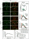
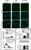
Figure 2. Cu-directed anterograde (TGN-to-apical) traffic occurs via TGN-derived vesicles that traverse an EEA1-positive compartment
A-B) WIF-B cells were incubated overnight in 10 μM BCS to stage ATP7B at the TGN region then C-D) switched to 10 μM CuCl2 for 15 minutes, or E-F) 10 μM CuCl2 for 60 minutes. Cells were fixed and double-stained with antibodies to ATP7B (green) and EEA1 (red) then imaged by confocal microscopy. A-F) depict single nuclear confocal planes. A'-F' depict single peripheral planes. Renderings of representative stacks from these time points are shown in the top row of Supplementary Figure S4. G) The number of ATP7B vesicles/cell was estimated using 3D objects-based analysis. H) Fraction of ATP7B fluorescence that was coincident with EEA1-positive small and large endosomes. Data shown in G and H represent the mean +/- SEM of at least 4 confocal stacks from a single experiment. I) Schematic cartoon of a WIF-B cell grown on a glass coverslip and rotated 90°. J) Confocal stacks were analyzed by displaying the normalized fractional distribution per slice of various organelle markers, to show the distribution of each marker along the z-plane of the cells. The Cu status of the cells had no effect on the distribution of these markers thus the data shown are pooled with no respect to Cu status, from 7-12 confocal stacks, from at least 2 independent experiments. EEA1-positive endosomes (red) distribute closer to the coverslip (∼2 μm above it) compared to the TGN38 (Golgi, green, ∼5 μm above coverslip) or APN (apical membrane, blue, ∼6 μm above coverslip). K) Cu-induced redistribution of ATP7B into a large basolateral EEA1 endosomes (15 minutes, red profile).
Figure 3
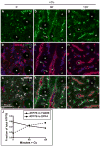
Figure 3. Cu-directed TGN-to-apical trafficking occurs via a basolateral compartment in hepatocytes in vivo
A-C) Liver section from a rat raised on a low-Cu diet for 38-45 days since birth. D-F) Liver section from a low copper rat given Cu then sacrificed after 60 minutes, or G-I) 120 minutes. Rat livers were fixed by immersion and cryosections were immunolabeled for ATP7B (green), TGN38 (red), or apical surface marker DPP4 (blue) then imaged by confocal microscopy. (G-I), Arrows indicate the transient appearance of ATP7B in the basolateral region 60 minutes after Cu injection. J) Quantification of Cu-induced trafficking in vivo. The graph depicts colocalization analysis on confocal stacks from liver sections stained above. Cu causes a decrease of ATP7B in the Golgi (solid line), with concomitant increase in ATP7B trafficking to the apical domain (dotted line).
Figure 4
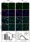
Figure 4. Treatment of WIF-B cells with Baf or weak bases prevents Cu-dependent delivery to the apical region
WIF-B cells were incubated overnight in 10 μM BCS to stage ATP7B at the TGN region. A-C) DMSO or G-I) 50 nM Baf were added for the last 30 minutes of BCS-treatment to deacidify compartments before Cu was added to induce ATP7B trafficking. Cells were then rinsed and transferred to 10 μM CuCl2 for 60 minutes in the continued presence of D-F) DMSO, or J-L) 50 nM Baf. Some of the cells were fixed at each step and triple-stained with antibodies to ATP7B (green), TGN38 (red) and APN (blue) then analyzed by confocal microscopy. A-L) Single planes from the nuclear region. C', F', I' and L') Single planes from the peripheral region. M) The overlap between ATP7B and TGN38 or APN was quantified by image analysis of the confocal stacks. Neither Baf nor the weak bases (300 μM CQ or 30 mM NH4Cl) affected ATP7B localization at the TGN region in BCS-treated cells (left bars), but all three drugs severely impaired traffic to the apical region (right bars). N) Applying the distribution-in-z analysis used in Figure 2, we observed a distribution of ATP7B in Cu plus Baf that indicates protracted accumulation of ATP7B in a peripheral/basolateral site, presumably the structures localized at the peripheral region in the presence of Baf (compare F' and L').
Figure 5
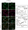
Figure 5. Baf plus Cu causes ATP7B to accumulate in large EEA1-positive endosomes located predominantly at the peripheral region of WIF-B cells
Cells were treated same as Figure 4 but fixed at 15 minute intervals during the 60 minutes of Cu-treatment plus DMSO or 50 nM Baf, then double-stained for ATP7B (green) and EEA1 (red). Substantial overlap between A) ATP7B and B) EEA1 was seen in large clusters at the peripheral region of cells as early as 15 minutes after Cu plus Baf. C) Magnification of a group of typical Baf-induced clusters (each ∼5 μm across), with ATP7B concentrated in subdomains (arrows) of the large and more homogeneous EEA1-positive endosomes. D) 60 minutes after Cu plus DMSO or E) Cu plus Baf. Single confocal planes from the D-E) nuclear or D'-E') peripheral region show overlap between ATP7B and EEA1 persisted in Baf-treated cells. 3D renderings of confocal image stacks representing 0, 15 and 60- minute time points are shown in Supplementary Movie S4. F) The extent of overlap between ATP7B and EEA1 during 60 minutes of Cu plus Baf was quantified from at least 4 confocal stacks from a single experiment. The 0 and 60 minute time points represent the mean of 10 confocal stacks, obtained from 3 independent experiments. G) To characterize the effect of Baf on the EEA1-positive endosomes, the mean cross-sectional area of the large EEA1 structures was measured across all experimental conditions and plotted against the time that the cells were incubated in Baf or DMSO. H) The fraction of total EEA1 that was present in “large” EEA1 organelles, defined as having > 1 μm2 cross-sectional area in the 2D analysis.
Figure 6
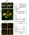
Figure 6. Detailed characterization of Baf-induced clusters of ATP7B and EEA1
WIF-B cells were treated with Cu in the presence of Baf, fixed after 15, 30, or 60 minutes, then immunolabled for EEA1 (red) and ATP7B (green). Confocal stacks were subjected to a blind deconvolution routine to enhance the signal-to-noise ratio. A) Example of a Baf-induced basolateral EEA structure from the 15 min time point (maximal Z-projection). B) Serial sections (0.25 μm steps) illustrating the close and often complementary relationship between EEA1 and ATP7B within the structure. C) Mean cross-sectional area of EEA1 channel measured at the 3 time points. D) Colocalization analysis measuring the percent of ATP7B fluorescence that was coincident with EEA1. Mean +/- S.E.M. N=22 structures analyzed.
Figure 7
Figure 7. Removal of Baf partially restores delivery of ATP7B to the apical region in Cu-treated WIF-B cells
A-B) WIF-B cells treated 60 minutes with Cu plus DMSO or C-D) Cu plus 50 nM Baf. E-F) A parallel set of Baf-treated cells was rinsed and incubated for an additional 180 minutes in 10 μM CuCl2 plus DMSO before fixation and double-stained with antibodies to ATP7B (green) and APN (blue). Cells were analyzed and imaged by confocal microscopy. A-F) Single planes from the nuclear region. B', D', F') Single planes from peripheral region. G) The extent of overlap between ATP7B and APN at the apical region indicates a partial restoration of apical targeting. H) Washout of Baf restores the number of ATP7B small vesicles to control levels. I) Axial-plane analysis of ATP7B distribution indicates a restoration of the peak ATP7B fluorescence to the typical apical region distribution. Measurements shown in graphs were performed on a minimum of 3 confocal stacks per condition, from a single experiment (approximately 84 cells). J) Western blots of ATP7B from duplicate coverslips in 4 separate experiments indicate no changes in ATP7B protein levels due to new synthesis or aberrant lysosomal targeting due to Baf during the course of the experiment.
Figure 8
Figure 8. Cu-dependent localization of ATP7B at the apical domain is maintained by local recycling through apical endosomes and is perturbed by Baf
A) WIF-B cells were incubated overnight in 10 μM BCS then switched to 10 μM CuCl2 for 60 minutes to stage ATP7B at the apical region, then fixed. B-G) Parallel sets of cells were maintained in Cu for an additional 30 minutes, or I-L) 180 minutes in the presence of B-D, I-J) DMSO, or E-G, K-L) 50 nM Baf. Cells were stained with antibodies to ATP7B (green), EEA1 (red) and APN (blue), and imaged by confocal microscopy. A-G and I-L) Single planes from the nuclear region. D', G', J' and L') Single planes from the peripheral region. H) Quantification of the extent of overlap between ATP7B and EEA1 or APN after 30 minutes or, M) 180 minutes in Cu ± Baf. Data shown in H are the mean of 7 confocal stacks from 2 independent experiments (∼196 cells); Data in M represent 3 stacks from a single experiment (∼84 cells). N) Western blots of ATP7B from duplicate coverslips treated as in A, I and K (in 3 separate experiments) indicate no significant changes in ATP7B protein levels.
Figure 9
Figure 9. Cu washout initiates retrograde traffic (apical to TGN) of ATP7B, which is largely unaffected by Baf
ATP7B was staged at the apical region of WIF-B cells with Cu (see Figure 7A). Cells were then pre-incubated for 30 minutes in DMSO or Baf to deacidify intracellular compartments [see Figure 7B (+DMSO) and Figure 7E (+Baf)]. Cells were thereafter rinsed and incubated for an additional 180 minutes in 10 μM BCS to initiate retrograde traffic of ATP7B. A-C) Kinetics of ATP7B retrograde trafficking in presence of DMSO or D-F), 50 nM Baf. Cells were fixed at various times after initiating retrograde trafficking then triple-stained with antibodies to ATP7B (green), EEA1 (red) and APN (blue), analyzed and imaged by confocal microscopy. A-F) Single planes from the nuclear region. C' and F') Single planes from the peripheral region. G) Image analysis was used to quantify the fraction of ATP7B that overlapped with EEA1, or H) TGN38. Data for each time point represent 4-7 confocal stacks from 1 or 2 independent experiments, depending on the time point.
Figure 10
Figure 10. Summary of results
A) When Cu levels are low, about 80% of ATP7B resides in the TGN, with the remainder in small vesicles that localize near substrate side of the cell, close to the coverslip. Treatment of WIF-B cells with Baf in low Cu does not perturb localization in the TGN. B) When Cu levels are increased, this induces a rapid doubling of ATP7B vesicles, presumably derived from the TGN. These vesicles transiently traverse a pool of large EEA1-positive endosomes located near the substrate. Segregation and exit from these “basal” endosomes is inhibited by loss of luminal acidification. A second site of Baf action was revealed to be rapid recycling through an endosomal pool that lies in close association with the apical plasma membrane. Treatment with Baf promotes accumulation in these endosomes. C) When Cu is removed to induce ATP7B trafficking back to the TGN, the process proceeds similarly in the absence or presence of Baf. Thus the role of acidification is selective for ATP7B trafficking routes induced by Cu.

Similar articles

Cited by

References

    1. Scheiber I, Dringen R, Mercer JF. Copper: effects of deficiency and overload. Metal Ions in Life Sciences. 2013;13:359–387. - PubMed
    1. Lutsenko S, Barnes NL, Bartee MY, Dmitriev OY. Function and regulation of human copper-transporting ATPases. Physiological Reviews. 2007;87(3):1011–1046. - PubMed
    1. Gupta A, Lutsenko S. Human copper transporters: mechanism, role in human diseases and therapeutic potential. Future Medicinal Chemistry. 2009;1(6):1125–1142. - PMC - PubMed
    1. Schaefer M, Hopkins RG, Failla ML, Gitlin JD. Hepatocyte-specific localization and copperdependent trafficking of the Wilson's disease protein in the liver. The American Journal of Physiology. 1999;276(3 Pt 1):G639–646. - PubMed
    1. Braiterman LT, Murthy A, Jayakanthan S, Nyasae L, Tzeng E, Gromadzka G, Woolf TB, Lutsenko S, Hubbard AL. Distinct phenotype of a Wilson disease mutation reveals a novel trafficking determinant in the copper transporter ATP7B. Proceedings of the National Academy of Sciences of the United States of America. 2014;111(14):E1364–1373. - PMC - PubMed

Publication types

LinkOut - more resources