. 2014 Oct;24(10):1270-3.
doi: 10.1038/cr.2014.123. Epub 2014 Sep 23.

Crystal structure of the novel di-nucleotide cyclase from Vibrio cholerae (DncV) responsible for synthesizing a hybrid cyclic GMP-AMP

Affiliations

Crystal structure of the novel di-nucleotide cyclase from Vibrio cholerae (DncV) responsible for synthesizing a hybrid cyclic GMP-AMP

Zhenhua Ming et al. Cell Res. 2014 Oct.
No abstract available


Figures

Figure 1
Figure 1
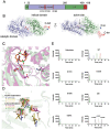
Structure of DncV from V. cholerae responsible for synthesizing a hybrid cGAMP molecule. (A) Schematic diagram of domain organization of DncV in primary sequence. The catalytic domain, in which the three catalytic residues are highlighted, and the helical domain (HD) followed by the C-terminal tail (CT) are colored as blue, green and red, respectively. The numbers for the residues are labeled in the upper panel. (B) The overall structure of DncV. The molecule for DncV is shown in a cartoon representation. The catalytic domain, helical domain and the C-terminal tail are colored in blue, green and red, respectively. Three key residues expected for catalytic reaction are shown as colored sticks. (C) Active site of DncV. Comparison of the active sites of DncV and active murine cGAS (PDB code: 4K9B). The polypeptides of DncV and active cGAS were shown as pale green and purple cartoons. The catalytic acidic residues from two structures were represented as colored sticks. A cGAMP molecule, which was modeled from the complex structure of cGAS, was shown as a colored stick diagram. Three secondary structures, α7, α8 and α9, of DncV were labeled out. (D) Comparison of the key residues of active sites from DncV, inactive (PDB code: 4K8V) and active (PDB code: 4K9B) murine cGAS, inactive (PDB code: 4JLX) and active porcine cGAS (PDB code: 4KB6), inactive porcine (PDB code: 1PX5) and active human OAS1 (PDB code: 4IG8), and inactive human cGAS (PDB code: 4KM5). (E) Key residues responsible for DncV activity. DncV-catalyzed reaction products and commercial standards of 3 cyclic di-nucleotides were fractionated by RP-HPLC. Wild-type (240 μM) and mutant DncVs (240 μM) were incubated with ATP (1 mM), GTP (1 mM), or ATP+GTP (1 mM for each) at 37 °C for 60 min and the products were fractionated by RP-HPLC. Red, blue and green denote products derived from reactions using ATP, GTP, and ATP+GTP as substrates, respectively. The elution peaks for chemical standards c-di-GMP, c-di-AMP and cGAMP are shown in the last panel. x axis represents the retention time in the C18 column. Please note that wt-DncV-catalyzed reaction products were uniformly eluted a bit earlier than the corresponding chemical standards, which might be caused by the heating treatment during the preparation of the product samples.

References

    1. Danilchanka O, Mekalanos JJ. Cell. 2013. pp. 962–970. - PMC - PubMed
    1. Davies BW, Bogard RW, Young TS, et al. Cell. 2012. pp. 358–370. - PMC - PubMed
    1. Sun L, Wu J, Du F, et al. Science. 2012. pp. 786–791. - PMC - PubMed
    1. Wu J, Sun L, Chen X, et al. Science. 2013. pp. 826–830. - PMC - PubMed
    1. Civril F, Deimling T, de Oliveira Mann CC, et al. Nature. 2013. pp. 332–337. - PMC - PubMed

Publication types

MeSH terms

Substances

LinkOut - more resources