. 2014 Oct:127:280-7.
doi: 10.1016/j.exer.2014.01.001.

A protective eye shield for prevention of media opacities during small animal ocular imaging

Affiliations

A protective eye shield for prevention of media opacities during small animal ocular imaging

Brent A Bell et al. Exp Eye Res. 2014 Oct.

Abstract

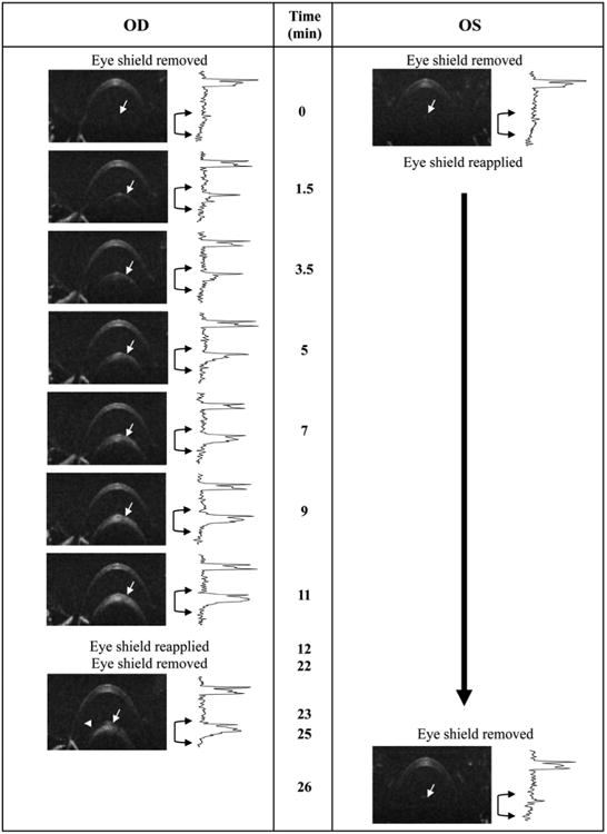
Optical coherence tomography (OCT), scanning laser ophthalmoscopy (SLO) and other non-invasive imaging techniques are increasingly used in eye research to document disease-related changes in rodent eyes. Corneal dehydration is a major contributor to the formation of ocular opacities that can limit the repeated application of these techniques to individual animals. General anesthesia is usually required for imaging, which is accompanied by the loss of the blink reflex. As a consequence, the tear film cannot be maintained, drying occurs and the cornea becomes dehydrated. Without supplemental hydration, structural damage to the cornea quickly follows. Soon thereafter, anterior lens opacities can also develop. Collectively these changes ultimately compromise image quality, especially for studies involving repeated use of the same animal over several weeks or months. To minimize these changes, a protective shield was designed for mice and rats that prevent ocular dehydration during anesthesia. The eye shield, along with a semi-viscous ophthalmic solution, is placed over the corneas as soon as the anesthesia immobilizes the animal. Eye shields are removed for only the brief periods required for imaging and then reapplied before the fellow eye is examined. As a result, the corneal surface of each eye is exposed only for the time required for imaging. The device and detailed methods described here minimize the corneal and lens changes associated with ocular surface desiccation. When these methods are used consistently, high quality images can be obtained repeatedly from individual animals.

Keywords: anterior segment; cataract; imaging; media; opacity; protective eye shield.


Figures

Figure 1
Figure 1
Corneal changes imaged by digital macro photography (Fig. 1a-c,f-g), SLO (Fig. 1d) and OCT (Fig 1e) in a Spraque-Dawley (SD) rat anesthetized using a combination of Ketamine (150mg/kg) and Xylazine (12mg/kg). Standard (no flash) macro photography of a recently anesthetized SD rat collected under ambient room lighting with normal cornea surface morphology (Fig. 1a). The two reflections present at 12 and 2 o'clock position are reflections from the fluorescent lamp fixtures located on the laboratory ceiling. Flash macro photography of the same rat before (Fig. 1b) and after (Fig. 1c) developing corneal perturbations (black arrows) several minutes after leaving the eye exposed to air (i.e. uncovered and unprotected against dehydration). The flash photography accentuates the corneal atrophy that occurs as a result of anesthesia induction and a lack of ocular protection against corneal tear film evaporation. This same phenomenon can be observed by infrared SLO imaging (Fig.1d) which appears as “pitting” of the corneal surface (white arrows). OCT imaging (1000A-scans/B-scan) of an affected eye (Fig. 1e) shows epithelial and stromal thinning (white arrows) which contributes to a non-uniform surface for light refraction. Imaging instruments like SLO and OCT are highly dependent on corneal refraction for generating images of the posterior segment. OCT image dimensions are 1.5mm (depth) × 6mm (width). Images from the eye of a separate rat (pigmented SD Zucker) showing the influence of corneal perturbations on retinal SLO image quality (Fig 1f-g). A cornea with perturbations causes a lack of image clarity (Fig 1f). After performing a “Refresh-Reset” (see Detailed Methods section) procedure the retinal image quality dramatically improves (Fig 1g) by rehydrating the corneal stroma and epithelial cells as well as smoothing out the tear film.
Figure 2
Figure 2
Engineering drawings and digital color photographs of eye shields designed for mice (Fig. 4a,b) and rats (Fig. 4c,d).
Figure 3
Figure 3
Representative demonstration of the utility of protective eye shields against opacity formation in an anesthetized wild type (C57BL/6J) mouse. In this example the right eye (OD) was exposed to room air for an extended period (∼10 minutes) without supplemental hydration, blinking or ocular protection, while the left eye (OS) remained protected by an eye shield and artificial tears (Systane Ultra). After ∼1.5 minutes of exposure, changes in the anterior lens (white arrows) can be seen in the right eye. More pronounced changes are observed after 3-5 minutes which continue to worsen as time elapses. At 12 minutes the right eye was covered using an eye shield in conjunction with Systane Ultra. Approximately 10 minutes later (22 min elapsed time), the eye shield was removed and the eye reimaged. For comparison, the left eye of the animal was covered with an eye shield throughout the entire duration of the imaging experiment with the right eye and then imaged after an elapsed time of 25 minutes. Beside each OCT image of the anterior segment is an intensity vs. depth profile obtained from the neighboring B-scan image. The profile reveals the scattering signal amplitude as a function of OCT imaging depth. In an opacity free animal, two primary peaks, one thick and the other narrow, are observed which originate from, and correspond to the cornea and lens capsule (white arrows), respectively. As the opacity begins to form an elevation in scattering signal amplitude begins to increase below the lens capsule boundary, which is apparent in the in-depth intensity profiles (black brackets with arrows). OCT B-scans (250A-scans/B-scan) are 1mm (depth) × 5mm (width). Experiment performed under lab environment conditions (75°F, 47% relative humidity) without any supplemental heating to maintain animal body temperature.
Figure 4
Figure 4
Media (anterior lens) opacity changes plotted as a function of time for protected and unprotected eyes in anesthetized wild type mice (n=4). Qualitative imaging data (OCT B-scans converted to in-depth intensity profiles) from Figure 3 was analyzed using an area-under-the-curve (AUC) algorithm to obtain opacity scattering magnitudes from the anterior lens region. Media scattering is shown as AUC magnitude (primary axis) and as percent change (secondary axis) relative to the first imaging time point (t0) collected immediately after removing the eye shield. A steep linear increase in scattering (∼25% min-1, P<0.0001) is observed in the anterior lens of the right eye (“OD exposed”) which stabilizes, and even begins to slightly resolve (n.s.), after an eye shield and artificial tears are applied (“OD covered”). In contrast, the left eye (“OS covered”) remains free of opacity for over 25 minutes when protected by an eye shield. On average, scattering magnitudes in the protected left eye improves by ∼35%, but is a nonsignificant (n.s.) reduction in scattering magnitude relative to the initially recorded baseline levels. Data shown as Mean +/- SD. Ordinary one-way Anova with a Dunnett's multiple comparisons test.
Figure 5
Figure 5
Representative digital color photographs (oblique view taken from the nasal direction) from the eyes of two mice immediately after undergoing the experiment shown in Fig. 3. Right eyes show evidence (blue-gray hue) of media opacities. In contrast, left eyes are completely free (dark with no evidence of the color hue) of opacity.

References

    1. Ocuscience. Electrodes and Accessories: Mini Contact Lenses. Xenotec, Inc; Rolla, MO: 2013.
    1. Bermudez MA, Vicente AF, Romero MC, Arcos MD, Abalo JM, Gonzalez F. Time Course of Cold Cataract Development in Anesthetized Mice. Current eye research. 2011;36:278–284. - PubMed
    1. Calderone L, Grimes P, Shalev M. Acute reversible cataract induced by xylazine and by ketamine-xylazine anesthesia in rats and mice. Experimental eye research. 1986;42:331–337. - PubMed
    1. Charbel Issa P, Singh MS, Lipinski DM, Chong NV, Delori FC, Barnard AR, MacLaren RE. Optimization of in vivo confocal autofluorescence imaging of the ocular fundus in mice and its application to models of human retinal degeneration. Invest Ophthalmol Vis Sci. 2012;53:1066–1075. - PMC - PubMed
    1. Ding C, Wang P, Tian N. Effect of general anesthetics on IOP in elevated IOP mouse model. Experimental eye research. 2011;92:512–520. - PMC - PubMed

Publication types

MeSH terms

LinkOut - more resources