Effect of Class III bone anchor treatment on airway
- PMID: 25245416
- PMCID: PMC8611741
- DOI: 10.2319/041614-282.1
Effect of Class III bone anchor treatment on airway
Abstract
Objectives: To compare airway volumes and minimum cross-section area changes of Class III patients treated with bone-anchored maxillary protraction (BAMP) versus untreated Class III controls.
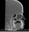
Materials and methods: Twenty-eight consecutive skeletal Class III patients between the ages of 10 and 14 years (mean age, 11.9 years) were treated using Class III intermaxillary elastics and bilateral miniplates (two in the infra-zygomatic crests of the maxilla and two in the anterior mandible). The subjects had cone beam computed tomographs (CBCTs) taken before initial loading (T1) and 1 year out (T2). Twenty-eight untreated Class III patients (mean age, 12.4 years) had CBCTs taken and cephalograms generated. The airway volumes and minimum cross-sectional area measurements were performed using Dolphin Imaging 11.7 3D software. The superior border of the airway was defined by a plane that passes through the posterior nasal spine and basion, while the inferior border included the base of the epiglottis to the lower border of C3.
Results: From T1 to T2, airway volume from BAMP-treated subjects showed a statistically significant increase (1499.64 mm(3)). The area in the most constricted section of the airway (choke point) increased slightly (15.44 mm(2)). The airway volume of BAMP patients at T2 was 14136.61 mm(3), compared with 14432.98 mm(3) in untreated Class III subjects. Intraexaminer correlation coefficients values and 95% confidence interval values were all greater than .90, showing a high degree of reliability of the measurements.
Conclusion: BAMP treatment did not hinder the development of the oropharynx.
Keywords: Airway; Class III; Skeletal anchorage.
Figures


References
-
- Ishii H, Morita S, Takeuchi Y, et al. Treatment effect of combined maxillary protraction and chincap appliance in severe skeletal Class III cases. Am J Orthod Dentofacial Orthop. 1987;92:304–312. - PubMed
-
- Allwright WC, Burndred WH. A survey of handicapping dentofacial anomalies among Chinese in Hong Kong. Int Dent J. 1964;14:505–519.
-
- Chen L, Chen R, Yang Y, Ji G, Shen G. The effects of maxillary protraction and its long-term stability—a clinical trial in Chinese adolescents. Eur J Orthod. 2012;34:88–95. - PubMed
Publication types
MeSH terms
Grants and funding
LinkOut - more resources
Full Text Sources
Other Literature Sources
Miscellaneous

