. 2014 Oct 15;5(19):8893-905.
doi: 10.18632/oncotarget.2360.

MUC1-C confers EMT and KRAS independence in mutant KRAS lung cancer cells

Affiliations

MUC1-C confers EMT and KRAS independence in mutant KRAS lung cancer cells

Akriti Kharbanda et al. Oncotarget. .

Abstract

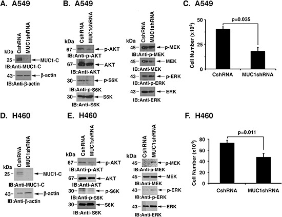
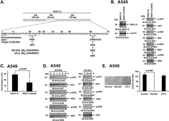
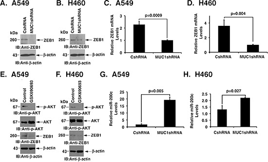
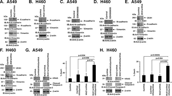
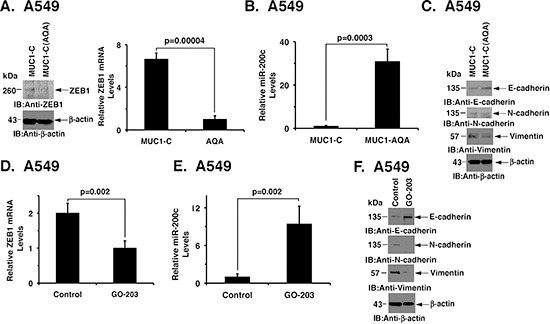
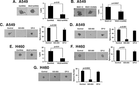
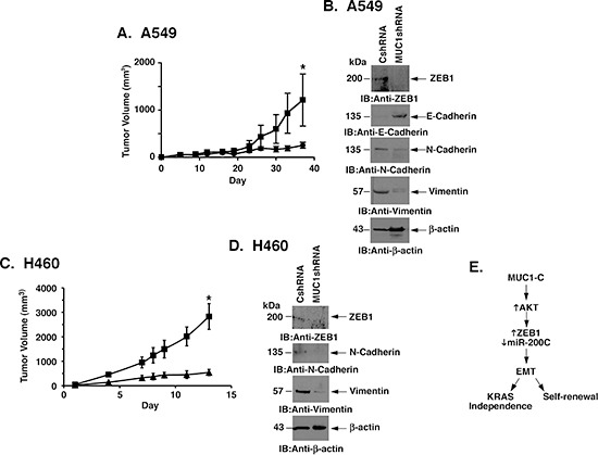
Non-small cell lung cancers (NSCLCs) that harbor an oncogenic KRAS mutation are often associated with resistance to targeted therapies. The MUC1-C transmembrane protein is aberrantly overexpressed in NSCLCs and confers a poor outcome; however, the functional role for MUC1-C in mutant KRAS NSCLC cells has remained unclear. The present studies demonstrate that silencing MUC1-C in A549/KRAS(G12S) and H460/KRAS(Q61H) NSCLC cells is associated with downregulation of AKT signaling and inhibition of growth. Overexpression of a MUC1-C(CQC→AQA) mutant, which inhibits MUC1-C homodimerization and function, suppressed both AKT and MEK activation. Moreover, treatment with GO-203, an inhibitor of MUC1-C homodimerization, blocked AKT and MEK signaling and decreased cell survival. The results further demonstrate that targeting MUC1-C suppresses expression of the ZEB1 transcriptional repressor by an AKT-mediated mechanism, and in turn induces miR-200c. In concert with these effects on the ZEB1/miR-200c regulatory loop, targeting MUC1-C was associated with reversal of the epithelial-mesenchymal transition (EMT) and inhibition of self-renewal capacity. Loss of MUC1-C function also attenuated KRAS independence and inhibited growth of KRAS mutant NSCLC cells as tumors in mice. These findings support a model in which targeting MUC1-C inhibits mutant KRAS signaling in NSCLC cells and thereby reverses the EMT phenotype and decreases self-renewal.


Conflict of interest statement

Conflict of Interest Disclosure

D.K. holds equity in Genus Oncology and is a consultant to the company. The other authors disclosed no potential conflicts of interest.

Figures

Figure 1
Figure 1. Silencing MUC1-C downregulates AKT and inhibits NSCLC cell growth
(A) A549 cells were stably infected with lentiviruses expressing a control scrambled shRNA (CshRNA) or a MUC1 shRNA. Lysates were immunoblotted with the indicated antibodies. (B) Lysates from A549/CshRNA and A549/MUC1shRNA cells were immunoblotted with the indicated antibodies. (C) A549/CshRNA and A549/MUC1shRNA cells were plated at 5 × 10⁴ cells/well. The results (mean±SD of three replicates) are expressed as cell number on day 4. (D) H460 cells were stably infected with lentiviruses expressing CshRNA or MUC1shRNA. Lysates were immunoblotted with the indicated antibodies. (E) Lysates from H460/CshRNA and H460/MUC1shRNA cells were immunoblotted with the indicated antibodies. (F) H460/CshRNA and H460/MUC1shRNA cells were plated at 5 × 10⁴ cells/well. The results (mean±SD of three replicates) are expressed as cell number on day 4.
Figure 2
Figure 2. Targeting MUC1-C function suppresses AKT and MEK→ERK signaling
(A) Schema of the MUC1-C subunit with the 58 aa extracellular domain (ED), 28 aa transmembrane domain (TM) and sequence of the 72 aa cytoplasmic domain (CD). The CQC motif is necessary for MUC1-C homodimerization and is the target for GO-203 treatment. Also highlighted are the binding sites that link the MUC1-C cytoplasmic domain to activation of the PI3K→AKT and MEK→ERK pathways. (B) A549 cells were stably transfected with vectors expressing MUC1-C or the MUC1-C(CQC→AQA) mutant [designated MUC1-C(AQA)]. Lysates were immunoblotted with the indicated antibodies. (C) A549/MUC1-C and A549/MUC1-C(AQA) cells were plated at 5 × 10⁴ cells/well. The results (mean±SD of three replicates) are expressed as cell number on day 4. (D) A549 cells were treated with 5 μM GO-203 at 0 and 24 h. Lysates were immunoblotted with the indicated antibodies (left and right). (E) A549 cells were seeded at 1000 cells/well in 6-well plates and left untreated (Control) or treated with 5 μM GO-203 or 5 μM CP-2 each day for 4 days. Colonies were stained with crystal violet on day 15 after treatment (left). Colony number (>30 cells) is expressed as the mean±SD of three replicates (right).
Figure 3
Figure 3. Silencing MUC1-C confers the coordinate downregulation of ZEB1 and induction of miR-200c expression
(A and B) Lysates from A549 (A) and H460 (B) cells expressing CshRNA or MUC1shRNA were immunoblotted with the indicated antibodies. (C and D) ZEB1 mRNA levels for the indicated A549 (C) and H460 (D) cells were determined by qRT-PCR. The results are expressed as relative ZEB1 mRNA levels (mean±SD of three determinations) as compared to that obtained for GAPDH as a control. (E and F) A549 (E) and H460 (F) cells were left untreated or treated with 10 μM GSK690693 for 48 h. Lysates were immunoblotted with the indicated antibodies. (G and H) Relative miR-200c levels in the indicated A549 (G) and H460 (H) cells were determined by qRT-PCR. The results are expressed as relative miR-200c levels (mean±SD of three determinations) as compared to that obtained for U6 as a control.
Figure 4
Figure 4. Silencing MUC1-C reverses EMT and KRAS independence
(A and B) Lysates from A549 (A) and H460 (B) cells expressing CshRNA or MUC1shRNA were immunoblotted with the indicated antibodies. (C and D) A549 and H460 cells were left untreated or treated with 10 μM GSK690693 for 48 h. Lysates were immunoblotted with the indicated antibodies. (E and F) Lysates from A549 (E) and H460 (F) cells expressing CshRNA or MUC1shRNA were immunoblotted with the indicated antibodies. (G and H) A549/CshRNA and A549/MUC1shRNA (G) or H460/CshRNA and H460/MUC1shRNA (H) cells were infected twice over 24 h with lentivirus expressing a KRAS shRNA. At 48 h post-infection, cells were (i) collected for immunoblotting with the indicated antibodies (left) or (ii) plated at a density of 5 × 10⁴ in a 6-well plate. The results (mean±SD of three replicates) are expressed as percent cell death as determined by trypan blue exclusion on day 4 (right).
Figure 5
Figure 5. Targeting MUC1-C induces MET
(A) Lysates from A549/MUC1-C and A549/MUC1-C(AQA) cells were immunoblotted with the indicated antibodies (left). ZEB1 mRNA levels were determined by qRT-PCR. The results are expressed as relative ZEB1 mRNA levels (mean±SD of three determinations) as compared to that obtained for GAPDH as a control (right). (B) miR-200c levels in A549/MUC1-C and A549/MUC1-C(AQA) cells were determined by qRT-PCR. The results are expressed as relative miR-200c levels (mean±SD of three determinations) as compared to that obtained for U6 as a control. (C) Lysates from A549/MUC1-C and A549/MUC1-C(AQA) were immunoblotted with the indicated antibodies. (D) A549 cells were left untreated or treated with 5 μM GO-203 for 48 h. ZEB1 mRNA levels were determined by qRT-PCR. The results are expressed as relative ZEB1 mRNA levels (mean±SD of three determinations) as compared to that obtained for GAPDH as a control. (E) A549 cells were left untreated or treated with 5 μM GO-203 for 48 h. Relative miR-200c levels were determined by qRT-PCR. The results are expressed as relative miR-200c levels (mean±SD of three determinations) as compared to that obtained for U6 as a control. (F) A549 cells were left untreated or treated with 5 μM GO-203 for 48 h. Lysates were immunoblotted with the indicated antibodies.
Figure 6
Figure 6. MUC1-C is necessary for self-renewal
(A and B) Representative images are shown for the indicated A549 cells plated at 2000 cells/well and grown for 5 days in sphere culture (left). Bar represents 100 microns. The percentage SFE is expressed as the mean±SD of three determinations (right). (C) A549 cells were plated at 2000 cells/well in sphere culture and left untreated (Control) or treated with 5 μM GO-203 or CP-2 for 3 days. Representative images on day 5 are shown for the indicated A549 cells (left). The percentage SFE is expressed as the mean±SD of three determinations (right). (D) A549 cells were plated at 2000 cells/well and cultured for 5 days. The established spheres were then left untreated (Control) or treated with 5 μM GO-203 or CP-2 for 3 days. Representative images are shown for the indicated A549 cells (left). The percentage SFE is expressed as the mean±SD of three determinations (right). (E) Representative images are shown for the indicated H460 cells plated at 1500 cells/well and grown for 5 days in sphere culture (left). Bar represents 100 microns. The percentage SFE is expressed as the mean±SD of three determinations (right). (F) H460 cells were plated at 1500 cells/well in sphere culture and left untreated (Control) or treated with 5 μM GO-203 or CP-2 for 3 days. Representative images on day 5 are shown for the indicated H460 cells (left). The percentage SFE is expressed as the mean±SD of three determinations (right). (G) H460 cells were plated at 1500 cells/well and cultured for 5 days. The established spheres were then left untreated (Control) or treated with 5 μM GO-203 or CP-2 for 3 days. Representative images are shown for the indicated H460 cells (left). The percentage SFE is expressed as the mean±SD of three determinations (right).
Figure 7
Figure 7. MUC1-C promotes mutant KRAS NSCLC cell tumorigenicity
(A) A549/CshRNA (squares) and A549/ MUC1shRNA (circles) cells (4 × 10⁶) were injected subcutaneously in the flanks of female nude mice. Tumor volumes were determined on the indicated days after injection. The results are expressed as tumor volumes (mean±SEM for 3 mice). The asterisk denotes a significant difference (p=0.02) between growth of the A549/CshRNA and A549/MUC1shRNA tumors on day 38. (B) Lysates from tumors isolated on day 30 from mice in the different treatment groups were immunoblotted with indicated antibodies. (C) H460/CshRNA (squares) and H460/MUC1shRNA (triangles) cells (4 × 10⁶) were injected subcutaneously in the flanks of female nude mice. Tumor volumes were determined on the indicated days after injection. The results are expressed as tumor volumes (mean±SEM for 3 mice). The asterisk denotes a significant difference (p=0.013) between growth of the H460/CshRNA and H460/MUC1shRNA tumors on day 13. (D) Lysates from tumors isolated on day 13 from mice in the different treatment groups were immunoblotted with indicated antibodies. (E) Schema depicting the proposed pathway in which MUC1-C activates AKT and thereby the coordinate induction of ZEB1 and suppression of miR-200c. In turn, MUC1-C drives EMT, self-renewal and KRAS independence.

References

    1. Pylayeva-Gupta Y, Grabocka E, Bar-Sagi D. RAS oncogenes: weaving a tumorigenic web. Nat Rev Cancer. 2011;11:761–774. - PMC - PubMed
    1. Engelman JA, Chen L, Tan X, Crosby K, Guimaraes AR, Upadhyay R, Maira M, McNamara K, Perera SA, Song Y, Chirieac LR, Kaur R, Lightbown A, et al. Effective use of PI3K and MEK inhibitors to treat mutant Kras G12D and PIK3CA H1047R murine lung cancers. Nat Med. 2008;14:1351–1356. - PMC - PubMed
    1. Barbie DA, Tamayo P, Boehm JS, Kim SY, Moody SE, Dunn IF, Schinzel AC, Sandy P, Meylan E, Scholl C, Frohling S, Chan EM, Sos ML, et al. Systematic RNA interference reveals that oncogenic KRAS-driven cancers require TBK1. Nature. 2009;462:108–112. - PMC - PubMed
    1. Kumar MS, Hancock DC, Molina-Arcas M, Steckel M, East P, Diefenbacher M, Armenteros-Monterroso E, Lassailly F, Matthews N, Nye E, Stamp G, Behrens A, Downward J. The GATA2 transcriptional network is requisite for RAS oncogene-driven non-small cell lung cancer. Cell. 2012;149:642–855. - PubMed
    1. Puyol M, Martin A, Dubus P, Mulero F, Pizcueta P, Khan G, Guerra C, Santamaria D, Barbacid M. A synthetic lethal interaction between K-Ras oncogenes and Cdk4 unveils a therapeutic strategy for non-small cell lung carcinoma. Cancer Cell. 2010;18:63–73. - PubMed

Publication types

MeSH terms