. 2014 Sep 23:11:165.
doi: 10.1186/s12974-014-0165-8.

α1-antitrypsin modulates microglial-mediated neuroinflammation and protects microglial cells from amyloid-β-induced toxicity

α1-antitrypsin modulates microglial-mediated neuroinflammation and protects microglial cells from amyloid-β-induced toxicity

Maike Gold et al. J Neuroinflammation. .

Abstract

Background: One hallmark of Alzheimer disease is microglial activation. Therapeutic approaches for this neurodegenerative disease include the modulation of microglial cells. α1-antitrypsin (A1AT) has been shown to exert anti-inflammatory effects on macrophages and lung epithelial cells and an inhibition of calpain activity in neutrophil granulocytes. Nothing is known about the effect of A1AT on microglial-mediated neuroinflammation. Our aim was to investigate the effect of A1AT on amyloid-β (Aβ)- and LPS-treated microglial cells in vitro with respect to cytokine production, stress pathways, cell viability, phagocytotic abilities and the underlying mechanisms.

Methods: Primary microglial cells were isolated from Swiss Webster mouse embryos on embryonic day 13.5. Cytokines in the supernatants of treated primary microglial cells were analyzed with ELISAs, and accumulated nitrite was detected with Griess reagents. Intracellular stress pathways were investigated in cell lysates using western blotting. Intracellular calcium levels were detected in BV-2 microglial cells loaded with the Ca2+-sensitive (fluorescent) dye Fluo-4. Calpain activity in primary microglial cells was assessed by using a calpain activity assay. Cell viability of Aβ-treated microglial cells was analyzed using MTT assay. Phagocytosis of Aβ was evaluated with western blot analysis.

Results: Upon co-administration, A1AT reduced pro-inflammatory mediators induced by LPS or Aβ. Interestingly, we detected a reduction in calpain activity and in the concentration of intracellular calcium that might mediate the anti-inflammatory effects of A1AT. Inhibition of the classic activation pathways, such as phosphorylation of mitogen-activated protein kinases or activation of protein kinase A were excluded as a mechanism of A1AT-mediated effects. In addition, A1AT increased the viability of Aβ-treated microglial cells and reduced Aβ phagocytosis.

Conclusions: We provide evidence on the mechanism of action of A1AT on microglial-mediated neuroinflammation in vitro. Our in vitro data indicate that A1AT treatment modulates microglial cells in inflammatory conditions and that this modulation is due to an inhibition of calpain activity and intracellular calcium levels. The underlying mechanisms of the effects observed here are promising for future therapeutic strategies and should thus be further pursued in transgenic mouse models of Alzheimer disease.


Figures

Figure 1
Figure 1
α 1 -antitrypsin (A1AT) reduces lipopolysaccharide (LPS)-induced release of pro-inflammatory mediators. A-C. Cell culture supernatants of LPS and/or A1AT co-treated cells were subjected to ELISA to detect the levels of pro-inflammatory cytokines. Therefore, primary microglial cells were treated with 0.1 μg/ml LPS and/or 2 and 4 mg/ml A1AT for 24 hours, followed by quantification of TNF-α, IL-6, and IL-1β levels. Values are given in ng/ml. Means and standard deviations of the mean of three independent experiments are shown (*P value ≤ 0.05, **P value ≤ 0.01, n.s., not significant). D. Nitric oxide (NO) was measured in the same cell culture supernatants using Griess reagents. Values are given in μM. Means and standard deviations of the mean of three independent experiments are shown (*P value ≤ 0.05, **P value ≤ 0.01).
Figure 2
Figure 2
α 1 -antitrypsin (A1AT)’s reduction of pro-inflammatory mediators is independent of MAPK and PKA activation. A. To detect the phosphorylation state of the MAPKs p38, p44/42 and JNK upon co-treatment with A1AT, primary microglial cells were incubated with 0.1 μg/ml lipopolysaccharide (LPS) and 2 and 4 mg/ml A1AT for 20 minutes, and cell lysates of the treated cells were subjected to western blotting. Antibodies against phospho-p38, phospho-p44/42 and phospho-JNK were used. GAPDH served as a loading control. One representative western blot out of three is shown. B. Band intensities of phosphorylated MAPK were measured by a densitometer and normalized with that of GAPDH. LPS-treated cells were set to 1. Means and standard deviations of the mean of three western blots are shown (*P value ≤ 0.05, **P value ≤ 0.01) C. To detect the effect of A1AT on the phosphorylation state of CREB, cells were pretreated with the PKA inhibitor H89 for 30 minutes and then treated with 4 mg/ml A1AT for 15 minutes or co-treated with 0.1 μg/ml LPS and 4 mg/ml A1AT for 15 minutes. Cell lysates were subjected to western blotting, and phospho-CREB was detected with a monoclonal antibody. Vinculin was used as a loading control. One representative western blot out of three is shown. D. To measure the impact on the release of the pro-inflammatory cytokine TNF-α, cells were pretreated with different concentrations of H89 and then treated with 0.1 μg/ml LPS and/or 2 mg/ml A1AT for 24 hours. TNF-α levels in cell culture supernatants of primary microglial cells were quantified with ELISA. Results were normalized to LPS-treated cells. Means and standard deviations of the mean of three independent experiments are shown.
Figure 3
Figure 3
α 1 -antitrypsin (A1AT) reduces intracellular calcium levels and calpain activity in microglial cells. To detect intracellular calcium levels, BV-2 microglial cells were loaded with Fluo-4 and baseline calcium levels for each well were measured and subtracted from the values. Cells were treated with 0.1 μg/ml lipopolysaccharide (LPS) and/or 2 and 4 mg/ml A1AT, and the course of intracellular calcium was measured over 200 minutes. RFU (relative fluorescence units) were normalized to time point zero. A shows the course of the values for LPS and LPS + 4 mg/ml A1AT co-treated cells. One representative experiment out of three is shown. B shows the mean values and standard deviations of the mean of the RFU of six wells after 100 minutes for LPS and/or A1AT-treated cells (***P value ≤ 0.001). C. Calpain activity was detected 24 hours after A1AT treatment with different concentrations of A1AT by using a calpain activity assay. Calpain activity of untreated cells was referred to as 100% activity. Means and standard deviations of the mean of three independent experiments are shown (*P value ≤ 0.05, **P value ≤ 0.01).
Figure 4
Figure 4
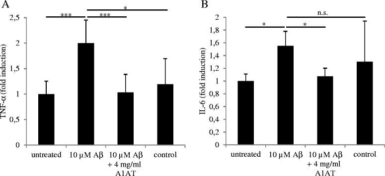
α 1 -antitrypsin (A1AT) reduces Aβ-induced release of TNF-α. ELISA measurements were performed to detect the amount of the pro-inflammatory cytokines TNF-α (A) and IL-6 (B) in supernatants of Aβ1-42 oligomer and/or A1AT co-treated primary microglial cells 24 hours after treatment. Results were normalized to basal cytokine levels of untreated cells. Experiments were performed at least three times independently. Means and standard deviations of the mean of three independent experiments are shown (*P value ≤0.05, ***P value ≤0.001, n.s., not significant).
Figure 5
Figure 5
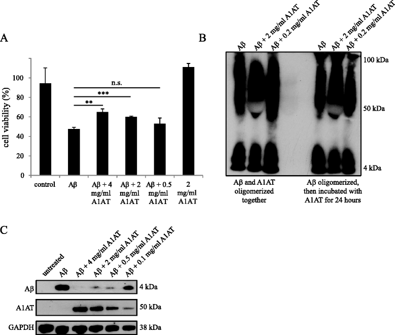
α 1 -antitrypsin (A1AT) treatment reduces Aβ oligomer-induced cytotoxicity on primary microglial cells. A. Primary microglial cells were treated with 2 μM Aβ1-42 oligomers and/or different concentrations of A1AT for 24 hours. An MTT-assay was performed to detect the cell viability. Viability of untreated cells was referred to as 100% viability. Means and standard deviations of the mean of three independent experiments are shown (**P value ≤ 0.01, ***P value ≤ 0.001, n.s., not significant). B. 58 μM Aβ preparations were oligomerized and the effect of A1AT on the oligomerization process was assessed by western blotting with the monoclonal Aβ antibody 6E10. One representative western blot out of three is shown. C. Uptake of Aβ1-42 oligomers under the influence of different concentrations of A1AT was investigated by western blotting. Cells were treated with 2 μM Aβ and different concentrations of A1AT (4 mg/ml to 0.1 mg/ml) for three hours. We used the monoclonal antibody 6E10 to detect Aβ and a polyclonal antibody to detect A1AT in cell lysates. GAPDH was used as loading control. One representative western blot out of three is shown.

References

    1. Braak H, Braak E. Alzheimer’s disease: striatal amyloid deposits and neurofibrillary changes. J Neuropathol Exp Neurol. 1990;49:215–224. doi: 10.1097/00005072-199005000-00003. - DOI - PubMed
    1. Walter S, Letiembre M, Liu Y, Heine H, Penke B, Hao W, Bode B, Manietta N, Walter J, Schulz-Schuffer W, Fassbender K. Role of the toll-like receptor 4 in neuroinflammation in Alzheimer's disease. Cell Physiol Biochem. 2007;20:947–956. doi: 10.1159/000110455. - DOI - PubMed
    1. Cunningham C. Microglia and neurodegeneration: the role of systemic inflammation. Glia. 2013;61:71–90. doi: 10.1002/glia.22350. - DOI - PubMed
    1. Cho S, Kim Y, Cruz MO, Park EM, Chu CK, Song GY, Joh TH. Repression of proinflammatory cytokine and inducible nitric oxide synthase (NOS2) gene expression in activated microglia by N-acetyl-O-methyldopamine: protein kinase A-dependent mechanism. Glia. 2001;33:324–333. doi: 10.1002/1098-1136(20010315)33:4<324::AID-GLIA1031>3.0.CO;2-M. - DOI - PubMed
    1. Akiyama H, Barger S, Barnum S, Bradt B, Bauer J, Cole GM, Cooper NR, Eikelenboom P, Emmerling M, Fiebich BL, Finch CE, Frautschy S, Griffin WS, Hampel H, Hull M, Landreth G, Lue L, Mrak R, Mackenzie IR, McGeer PL, O'Banion MK, Pachter J, Pasinetti G, Plata-Salaman C, Rogers J, Rydel R, Shen Y, Streit W, Strohmeyer R, Tooyoma I, et al. Inflammation and Alzheimer's disease. Neurobiol Aging. 2000;21:383–421. doi: 10.1016/S0197-4580(00)00124-X. - DOI - PMC - PubMed