. 2015:1189:135-62.
doi: 10.1007/978-1-4939-1164-6_10.

3D culture assays of murine mammary branching morphogenesis and epithelial invasion

Affiliations

3D culture assays of murine mammary branching morphogenesis and epithelial invasion

Kim-Vy Nguyen-Ngoc et al. Methods Mol Biol. 2015.

Abstract

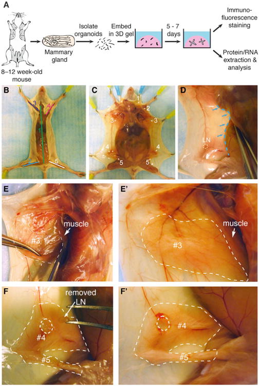
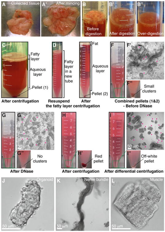
Epithelia are fundamental tissues that line cavities, glands, and outer body surfaces. We use three-dimensional (3D) embedded culture of primary murine mammary epithelial ducts, called "organoids," to recapitulate in days in culture epithelial programs that occur over weeks deep within the body. Modulating the composition of the extracellular matrix (ECM) allows us to model cell- and tissue-level behaviors observed in normal development, such as branching morphogenesis, and in cancer, such as invasion and dissemination. Here, we describe a collection of protocols for 3D culture of mammary organoids in different ECMs and for immunofluorescence staining of 3D culture samples and mammary gland tissue sections. We illustrate expected phenotypic outcomes of each assay and provide troubleshooting tips for commonly encountered technical problems.


Figures

Fig. 1
Fig. 1
Collection of mouse mammary glands for organoid isolation and 3D culture. (a) Schematic description of isolation and 3D culture of mouse mammary organoids. (b) Scheme for surgically accessing the mammary glands. Numbers indicate the order of cuts. (c) Locations of the ten mammary glands. (d) Expose glands #3, #4, and #5 by pushing back the abdomen (blue dotted line) with the back of the Graefe forceps. (ee′) A thin layer of muscle partially covers gland #3 (e) and should be pushed back before dissection (e′). Dotted line in (e′) indicates the region of gland #3 to be collected. (f) Use the Graefe forceps to pluck out the lymph node in gland #4. Dotted line in (f′) indicates the approximate region of glands #4 and #5 to be collected (Color figure online)
Fig. 2
Fig. 2
Mammary organoid isolation. (aa′) Collected mammary glands are pooled in a Petri dish (a) and minced until the tissue relaxes, typically 25–50 cuts (a′). (bb″) Incubation in collagenase solution breaks up the fat pad (b) into smaller pieces that are relatively dispersed (b′). Too long of a digestion (b″) will cause organoids to be too small and not grow well. (c) Following incubation in collagenase solution, centrifugation separates the suspension into three layers, with a top opaque layer of fat and a pellet (#1) of epithelium and stroma. (d) The fatty layer is transferred to a new tube and resuspended in 10 mL DMEM/F12. (e) Centrifugation of the dispersed fatty layer recovers additional epithelium in the pellet (#2). (ff″) The combined pellets from (c) and (e) are resuspended in 4 mL DMEM/F12 with DNase (f). Before DNase treatment, organoids (pink arrowheads) are loosely attached to each other and to stromal cells (f′), forming visible clusters in the tube (f″). (gg″) DNase treatment causes organoids (pink arrowheads) to detach from one another (g′) and the clusters to disappear (g″). (hh′) Centrifugation of the suspension in (g) results in a compact red pellet (h′). (ii″) Differential centrifugation removes single cells from the suspension (i′) and results in an off-white pellet of purified epithelial organoids (i″). Organoids (pink arrowheads) may appear rounded and small or more elongated and even branched (i′). Larger organoids typically survive and branch more efficiently in our assays. (j) Close-up view of an organoid. (kl) Non-epithelial tissues can be observed in the final suspension, including nerve bundles (k) and muscle (l) (Color figure online)
Fig. 3
Fig. 3
Precoating tubes and pipette tips with BSA. Fresh tissue can adhere to uncoated plastic surfaces, and this protocol involves many pipetting steps. Accordingly, it is essential to precoat the plastic surfaces with BSA solution to maximize final organoid yield. (a) Precoat a 15 mL tube by filling the tube with BSA solution, inverting the tube to precoat the cap, and removing the BSA solution. (b) Precoat a 10 mL pipette tip by taking up BSA solution to fill the entire pipette and ejecting back out. (c, d) Use the same approach to precoat a microcentrifuge tube (c) and a small pipette tip (d) with BSA solution
Fig. 4
Fig. 4
Setting up the tissue culture hood for plating. (a) Sample layout of reagents, tools, and equipment used for plating 3D culture samples. (b) Heating block setup for plating in 2-well or 4-well chambers. (c, c′) To plate in a 24-well dish, remove one of the blocks from the heating block (c) to establish direct contact between the remaining block and the plate bottom (c′)
Fig. 5
Fig. 5
Plating organoids in 3D Matrigel and collagen I. (a) Schematic description of plating organoids in Matrigel. (bb′) Schematic description of preparing preassembled collagen I (b), which can be used alone or mixed with Matrigel (b′). (c1c7) Color indicators for the pH of the collagen I solution during neutralization. (d1d6) Decreasing transparency of the collagen I solution during preincubation on ice. (ee′) Schematic description of plating organoids in 3D collagen I or in a mixture of Matrigel and collagen I. (e) Shows a top view for making an underlay on the cover glass. (e′) Shows a side view of how to plate the organoid/collagen I suspension on top of the gelled underlay. (ff′) Representative DIC images of collagen I fibers at low (f) and high (f′) magnification (Color figure online)
Fig. 6
Fig. 6
3D organotypic culture assays. (a) Schematic description of four assays that use different extracellular matrix compositions to model specific epithelial behaviors. (be) Representative frames of DIC time-lapse movies showing cyst formation in Matrigel in basal medium (b), branching morphogenesis in Matrigel induced by FGF2 (c), branching morphogenesis in a mixture of Matrigel and collagen I induced by FGF2 (d), and epithelial cell invasion into pure collagen I induced by FGF2 (e)
Fig. 7
Fig. 7
Phenotypic variability in assay outcomes. (a) Schematic description of a cyst. (a1a4) DIC images showing variation in cyst morphology. (a4′) An inset of (a4) showing a smooth basal surface with Matrigel. (b) Schematic description of a stratified, unbranched organoid. (b1b4) DIC images showing examples of stratified, unbranched organoids in Matrigel. (b4′) An inset of (b4) showing a smooth basal surface with Matrigel. (c) Schematic description of a branched organoid in Matrigel. (c1c4) DIC images showing variation in branching morphology. (c4′) An inset of (c4) showing a smooth basal surface with Matrigel. (d) Schematic description of a branched organoid in a mixture of Matrigel and collagen I. (d1d4) DIC images showing variation in branching morphology. (d4′) An inset of (d4) showing a smooth basal surface with the mixed matrix. (e) Schematic description of an organoid with protrusive tips in collagen I. (e1e4) DIC images showing variation in protrusive invasion. (e4′) An inset of (e4) showing protrusive tips into collagen I. (f) DIC images showing commonly observed technical issues. (f1f2) Organoids lose their 3D organization in Matrigel (f1) and collagen I gels (f2) when they make contact with the cover glass. (f3) Non-epithelial species (red arrowheads) attached to organoids may appear elongated and mesenchymal (ECM: Matrigel). (f4) A group of dead cells beside a branching organoid (ECM: collagen I). (f5) A cluster of elongated, non-epithelial cells (ECM: Matrigel). (f6) A nerve bundle disseminating single cells into the surrounding matrix (ECM: Matrigel) (Color figure online)
Fig. 8
Fig. 8
Correlation between epithelial morphologies in 3D organotypic assays and in vivo. (ac) Representative confocal images of a cyst in Matrigel (a), branched buds in Matrigel (b), and a stratified, elongating bud in a mixture of Matrigel and collagen I (c). (df) Representative confocal images from mammary gland tissue sections of a bilayered duct (d), a side branch (e), and a terminal end bud (f)

Similar articles

Cited by

References

    1. Sternlicht MD. Key stages in mammary gland development: the cues that regulate ductal branching morphogenesis. Breast Cancer Res. 2006;8:201. - PMC - PubMed
    1. Hogg NA, Harrison CJ, Tickle C. Lumen formation in the developing mouse mammary gland. J Embryol Exp Morphol. 1983;73:39–57. - PubMed
    1. Williams JM, Daniel CW. Mammary ductal elongation: differentiation of myoepithelium and basal lamina during branching morphogenesis. Dev Biol. 1983;97:274–290. - PubMed
    1. Hinck L, Silberstein GB. Key stages in mammary gland development: the mammary end bud as a motile organ. Breast Cancer Res. 2005;7:245–251. - PMC - PubMed
    1. Sternlicht MD, Kouros-Mehr H, Lu P, Werb Z. Hormonal and local control of mammary branching morphogenesis. Differentiation. 2006;74:365–381. - PMC - PubMed

Publication types

MeSH terms