. 2014 Dec;82(12):5086-98.
doi: 10.1128/IAI.02315-14. Epub 2014 Sep 22.

FNR regulates expression of important virulence factors contributing to pathogenicity of uropathogenic Escherichia coli

Affiliations

FNR regulates expression of important virulence factors contributing to pathogenicity of uropathogenic Escherichia coli

Nicolle L Barbieri et al. Infect Immun. 2014 Dec.

Abstract

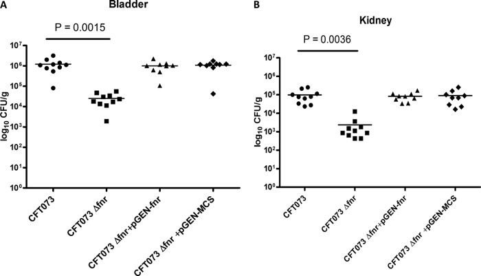
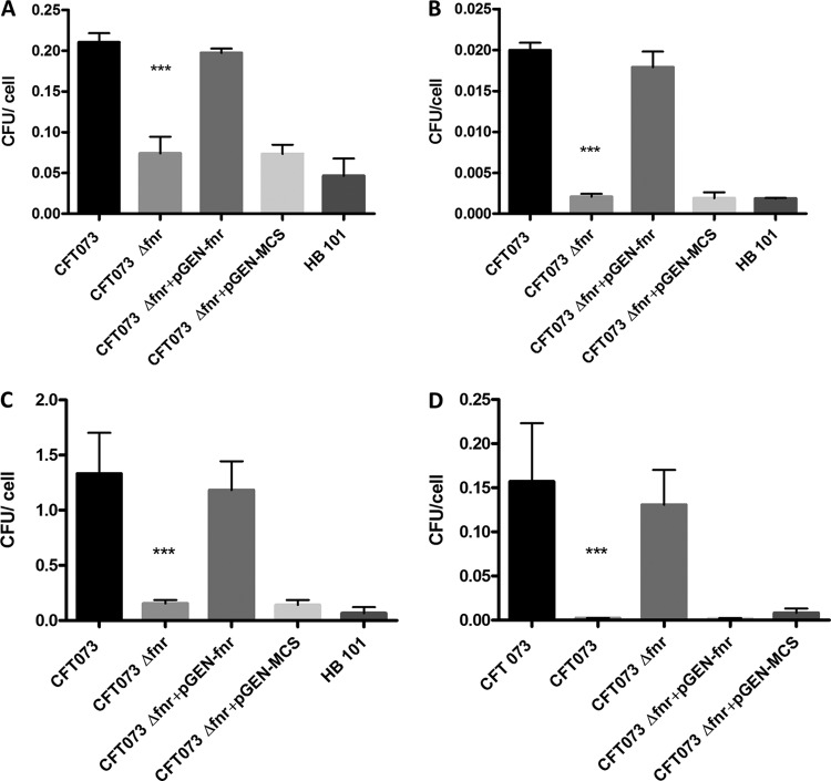
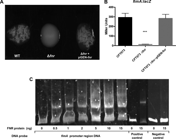
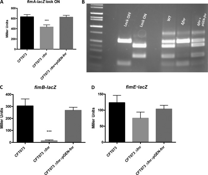
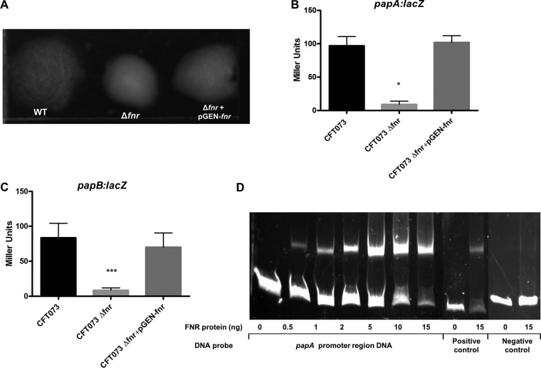
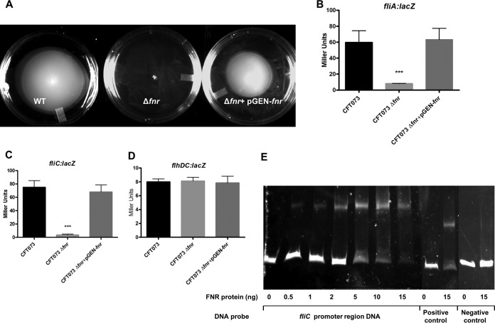
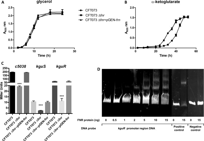
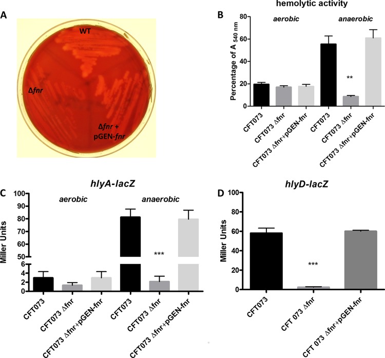
Uropathogenic Escherichia coli (UPEC) is responsible for the majority of urinary tract infections (UTIs), which are some of the world's most common bacterial infections of humans. Here, we examined the role of FNR (fumarate and nitrate reduction), a well-known global regulator, in the pathogenesis of UPEC infections. We constructed an fnr deletion mutant of UPEC CFT073 and compared it to the wild type for changes in virulence, adherence, invasion, and expression of key virulence factors. Compared to the wild type, the fnr mutant was highly attenuated in the mouse model of human UTI and showed severe defects in adherence to and invasion of bladder and kidney epithelial cells. Our results showed that FNR regulates motility and multiple virulence factors, including expression of type I and P fimbriae, modulation of hemolysin expression, and expression of a novel pathogenicity island involved in α-ketoglutarate metabolism under anaerobic conditions. Our results demonstrate that FNR is a key global regulator of UPEC virulence and controls expression of important virulence factors that contribute to UPEC pathogenicity.


Figures

FIG 1
FIG 1
Deletion of fnr attenuates virulence in the mouse model of UTI by UPEC CFT073. The WT and fnr mutant strains, the wild-type strain containing the empty vector (pGEN-MCS), and the mutant strain containing the complementation plasmid (pGEN-fnr) were mixed in a 1:1 ratio and approximately 2 × 10⁹ CFU was transurethrally inoculated into female mice. Two days after infection, the mice were sacrificed and their bladders (A) and kidneys (B) were aseptically removed. WT and fnr mutant bacteria were recovered by plating homogenized tissue samples on LB medium or LB medium containing kanamycin, and CFU counts were determined. The wild-type strain containing the empty vector (pGEN-MCS) and the mutant strain containing the complementation plasmid (pGEN-fnr) were recovered by plating homogenized tissue samples on LB medium with ampicillin or LB medium with both ampicillin and kanamycin. Each dot represents the log10 number of CFU/g in the bladder or kidney from an individual animal, and the detection limit was 1,000 CFU/g. Bars indicate the median log10 number of CFU/g. A two-tailed Wilcoxon matched-pairs test was performed, and the difference in the colonization levels of the WT and mutants was considered statistically significant if P was <0.05.
FIG 2
FIG 2
Adherence to and invasion of the bladder J82 cell line and A498 kidney cell line by CFT073 and its mutants. J82 cells (A and B) and A498 cells (C and D) were infected at an MOI of 10 CFU/cell, as described in Materials and Methods. For the association assays (A and C), cells were lysed at 1 h postinfection, and the extracts were plated onto LB agar for enumeration, For the invasion assays (B and D), at 1 h postinfection, cells were washed with PBS and incubated for a further 3 h in the presence of gentamicin. The cells were then lysed, and the extracts were plated onto LB agar for counting of the CFU. E. coli HB101 was used as a negative control. The values shown are means plus standard deviations for quadruplicate samples from four independent experiments. Significant differences are indicated by asterisks (***, P < 0.0001 compared to the WT and mutant).
FIG 3
FIG 3
UPEC type I fimbria regulation. (A) Yeast agglutination assay with dilutions of the WT (1:16); Δfnr (1:1), and Δfnr/pGEN-fnr (1:16) strains. Bacteria were grown statically for 48 h, agglutination was read after 10 min at room temperature, and the strength of the agglutination was determined by measurement of the titer of serial 2-fold dilutions of the bacterial suspensions in PBS. The experiments were performed four times in quadruplicate. (B) β-Galactosidase activity assay for expression of fimA. fimA-lacZ transcriptional fusion strains were grown statically in LB medium for 48 h at 37°C. β-Galactosidase activity was measured, and the values shown are means plus standard deviations for triplicate samples from three independent experiments. Significant differences are indicated by asterisks (***, P < 0.0001 compared to the WT and mutant). (C) Nonradioactive EMSA of binding of (FnrD154A)2-His6 to the promoter regions. The PCR product of the fimA promoter region was used as the probe at 300 ng per reaction mixture. Purified (FnrD154A)2-His6 fusion protein was added to each reaction mixture at different concentrations, as indicated; ydfZ promoter region DNA probes with and without the FNR protein were used as positive controls, and fimA coding region DNA probes with and without the FNR protein were used as negative controls. DNA fragments were stained with SYBR green.
FIG 4
FIG 4
UPEC type 1 fimbria phase variation. (A) β-Galactosidase activity assay for expression of fimA in lock ON strains. fimA-lacZ transcriptional fusion strains were grown statically in LB medium for 48 h at 37°C. β-Galactosidase activity was measured, and the values shown are means plus standard deviations for triplicate samples from three independent experiments. (B) Phase variation electrophoresis. PCR products were purified, digested with HinfI for 4 h, and analyzed on a 2% agarose gel. fimB-lacZ and fimE-lacZ transcriptional fusion strains were grown statically in LB medium for 48 h at 37°C. (C, D) β-Galactosidase activity assay for expression of fimB (C) and fimE (D). β-Galactosidase activity was measured, and the values shown are means plus standard deviations for triplicate samples from three independent experiments. Significant differences are indicated by asterisks (***, P < 0.0001 compared to the WT and mutant).
FIG 5
FIG 5
UPEC type P fimbria regulation. (A) RBC agglutination assay with dilutions of the WT (1:32); Δfnr (1:1), and Δfnr/pGEN-fnr (1:32) strains. Bacteria were grown statically for 48 h, agglutination was read after 10 min at room temperature, and the strength of the agglutination was determined by measurement of the titer of serial 2-fold dilutions of the bacterial suspensions in PBS. The experiments were performed four times in quadruplicate. (B, C) β-Galactosidase activity assay for expression of papA (B) and papB (C). papA-lacZ and papB-lacZ transcriptional fusion strains were grown in CFA medium overnight without shaking at 37°C. β-Galactosidase activity was measured, and the values shown are means plus standard deviations for triplicate samples from three independent experiments. Significant differences are indicated by asterisks (*, P < 0.05 compared to the WT and mutant; ***, P < 0.0001 compared to the WT and mutant). (D) Nonradioactive EMSA of binding of (FnrD154A)2-His6 to the promoter regions. PCR products of the papA promoter region were used as probes at 300 ng per each reaction mixture. Purified (FnrD154A)2-His6 fusion protein was added at different concentrations to each reaction mixture, as indicated; ydfZ promoter region DNA probes with and without the FNR protein were used as positive controls, and papA coding region DNA probes with and without the FNR protein were used as negative controls. DNA fragments were stained with SYBR green.
FIG 6
FIG 6
Motility regulation. (A) Soft-agar motility assay. Bacterial cultures were stabbed in the middle of each soft-agar plate and incubated at 37°C for 16 h. The experiments were performed four times in quadruplicate. (B to D) β-Galactosidase activity assay for expression of fliA (B), fliC (C), and flhDC (D). fliA-lacZ, fliC-lacZ, and flhDC-lacZ transcriptional fusion strains were grown at 37°C in LB medium with shaking until the OD reached 0.5. β-Galactosidase activity was measured. The values shown are means plus standard deviations for triplicate samples from three independent experiments. Significant differences are indicated by asterisks (***, P < 0.0001 compared to the WT and mutant). (E) Nonradioactive EMSA of binding of (FnrD154A)2-His6 to the promoter regions. PCR products of the fliC promoter region were used as probes at 300 ng per reaction mixture. Purified (FnrD154A)2-His6 fusion protein was added to each reaction mixture at different concentrations, as indicated; ydfZ promoter region DNA probes with and without the FNR protein were used as positive controls, and fliC coding region DNA probes with and without the FNR protein were used as negative controls. DNA fragments were stained with SYBR green.
FIG 7
FIG 7
UPEC α-ketoglutarate metabolism regulation. (A, B) In vitro growth of fnr mutants in M9 medium containing α-ketoglutarate as the sole carbon source (A) or M9 medium containing glycerol as the sole carbon source (B). The optical density of the UPEC CFT073 WT and mutants during growth in M9 medium containing α-ketoglutarate or glycerol as the sole carbon source under anaerobic conditions was determined. Growth curves represent the average measurement at each time point in duplicate from three independent experiments. (C) β-Galactosidase activity assay for expression of c5038, kguS, and kguR (C). c5038-lacZ, kguS-lacZ, and kguR-lacZ transcriptional fusion strains were grown anaerobically at 37°C in M9 medium with α-ketoglutarate overnight. β-Galactosidase activity was measured, and the values shown are means plus standard deviations for triplicate samples from three independent experiments. Significant differences are indicated by asterisks (***, P < 0.0001 compared to the WT and mutant). (D) Nonradioactive EMSA of binding of (FnrD154A)2-His6 to the promoter regions. PCR products of the kguR promoter region were used as probes at 300 ng per reaction mixture. Purified (FnrD154A)2-His6 fusion protein was added to each reaction mixture at different concentrations, as indicated; ydfZ promoter region DNA probes with and without the FNR protein were used as positive controls, and fliC coding region DNA probes with and without the FNR protein were used as negative controls. DNA fragments were stained with SYBR green.
FIG 8
FIG 8
UPEC hemolysin regulation. (A) Blood agar hemolytic assay. Bacteria were inoculated into blood agar plates and grown overnight at 37°C under anaerobic conditions. (B) For the hemolytic activity quantification, bacterial strains were grown in the presence of RBCs overnight at 37°C under aerobic and anaerobic conditions, and the OD540 was recorded; a value of 100% was assigned to RBCs lysed with 1% Triton X-100. The values shown are means plus standard deviations for quadruplicate samples from four independent experiments. (C, D) β-Galactosidase activity assay for expression of hlyA (C) and hlyD (D). hlyA-lacZ strains were grown on blood agar plates overnight at 37°C under aerobic and anaerobic conditions, and hlyD-lacZ transcriptional fusion strains were grown on blood agar plates overnight at 37°C under anaerobic conditions. β-Galactosidase activity was measured, and the values shown are means plus standard deviations for triplicate samples from three independent experiments. Significant differences are indicated by asterisks (**, P < 0.001 compared to the WT and mutant; ***, P < 0.0001 compared to the WT and mutant).

Similar articles

Cited by

References

    1. Lambden PR, Guest JR. 1976. Mutants of Escherichia coli K12 unable to use fumarate as an anaerobic electron acceptor. J. Gen. Microbiol. 97:145–160. 10.1099/00221287-97-2-145. - DOI - PubMed
    1. Guest JR, Green J, Irvine AS, Spiro S. 1996. The FNR modulon and FNR-regulated gene expression, p 317–342 In Lin ECC, Lynch AS. (ed), Regulation of gene expression in Escherichia coli. R. G. Landes & Co, Austin, TX.
    1. Kiley PJ, Beinert H. 1999. Oxygen sensing by the global regulator, FNR: the role of the iron-sulfur cluster. FEMS Microbiol. Rev. 22:341–352. - PubMed
    1. Crack JC, Green J, Cheesman MR, Le Brun NE, Thomson AJ. 2007. Superoxide-mediated amplification of the oxygen-induced switch from [4Fe-4S] to [2Fe-2S] clusters in the transcriptional regulator FNR. Proc. Natl. Acad. Sci. U. S. A. 104:2092–2097. 10.1073/pnas.0609514104. - DOI - PMC - PubMed
    1. Lazazzera BA, Bates DM, Kiley PJ. 1993. The activity of the Escherichia coli transcription factor Fnr is regulated by a change in oligomeric state. Genes Dev. 7:1993–2005. 10.1101/gad.7.10.1993. - DOI - PubMed

Publication types

MeSH terms