. 2014 Sep 22:14:33.
doi: 10.1186/1471-2342-14-33.

Interactive neonatal gastrointestinal magnetic resonance imaging using fruit juice as an oral contrast media

Affiliations

Interactive neonatal gastrointestinal magnetic resonance imaging using fruit juice as an oral contrast media

Owen J Arthurs et al. BMC Med Imaging. .

Abstract

Background: The objective was to evaluate the use of fruit juice with an interactive inversion recovery (IR) MR pulse sequence to visualise the gastrointestinal tract.

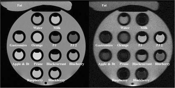
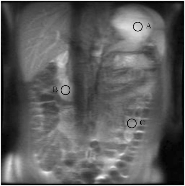
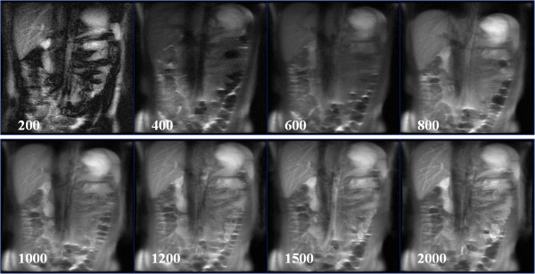
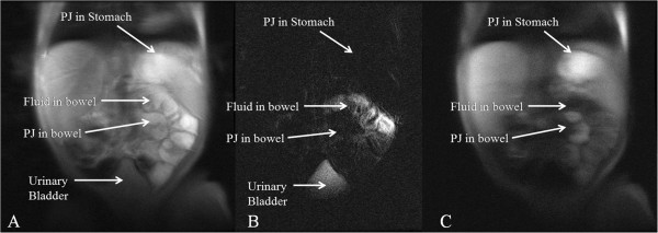
Methods: We investigated the relaxation properties of 12 different natural fruit juices in vitro, to identify which could be used as oral contrast. We then describe our initial experience using an interactive MR pulse sequence to allow optimal visualisation after administering pineapple juice orally, and suppressing pre-existing bowel fluid contents, with variable TI in three adult and one child volunteer.

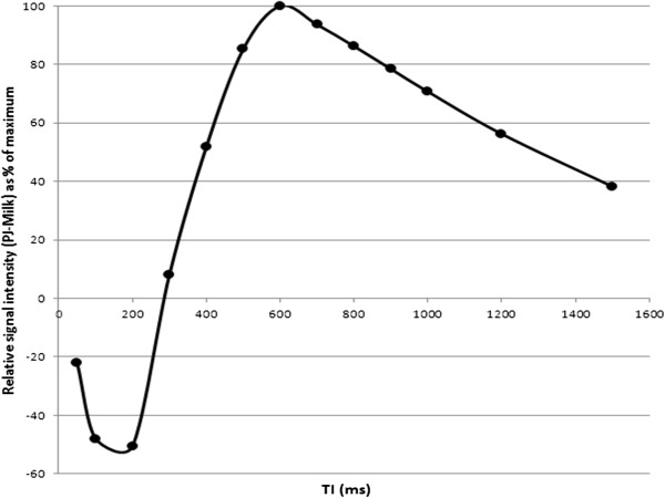
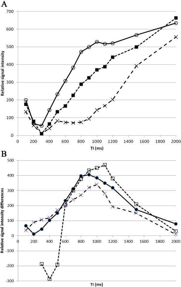
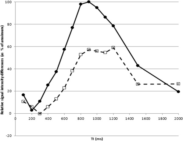
Results: Pineapple juice (PJ) had both the shortest T1 (243 ms) and shortest T2 (48 ms) of the fruit juices tested. Optimal signal differentiation between pre-existing bowel contents and oral PJ administration was obtained with TIs of between 900 and 1100 ms.

Conclusion: The use of an inversion recovery preparation allowed long T1 pre-existing bowel contents to be suppressed whilst the short T1 of fruit juice acts as a positive contrast medium. Pineapple juice could be used as oral contrast agent for neonatal gastrointestinal magnetic resonance imaging.


Figures

Figure 1
Figure 1
12 different contrast media assessed in-vitro. Pineapple juice (PJ and PJ2) have the lowest signal on T2w imaging (left : PDw SSFSE; TR 2500), but highest signal recovery on T1w imaging (right: IR-FSPGR with TI = 640 ms).
Figure 2
Figure 2
Coronal IR-SSFSE images through the abdomen in an adult following oral pineapple juice administration. Signal intensity was measured in 3 regions of interest: the stomach (A), proximal small bowel (duodenum; B) and mid-small bowel (jejunum; C).
Figure 3
Figure 3
By subtracting the signal intensity of PJ and milk, we were able to plot the relative differences in signal (plotted as a % of maximum) between the two contrast media at different TIs. Maximal contrast differences are seen with TI between 500 – 900 ms.
Figure 4
Figure 4
Coronal IR-SSFSE images (TI = 1200 ms) through the abdomen in an adult following oral pineapple juice administration. Examples of different TI times (200 – 2000 ms) are given. Relative signal intensity from this study is given in Figure  5.
Figure 5
Figure 5
A. Relative signal intensity in three gut regions (A, B and C in Figure2) following oral pineapple juice administration. ROIs were A: stomach (open circles, solid line), B : proximal small bowel (filled squares, dashed line), and C: more distal small bowel (cross, dotted line). B. Relative signal intensity differences (as a % of maximum) between stomach and distal small bowel (A – C; filled circles, solid line), and between proximal and more distal small bowel (B – C; open squares, dashed line).
Figure 6
Figure 6
Relative signal intensity differences between stomach and proximal small bowel, with maximum differences observed at TIs of 1000 ms, 1100 ms, and 900 ms in three individual adult volunteers. The shape of each graph was similar, with signal brighter in small bowel than stomach in one individual, which may suggest rapid transit time. The consistent maximum differences at TI of 900 – 1100 ms are roughly in keeping with the in-vitro results.
Figure 7
Figure 7
Coronal IR-SSFSE of neonatal GI tract. A 2 week old 3 kg male infant underwent MRI following PJ administration. A. Conventional SSFSE (TR 2500 ms) imaging. It is not possible to differentiate between PJ in the stomach and proximal small bowel from fluid already in the small bowel on conventional T2w imaging. B. T2w hydrographic SSFSE image (TR of 4000 ms) demonstrating location of pre-existing bowel fluid. The short T2 PJ in the stomach and proximal bowel is low signal. C. IR-SSFSE PDw (TI = 1000 ms) that nulls the signal from pre-existing bowel fluid, and demonstrates PJ as high signal in stomach and proximal bowel.

References

    1. Long FR, Kramer SS, Markowitz RI, Taylor GE. Radiographic patterns of intestinal malrotation in children. Radiographics. 1996;16:547–556. doi: 10.1148/radiographics.16.3.8897623. - DOI - PubMed
    1. Sizemore AW, Rabbani KZ, Ladd A, Applegate KE. Diagnostic performance of the upper gastrointestinal series in the evaluation of children with clinically suspected malrotation. Pediatr Radiol. 2008;38:518–528. - PubMed
    1. Daneman A. Malrotation: the balance of evidence. Pediatr Radiol. 2009;39:S164–S166. - PubMed
    1. Taylor GA. CT appearance of the duodenum and mesenteric vessels in children with normal and abnormal gut rotation. Pediatr Radiol. 2011;41:1378–1383. doi: 10.1007/s00247-011-2118-z. - DOI - PubMed
    1. Graves MJ, Wong P, Black RT, Lomas DJ. MR fluoroscopy employing interactive pulse sequence switching. Proc ISMRM. 2007;15:1654.

Publication types