. 2014 Dec;26(12):3027-35.
doi: 10.1016/j.cellsig.2014.09.010. Epub 2014 Sep 19.

Angiotensin-(1-7) abolishes AGE-induced cellular hypertrophy and myofibroblast transformation via inhibition of ERK1/2

Affiliations

Angiotensin-(1-7) abolishes AGE-induced cellular hypertrophy and myofibroblast transformation via inhibition of ERK1/2

Ebaa M Alzayadneh et al. Cell Signal. 2014 Dec.

Abstract

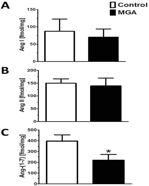
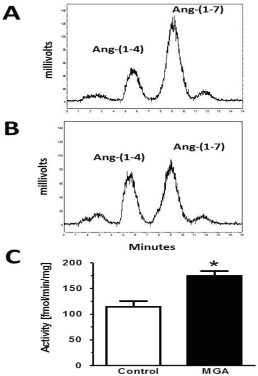
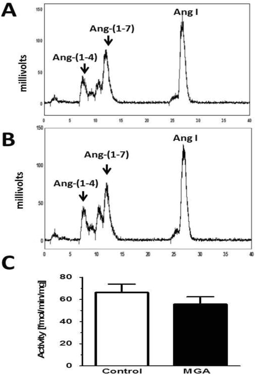
Angiotensin-(1-7) (Ang-(1-7))/AT7-Mas receptor axis is an alternative pathway within the renin-angiotensin system (RAS) that generally opposes the actions of Ang II/AT1 receptor pathway. Advanced glycated end product (AGEs) including glucose- and methylglyoxal-modified albumin (MGA) may contribute to the development and progression of diabetic nephropathy in part through activation of the Ang II/AT1 receptor system; however, the influence of AGE on the Ang-(1-7) arm of the RAS within the kidney is unclear. The present study assessed the impact of AGE on the Ang-(1-7) axis in NRK-52E renal epithelial cells. MGA exposure for 48 h significantly reduced the intracellular levels of Ang-(1-7) approximately 50%; however, Ang I or Ang II expression was not altered. The reduced cellular content of Ang-(1-7) was associated with increased metabolism of the peptide to the inactive metabolite Ang-(1-4) [MGA: 175±9 vs.

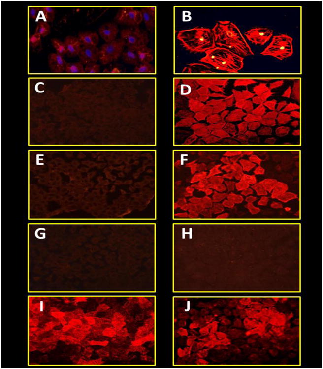
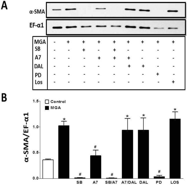
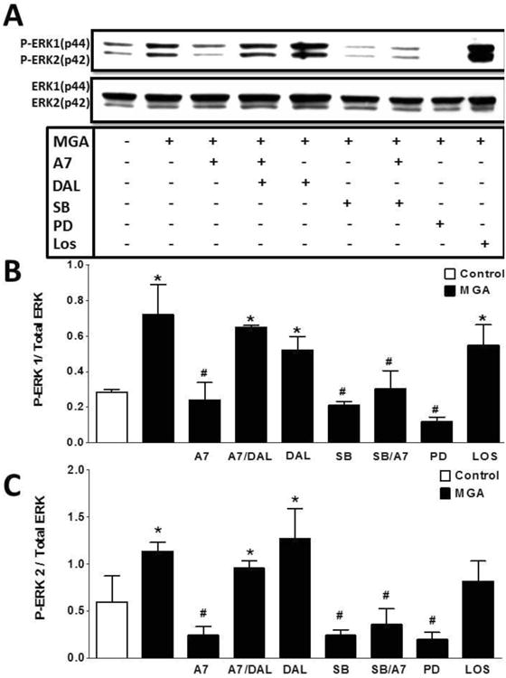
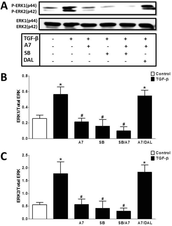
Control: 115±11 fmol/min/mg protein, p<0.05, n=3] but no change in the processing of Ang I to Ang-(1-7). Treatment with Ang-(1-7) reversed MGA-induced cellular hypertrophy and myofibroblast transition evidenced by reduced immunostaining and protein expression of α-smooth muscle actin (α-SMA) [0.4±0.1 vs. 1.0±0.1, respectively, n=3, p<0.05]. Ang-(1-7) abolished AGE-induced activation of the MAP kinase ERK1/2 to a similar extent as the TGF-β receptor kinase inhibitor SB58059; however, Ang-(1-7) did not attenuate the MGA-stimulated release of TGF-β. The AT7-Mas receptor antagonist D-Ala(7)-Ang-(1-7) abolished the inhibitory actions of Ang-(1-7). In contrast, AT1 receptor antagonist losartan did not attenuate the MGA-induced effects. We conclude that Ang-(1-7) may provide an additional therapeutic approach to the conventional RAS blockade regimen to attenuate AGE-dependent renal injury.

Keywords: Advanced glycation endproducts; Angiotensin; EMT; ERK1/2; TGF-B.


Conflict of interest statement

Conflict of Interests: None declared

Figures

Figure 1
Figure 1. MGA exposure reduces intracellular levels of Ang-(1-7) in the in the NRK-52E cells
Panel A: Ang I peptide levels are not different between control and MGA treated cells. Panel B: There are no difference in Ang II peptide levels between MGA treated cells and control cells. Panel C: Ang-(1-7) peptide levels were significantly lower in the MGA treated cells than control cells. Intracellular peptide content was quantified by separate RIAs to Ang I, Ang II and Ang-(1-7). Data are the mean ± SEM from 3 different cell passages. *P< 0.05.
Figure 2
Figure 2. MGA exposure increases Ang-(1-7) metabolism in the NRK-52E cells
Peptide metabolism was assessed in the 100,000 × g supernatant fraction from control and MGA-treated (48 hours) NRK-52E cells for 30 minutes at 37°C. Peptide products were determined by HPLC separation with isocratic elution conditions (38). Panel A: Ang-(1-7) conversion to Ang-(1-4) in control cells. Panel B: Ang-(1-7) conversion to Ang-(1-4) in MGA-treated cells. Panel C: Quantification of Ang-(1-4) generation from Ang-(1-7) revealed a higher rate of Ang-(1-7) conversion with MGA exposure. Data are the mean ± SEM of 3 different cell passages. *P< 0.05 vs. control.
Figure 3
Figure 3. MGA exposure does not influence Ang I to Ang-(1-7) formation in the NRK-52E cells
Peptide metabolism was assessed in the 100,000 × g supernatant fraction from control and MGA-treated (48 hours) NRK-52E cells for 30 minutes at 37°C. Peptide products were determined by HPLC separation under gradient conditions (1). Panel A: Ang I conversion to Ang-(1-7) in control cells. Panel B: Ang I conversion to Ang-(1-7) in MGA treated cells. Panel C: Quantification of Ang-(1-7) generation from Ang I revealed no differences between control and MGA treated cells. Data are the mean ± SEM of 3 different cell passages.
Figure 4
Figure 4. Ang-(1-7) attenuates MGA-induced cell hypertrophy in the NRK-52E cells
Left: MGA treatment for 48 hours significantly increased 3H-leucine incorporation approximately 180% of control. Ang-(1-7) (A7, 100 nM) significantly reduced the extent of hypertrophy induced by MGA. The Ang-(1-7) receptor antagonist D-Ala7-Ang-(1-7) (DAL, 10 μM), reversed the inhibitory effects of Ang-(1-7) on MGA-induced hypertrophy. The ERK 1/2 inhibitor PD98059 (PD, 1 μM), abolished the cellular hypertrophy induced by MGA. Ang-(1-7) and PD98059 exhibit no additive effects on hypertrophy. The DAL antagonist alone or the AT1 antagonist losartan (LOS) alone did not influence the MGA-dependent hypertrophy. 3H-leucine incorporation is expressed as percentage (%) from control cells for each experiment. Data are the mean ± SEM from 5 different cell passages. Inset: MGA treatment for 48 hours increased cell area as measured in immunofluorescent images. Ang-(1-7) (A7, 100 nM) and PD98059 (PD, 1 μM), reversed the increase in cell area induced by MGA.*P < 0.05 vs. control, #P < 0.05 vs. MGA-treated cells.
Figure 5
Figure 5. Ang-(1-7) appears to inhibit MGA-induced fluorescent staining for de novo expression of α-SMA in NRK 52-E cells
Cells were incubated with serum free media (A and C) or MGA (100 μM, B, D-I) or TGF-β (5 ng, J) for 72 hrs. MGA-treated cells were treated with either Ang-(1-7) (100 nM, E), Ang-(1-7) and DAL (10 μM, F), ERK1/2 inhibitor PD98059 (1 μM, G), TGF-β receptor kinase inhibitor SB525334 (1 μM, H) or AT1 receptor antagonist losartan (1 μM, I). The immunofluorescent images are representative of 3 different cell passages.
Figure 6
Figure 6. Ang-(1-7) inhibits MGA-induced protein expression of expression of α-SMA in NRK 52-E cells
Western blot analysis of MGA (100 μM) induced α-SMA expression after 48 hours. Ang-(1-7) (A7, 100 nM) attenuated the increase in α-SMA, which was blocked by the Mas receptor antagonist D-Ala7-Ang-(1-7) (DAL, 10 μM). Both the ERK1/2 inhibitor PD98059 (PD, 1 μM) and TGF-β receptor kinase inhibitor SB525334 (SB, 1 μM) abolished the increase in α-SMA expression. The AT1 antagonist losartan (LOS, 1 μM) did not influence the α-SMA expression. Data are the mean ± SEM from 3-4 different cell passages. *P < 0.05 vs. control, #P < 0.05 vs. MGA.
Figure 7
Figure 7. Ang-(1-7) does not influence MGA-induced release of TGF-β in NRK-52E cells
Cells were exposed to MGA for 48 hours and TGF-β in the cell media determined by ELISA. MGA exposure increased TGF-β release approximately 3-fold. There were no significant effects on MGA-induced TGF-β release by Ang-(1-7) (A7, 100 nM), D-Ala7-Ang-(1-7) (DAL, 10 μM), or the AT1 receptor antagonist losartan (LOS, 1 μM). TGF-β release was not significantly different between the ERK1/2 inhibitor PD98059 (PD, 1 μM) or combined Ang-(1-7) and PD (A7/PD) treatment to control. Data are the mean ± SEM from 4-5 different cell passages. *P < 0.05 vs. control.
Figure 8
Figure 8. Ang-(1-7) inhibits MGA-induced phosphorylation of ERK 1/2 in NRK 52-E cells
Western blot analysis of ERK1/2 with MGA treatment for 48 hours. MGA increased the phosphorylation of ERK1/2. Ang-(1-7) attenuated that MGA-induced phosphorylation which was blocked by D-Ala7-Ang-(1-7) (DAL, 10 μM). Inhibitors to TGF-β1 receptor kinase (SB, 1 μM) and ERK1/2 (PD, 1 μM) abolished ERK1/2 phosphorylation. The AT1 receptor antagonist losartan (LOS, 1 μM) did not inhibit the MGA-induced ERK1/2 activation. Data are the mean ± SEM from 3 different cell passages. *P < 0.05 vs. control, #P < 0.05 vs. MGA.
Figure 9
Figure 9. Ang-(1-7) inhibits TGF-β induced phosphorylation of ERK 1/2 in NRK-52E cells
Western blot analysis of TGF-β-induced ERK1/2 phosphorylation. TGF-β (5 ng/ml) stimulated ERK1/2 phosphorylation after a 15 min incubation. Ang-(1-7) (A7, 100 nM) pretreatment inhibited TGF-β induced ERK1/2 phosphorylation. The inhibitory effect of Ang-(1-7) was reversed by the antagonist D-Ala7-Ang-(1-7) (DAL, 10 μM). Pretreatment with TGF-β receptor kinase inhibitor SB525334 (SB, 1 μM) abolished ERK1/2 phosphorylation and there was no additive effect of Ang-(1-7) and SB. Data are the mean ± SEM from 3 different cell passages. *P < 0.05 vs. control, #P < 0.05 vs. MGA treated cells.

References

    1. Alzayadneh EM, Chappell MC. Nuclear expression of renin-angiotensin system components in NRK-52E renal epithelial cells. J Renin Angiotensin Aldosterone Syst. 2014 Epub. - PMC - PubMed
    1. Benter IF, Yousif MH, Dhaunsi GS, Kaur J, Chappell MC, Diz DI. Angiotensin-(1-7) prevents activation of NADPH oxidase and renal vascular dysfunction in diabetic hypertensive rats. Am J Nephrol. 2008;28:25–33. - PubMed
    1. Benter IF, Yousif MHM, Anim JT, Cojocel C, Diz DI. Angiotensin-(1-7) prevents development of severe hypertension and end-organ damage in spontaneously hypertensive rats treated with L-NAME. Am J Physiol Heart Circ Physiol. 2006;290:H684–H691. - PubMed
    1. Brenner BM, Cooper ME, de ZD, Keane WF, Mitch WE, Parving HH, Remuzzi G, Snapinn SM, Zhang Z, Shahinfar S. Effects of losartan on renal and cardiovascular outcomes in patients with type 2 diabetes and nephropathy. N Engl J Med. 2001;345:861–869. - PubMed
    1. Burns WC, Velkoska E, Dean R, Burrell LM, Thomas MC. Angiotensin II mediates epithelial-to-mesenchymal transformation in tubular cells by ANG 1-7/MAS-1-dependent pathways. Am J Physiol Renal Physiol. 2010;299:F585–F593. - PubMed

Publication types

MeSH terms