. 2014 Oct 7;111(40):14436-41.
doi: 10.1073/pnas.1409389111. Epub 2014 Sep 22.

Hematopoietic RIPK1 deficiency results in bone marrow failure caused by apoptosis and RIPK3-mediated necroptosis

Affiliations

Hematopoietic RIPK1 deficiency results in bone marrow failure caused by apoptosis and RIPK3-mediated necroptosis

Justine E Roderick et al. Proc Natl Acad Sci U S A. .

Abstract

Receptor-interacting serine/threonine-protein kinase 1 (RIPK1) is recruited to the TNF receptor 1 to mediate proinflammatory signaling and to regulate TNF-induced cell death. RIPK1 deficiency results in postnatal lethality, but precisely why Ripk1(-/-) mice die remains unclear. To identify the lineages and cell types that depend on RIPK1 for survival, we generated conditional Ripk1 mice. Tamoxifen administration to adult RosaCreER(T2)Ripk1(fl/fl) mice results in lethality caused by cell death in the intestinal and hematopoietic lineages. Similarly, Ripk1 deletion in cells of the hematopoietic lineage stimulates proinflammatory cytokine and chemokine production and hematopoietic cell death, resulting in bone marrow failure. The cell death reflected cell-intrinsic survival roles for RIPK1 in hematopoietic stem and progenitor cells, because Vav-iCre Ripk1(fl/fl) fetal liver cells failed to reconstitute hematopoiesis in lethally irradiated recipients. We demonstrate that RIPK3 deficiency partially rescues hematopoiesis in Vav-iCre Ripk1(fl/fl) mice, showing that RIPK1-deficient hematopoietic cells undergo RIPK3-mediated necroptosis. However, the Vav-iCre Ripk1(fl/fl) Ripk3(-/-) progenitors remain TNF sensitive in vitro and fail to repopulate irradiated mice. These genetic studies reveal that hematopoietic RIPK1 deficiency triggers both apoptotic and necroptotic death that is partially prevented by RIPK3 deficiency. Therefore, RIPK1 regulates hematopoiesis and prevents inflammation by suppressing RIPK3 activation.

Keywords: hematopoietic failure; programmed necrosis; tumor necrosis factor.


Conflict of interest statement

The authors declare no conflict of interest.

Figures
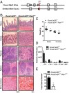
Fig. 1.
Fig. 1.
Acute ablation of RIPK1 results in intestinal epithelial and hematopoietic cell loss. (A) Schematic depiction of the gene-targeting strategy used to generate the conditional Ripk1 allele. (B) Representative H&E sections of intestines and hematopoietic organs from the indicated genotypes. (C) Total body weight of RosaCreERT2 (n = 10) and RosaCreERT2 Ripk1fl/fl (n = 9) mice. (D) Graph depicting absolute numbers of HSPCs from RosaCreERT2 (n = 6) and Rosa-CreERT2 Ripk1fl/fl (n = 6) mice. (E) Colony-forming assay of bone marrow from Rosa-CreERT2 (n = 2) and RosaCreERT2 Ripk1fl/fl (n = 2) mice. All data were collected 24 h after the third tamoxifen injection. Error bars represent SEM. *P < 0.05, **P < 0.01, ***P < 0.001.
Fig. 2.
Fig. 2.
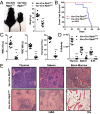
Deletion of RIPK1 in the hematopoietic lineage results in acute lethal bone marrow failure. (A) Macroscopic images of indicated genotypes (Left) and total body weight when killed (Right). (B) Kaplan–Meier survival curve of Vav-iCre Ripk1fl/fl mice (n = 14) and Vav-iCre Ripk1fl/+ (n = 11) controls (average latency 35 d, P < 0.0004). (C) Blood cell counts and hematocrits for Vav-iCre Ripk1fl/+ (n = 11) and Vav-iCre Ripk1fl/fl (n = 14) mice. (D) Graph showing thymus, spleen, and bone marrow cellularity for Vav-iCre Ripk1fl/+ (n = 11) and Vav-iCre Ripk1fl/fl (n = 14) mice. (E) Representative histological images of hematopoietic organs stained with H&E. Error bars represent SEM. ***P < 0.001.
Fig. 3.
Fig. 3.
Bone marrow failure arises in Vav-iCre Ripk1fl/fl mice as the result of HSPC loss. (A and B) Graphs depicting absolute numbers of HSPCs in Vav-iCre Ripk1fl/+ (n = 5) and Vav-iCre Ripk1fl/fl (n = 6) mice at 14 d old (A) or in Vav-iCre Ripk1fl/fl (n = 3) and Vav-iCre Ripk1fl/+ (n = 3) mice at the time of disease (B). (C) Graph depicting hematopoietic colony potential at 14 d old or at time of disease (n = 4 for each genotype). (D) Graph showing total colony number in the presence of vehicle, TRAIL, IFN-γ, IFN-α, or TNF-α for Vav-iCre Ripk1fl/+ (n = 3) and Vav-iCre Ripk1fl/fl (n = 3) mice. Total colony number was normalized to vehicle-treated bone marrow. Error bars represent SEM. *P < 0.05, **P < 0.01, ***P < 0.001.
Fig. 4.
Fig. 4.
RIPK1-deficient HSPCs fail to reconstitute lethally irradiated recipients. (A) Experimental design. (B and C) Graphs showing absolute number of donor-derived, CD45.2+ HSPCs from mice transplanted with Vav-iCre Ripk1fl/+ or Vav-iCre Ripk1fl/fl fetal liver cells at 2 wk (B) and 8 wk (C) posttransplantation. Error bars represent SEM. *P < 0.05, **P < 0.01.
Fig. 5.
Fig. 5.
RIPK3 deficiency partially rescues the lethal bone marrow failure of Vav-iCre Ripk1fl/fl mice. (A and B) Macroscopic images (A) and total body weight (B) of indicated genotypes at age 5 wk. (C) Blood cell counts and hematocrits of diseased Vav-iCre Ripk1fl/fl mice (n = 14) compared with age-matched Vav-iCre Ripk1fl/fl Ripk3 −/− mice (n = 5) and littermate controls Vav-iCre Ripk1fl/+ (n = 11) and Vav-iCre Ripk1fl/+Ripk3 −/− (n = 3). (D) Bone marrow cellularity for Vav-iCre Ripk1fl/+ (n = 11), Vav-iCre Ripk1fl/fl (n = 14), Vav-iCre Ripk1fl/+ Ripk3 −/− (n = 3), and Vav-iCre Ripk1fl/fl Ripk3 −/− (n = 5) mice. (E) Representative images of hematopoietic organs stained with H&E. (F and G) Graphs showing serum chemokine (F) and cytokine (G) levels for Vav-iCre Ripk1fl/fl (n = 10) and Vav-iCre Ripk1fl/fl Ripk3 −/−(n = 5) mice. (H) Colony-forming assay of bone marrow from 5-wk-old Vav-iCre Ripk1fl/+ Ripk3−/− (n = 3) and Vav-iCre Ripk1fl/fl Ripk3−/− (n = 5) mice cultured in the presence of vehicle, TRAIL, IFN-γ, IFN-α, or TNF-α. Total colony number was normalized to vehicle-treated bone marrow. (I) Absolute numbers of HSPCs were determined by flow cytometry in 5-wk-old Vav-iCre Ripk1fl/+ Ripk3−/−(n = 3) and Vav-iCre Ripk1fl/fl Ripk3−/− (n = 5) mice. (J) Kaplan–Meier survival curve of lethally irradiated recipients transplanted with Vav-iCre Ripk1fl/+ Ripk3−/− (n = 5) or Vav-iCre Ripk1fl/fl Ripk3−/− (n = 6) bone marrow cells. Error bars represent SEM. *P < 0.05, **P < 0.01, ***P < 0.001 (Student’s t test in B, C, H, and I; Mann–Whitney test in F and G).

References

    1. Chen ZJ. Ubiquitination in signaling to and activation of IKK. Immunol Rev. 2012;246(1):95–106. - PMC - PubMed
    1. Vanden Berghe T, Linkermann A, Jouan-Lanhouet S, Walczak H, Vandenabeele P. Regulated necrosis: the expanding network of non-apoptotic cell death pathways. Nat Rev Mol Cell Biol. 2014;15(2):135–147. - PubMed
    1. Lee TH, Shank J, Cusson N, Kelliher MA. The kinase activity of Rip1 is not required for tumor necrosis factor-alpha-induced IkappaB kinase or p38 MAP kinase activation or for the ubiquitination of Rip1 by Traf2. J Biol Chem. 2004;279(32):33185–33191. - PubMed
    1. Ikeda F, et al. SHARPIN forms a linear ubiquitin ligase complex regulating NF-κB activity and apoptosis. Nature. 2011;471(7340):637–641. - PMC - PubMed
    1. Wertz IE, et al. De-ubiquitination and ubiquitin ligase domains of A20 downregulate NF-kappaB signalling. Nature. 2004;430(7000):694–699. - PubMed

Publication types

MeSH terms