. 2014 Oct 7;111(40):14430-5.
doi: 10.1073/pnas.1322917111. Epub 2014 Sep 22.

Platelet mechanosensing of substrate stiffness during clot formation mediates adhesion, spreading, and activation

Affiliations

Platelet mechanosensing of substrate stiffness during clot formation mediates adhesion, spreading, and activation

Yongzhi Qiu et al. Proc Natl Acad Sci U S A. .

Abstract

As platelets aggregate and activate at the site of vascular injury to stem bleeding, they are subjected to a myriad of biochemical and biophysical signals and cues. As clot formation ensues, platelets interact with polymerizing fibrin scaffolds, exposing platelets to a large range of mechanical microenvironments. Here, we show for the first time (to our knowledge) that platelets, which are anucleate cellular fragments, sense microenvironmental mechanical properties, such as substrate stiffness, and transduce those cues into differential biological signals. Specifically, as platelets mechanosense the stiffness of the underlying fibrin/fibrinogen substrate, increasing substrate stiffness leads to increased platelet adhesion and spreading. Importantly, adhesion on stiffer substrates also leads to higher levels of platelet activation, as measured by integrin αIIbβ3 activation, α-granule secretion, and procoagulant activity. Mechanistically, we determined that Rac1 and actomyosin activity mediate substrate stiffness-dependent platelet adhesion, spreading, and activation to different degrees. This capability of platelets to mechanosense microenvironmental cues in a growing thrombus or hemostatic plug and then mechanotransduce those cues into differential levels of platelet adhesion, spreading, and activation provides biophysical insight into the underlying mechanisms of platelet aggregation and platelet activation heterogeneity during thrombus formation.

Keywords: biomaterials; cell mechanics; mechanotransduction; platelet cytoskeleton.


Conflict of interest statement

The authors declare no conflict of interest.

Figures
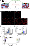
Fig. 1.
Fig. 1.
Differential platelet adhesion and spreading on immobilized fibrinogen is mediated by substrate stiffness. (A) Experimental setup illustrating how human platelets were incubated on polyacrylamide (PA) gels of varied stiffnesses but conjugated with the same fibrinogen concentration on the top surface. This creates a microenvironment in which the mechanical properties are uncoupled from the biochemical properties. (B) Confocal microscopy images of adherent platelets, stained with a fluorescent membrane dye, on PA gels with stiffnesses ranging from 0.25 to 100 kPa and glass. (C) The number of platelets adhered on PA gels of different stiffness and glass. (D) The spread surface area of adherent platelets on PA gels of different stiffness and glass. (E) Cumulative frequency of spread platelet surface area on substrates of different stiffnesses. (Scale bar: 10 μm.) (P < 0.05; n = 3 experiments; error bars indicate SD.)
Fig. 2.
Fig. 2.
Rac1 mediates platelet mechanosensing during adhesion, whereas Rac1, myosin, and actin polymerization mediate platelet mechanosensing during spreading. (A) Rac1 inhibition attenuates platelet mechanosensing during adhesion on fibrinogen-conjugated PA gels in a dose-dependent fashion. (B) Likewise, Rac1 inhibition also attenuates platelet mechanosensing during spreading on fibrinogen-conjugated PA gels in a dose-dependent fashion. (C) The number of platelets treated with vehicle (DMSO) or other cytoskeletal inhibitors after adhering onto PA gels of different stiffnesses. Inhibition of ROCK, MLCK, or actin polymerization does not alter the substrate stiffness-mediated effects on platelet adhesion. (D) On the other hand, inhibition of ROCK, MLCK, or actin polymerization all attenuate the substrate stiffness-mediated effects on platelet adhesion. (*P < 0.05; n = 3 experiments; error bars indicate SD.)
Fig. 3.
Fig. 3.
Platelet activation is mediated by substrate stiffness. (A) Increased substrate stiffness increases activation of the αIIbβ3 platelet integrin as measured by PAC-1–FITC immunostaining via confocal microscopy. When normalized to spread platelet surface area, the average intensity is higher for the fibrinogen-conjugated 5- and 50-kPa gels compared with 0.5-kPa gels. (B) Inhibition of Rac1, SFK, MLCK, and actin polymerization attenuates the substrate stiffness-mediated αIIbβ3 activation of adherent platelets as measured by the average intensity of PAC-1–FITC staining. (C) Increased substrate stiffness increases P-selectin expression on adherent platelets as measured by P-selectin immunostaining (red: fluorescent membrane dye; green: P-selectin immunostaining) via confocal microscopy. (D) Inhibition of Rac1, SFK, ROCK, and MLCK differentially attenuate the substrate stiffness-mediated P-selectin expression on adherent platelets. (E) PS exposure was found only on platelets adhered onto stiffer gels of 5 and 50 kPa as measured as measured by Annexin V staining via confocal microscopy. (F) Inhibition of Rac1, SFK, MLCK, and actin polymerization abolished the substrate stiffness-mediated PS exposure on adherent platelets. (P < 0.05; n = 3 experiments; error bars indicate SD.) (Scale bar: 10 μm.)
Fig. 4.
Fig. 4.
Proposed mechanisms of how substrate stiffness affects platelet activation. Platelet mechanosensing of the microenvironment, which includes heterogeneous fibrin polymer networks of differing mechanical properties, likely starts with the initial adhesive event, during which the resistant substrate force balances the inertial movement of the platelet. (A) A platelet engaging with a relatively soft substrate results in a weak αIIbβ3–fibrinogen interaction and short bond lifetime. (B) A stiffer substrate, however, would provide more resistant force leading to more platelet adhesion and outside-in αIIbβ3 signaling. This in turn, would generate a high acto-myosin–mediated internal balancing force, resulting in more platelet spreading on stiffer substrate. Downstream signals will then further trigger platelet activation, thus up-regulating more αIIbβ3 activation, granule secretion, and PS exposure.

Similar articles

Cited by

References

    1. Jackson SP. Arterial thrombosis—insidious, unpredictable and deadly. Nat Med. 2011;17(11):1423–1436. - PubMed
    1. Brass LF, Wannemacher KM, Ma P, Stalker TJ. Regulating thrombus growth and stability to achieve an optimal response to injury. J Thromb Haemost. 2011;9(Suppl 1):66–75. - PMC - PubMed
    1. Coller BS, Shattil SJ. The GPIIb/IIIa (integrin alphaIIbbeta3) odyssey: A technology-driven saga of a receptor with twists, turns, and even a bend. Blood. 2008;112(8):3011–3025. - PMC - PubMed
    1. Ruggeri ZM. Platelets in atherothrombosis. Nat Med. 2002;8(11):1227–1234. - PubMed
    1. Shattil SJ, Kashiwagi H, Pampori N. Integrin signaling: The platelet paradigm. Blood. 1998;91(8):2645–2657. - PubMed

Publication types

MeSH terms