. 2014 Oct 7;111(40):E4274-83.
doi: 10.1073/pnas.1416598111. Epub 2014 Sep 22.

α-Synuclein assembles into higher-order multimers upon membrane binding to promote SNARE complex formation

Affiliations

α-Synuclein assembles into higher-order multimers upon membrane binding to promote SNARE complex formation

Jacqueline Burré et al. Proc Natl Acad Sci U S A. .

Abstract

Physiologically, α-synuclein chaperones soluble NSF attachment protein receptor (SNARE) complex assembly and may also perform other functions; pathologically, in contrast, α-synuclein misfolds into neurotoxic aggregates that mediate neurodegeneration and propagate between neurons. In neurons, α-synuclein exists in an equilibrium between cytosolic and membrane-bound states. Cytosolic α-synuclein appears to be natively unfolded, whereas membrane-bound α-synuclein adopts an α-helical conformation. Although the majority of studies showed that cytosolic α-synuclein is monomeric, it is unknown whether membrane-bound α-synuclein is also monomeric, and whether chaperoning of SNARE complex assembly by α-synuclein involves its cytosolic or membrane-bound state. Here, we show using chemical cross-linking and fluorescence resonance energy transfer (FRET) that α-synuclein multimerizes into large homomeric complexes upon membrane binding. The FRET experiments indicated that the multimers of membrane-bound α-synuclein exhibit defined intermolecular contacts, suggesting an ordered array. Moreover, we demonstrate that α-synuclein promotes SNARE complex assembly at the presynaptic plasma membrane in its multimeric membrane-bound state, but not in its monomeric cytosolic state. Our data delineate a folding pathway for α-synuclein that ranges from a monomeric, natively unfolded form in cytosol to a physiologically functional, multimeric form upon membrane binding, and show that only the latter but not the former acts as a SNARE complex chaperone at the presynaptic terminal, and may protect against neurodegeneration.

Keywords: Parkinson's disease; SNARE proteins; membrane fusion; synapse.


Conflict of interest statement

The authors declare no conflict of interest.

Figures

Fig. 1.
Fig. 1.
α-Synuclein multimerization upon lipid membrane binding activates SNARE complex assembly. (A) Diagram of the experimental design. (B–D) Binding of α-synuclein to both phospholipid membranes and synaptobrevin is required for α-synuclein to enhance SNARE complex assembly. Recombinant SNARE proteins were incubated with or without addition of α-synuclein in presence or absence of charged liposomes [composition: 30% phosphatidylserine (PS), 70% phosphatidylcholine (PC)]. SNARE complexes were immunoprecipitated with antibodies to syntaxin-1 or preimmune sera (“Pre-Imm,” as a control), and coimmunoprecipitated synaptobrevin-2 (Syb2), SNAP-25, and α-synuclein were analyzed by quantitative immunoblotting. (E and F) Independently, SNARE complex assembly in the same experiment as B–D was quantitated using SDS-resistant complexes on SDS/PAGE. For input protein levels, see Fig. S1. All data shown are means ± SEM (*P < 0.05; ***P < 0.001 by Student t test; n = 3).
Fig. 2.
Fig. 2.
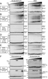
Native α-synuclein multimerizes on the surface of brain membranes. (A–E) Brain homogenates containing intact membranes (A and B; 900 µg of protein), brain cytosol (C; 150 µg of protein), brain homogenates whose membranes were solubilized by addition of Triton X-100 (D; 900 µg of protein), and brain cytosol with readdition of brain membranes (E; 150 µg of cytosolic protein plus 750 µg of membrane protein) were exposed to increasing concentrations of chemical cross-linking agents [Left, glutaraldehyde (0–0.01%); Right, dimethyl suberimidate (DMS) (0–25 mM)], and equal volumes were analyzed by immunoblotting (arrowheads, α-synuclein oligomers as determined by SDS/PAGE). For additional cross-linking experiments, quantitations, cross-linker concentrations used, size calibrations, and controls, see Fig. S2. (F) Control cross-linking experiments. Brain homogenates were exposed to same increasing concentrations of glutaraldehyde and analyzed as described above (GDI, guanine-nucleotide dissociation inhibitor; SGT, small glutamine-rich tetratricopeptide repeat-containing protein; Synt-1, syntaxin-1; arrowheads, protein monomers and homooligomers or heterooligomers as determined by SDS/PAGE migration analysis; asterisks indicate nonspecific immunosignals).
Fig. 3.
Fig. 3.
α-Synuclein forms multimers in brain slices. (A) Acute brain slices from wild-type mice were exposed to increasing concentrations of cross-linking agents [Left, glutaraldehyde (0–0.03%); Right, dimethyl suberimidate (DMS) (0–3 mM)]. Brain slices were homogenized in sample buffer after cross-linking, and equal volumes of cross-linked proteins were analyzed by immunoblotting (Upper, 15% gels; Lower, 10% gels). The arrowheads indicate α-synuclein multimers as determined by SDS/PAGE migration analysis; the asterisks indicate nonspecific immunosignals (means ± SEMs; n = 3). (B) Control cross-linking experiments. Acute brain slices were exposed to same increasing concentrations of the cross-linker DMS and analyzed as described above (GDI, guanine-nucleotide dissociation inhibitor; SGT, small glutamine-rich tetratricopeptide repeat-containing protein; Syb2, synaptobrevin-2; Synt-1, syntaxin-1).
Fig. 4.
Fig. 4.
Reconstitution of α-synuclein multimerization on charged liposomes. (A and B) Recombinant α-synuclein was incubated with negatively charged liposomes (composition: 30% PS, 70% PC) and exposed to increasing concentrations of the chemical cross-linkers glutaraldehyde (A; 0–0.05%) or DMS (B; 0–25 mM). Cross-linked species were analyzed by 15% and 10% SDS/PAGE (Top and Middle) or 6% Tris-acetate PAGE (Bottom). (C and D) Recombinant α-synuclein was incubated in buffer or with neutral liposomes (composition: 100% PC), and cross-linked as described above. Cross-linked species were analyzed by 15% SDS/PAGE. (E and F) Quantitations of dimerization and tetramerization of α-synuclein upon chemical cross-linking in buffer, on neutral or negatively charged liposomes, using glutaraldehyde (E) or DMS (F). Multimerization was plotted relative to α-synuclein input levels and was measured by quantitative immunoblotting (means ± SEMs; n = 3; *P < 0.05; ***P < 0.001).
Fig. 5.
Fig. 5.
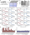
α-Synuclein multimerizes on the phospholipid membrane surface in a defined orientation. (A) Fluorescent α-synuclein labeling scheme. Single-cysteine substitutions were introduced into α-synuclein at position 1 (M1C), 8 (L8C), 41 (G41C), 96 (K96C), and 140 (A140C) for modification with Alexa 488- or Alexa 546-maleimide. (B) SDS/PAGE analysis of purified Alexa 488- or Alexa 546-labeled recombinant α-synuclein proteins (3 µg of protein per lane). (C) Liposome binding of wild-type and mutant α-synuclein. Recombinant α-synuclein was incubated with negatively charged liposomes (30% PS, 70% PC) and subjected to a flotation assay. Eight fractions were collected from top to bottom, and α-synuclein was quantified in each fraction (Fig. S4A). Liposome binding is expressed as the amount of α-synuclein present in the liposome fractions of the flotation gradient in percent of the total α-synuclein. (D and E) Fluorescence resonance energy transfer (FRET) between donor and acceptor α-synuclein. Alexa 488-labeled donor α-synuclein (2.5 µg) was incubated for 2 h with Alexa 546-labeled acceptor or unlabeled α-synuclein (2.5 µg) in the presence of 100 µg of charged (red spectra) or neutral liposomes (blue spectra). Emission spectra (D) were used for calculation of FRET signals (E). For further spectra, see Fig. S4B. Data in C and E are means ± SEM (*P < 0.05 by Student t test; n = 3).
Fig. 6.
Fig. 6.
α-Synuclein multimers on the phospholipid membrane surface adopt an antiparallel configuration. (A–D) FRET between donor and acceptor α-synuclein. Alexa 488-labeled donor α-synuclein L8C (A and B; 2.5 µg) or A140C (C and D; 2.5 µg) was incubated for 2 h in the presence of 100 µg of charged liposomes with the indicated increasing amounts of Alexa 546-labeled acceptor α-synuclein L8C (A and C) or A140C (B and D), or with unlabeled α-synuclein to balance total α-synuclein amounts. Emission spectra (Left) were used for calculation of FRET signals (Right; means ± SEM; *P < 0.05; n = 3). (E and F) Modeling of α-synuclein multimers based on FRET experiments. E shows schematic drawing of two α-synuclein proteins (red lines, positive FRET signals; blue lines, no FRET signals. F shows the model of a possible arrangement of α-synuclein multimers on the surface that would be consistent with the FRET signals.
Fig. 7.
Fig. 7.
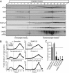
α-Synuclein associates with synaptic vesicles docked at the presynaptic plasma membrane. (A) Separation of synaptic vesicles by sucrose gradient centrifugation into free (fractions 6–13) and docked vesicle pools (fractions 23–32). Equal volumes of each fraction were analyzed by immunoblotting (Syb2, synaptobrevin-2; α-Syn, α-synuclein; Synt1, syntaxin-1; Syp1, synaptophysin 1). (B and C) Quantitation of the distribution of the indicated proteins across the sucrose gradient (B) and of relative protein levels in the free vs. docked synaptic vesicle pool (C; means ± SEM; *P < 0.05; **P < 0.01; ***P < 0.001 by Student t test; n = 3).
Fig. 8.
Fig. 8.
Scheme of a physiological folding pathway for α-synuclein. Soluble α-synuclein is natively unstructured and monomeric (Left). Upon binding to synaptic vesicles during docking and priming of the vesicles, which involves partial SNARE complex assembly, α-synuclein undergoes a conformational change, folds into a “broken” amphipathic α-helix and multimerizes. As a result of membrane binding, α-synuclein becomes competent to promote SNARE complex assembly during docking and priming of synaptic vesicles.

References

    1. George JM, Jin H, Woods WS, Clayton DF. Characterization of a novel protein regulated during the critical period for song learning in the zebra finch. Neuron. 1995;15(2):361–372. - PubMed
    1. Iwai A, et al. The precursor protein of non-A beta component of Alzheimer’s disease amyloid is a presynaptic protein of the central nervous system. Neuron. 1995;14(2):467–475. - PubMed
    1. Burré J, et al. α-synuclein promotes SNARE-complex assembly in vivo and in vitro. Science. 2010;329(5999):1663–1667. - PMC - PubMed
    1. Polymeropoulos MH, et al. Mutation in the alpha-synuclein gene identified in families with Parkinson’s disease. Science. 1997;276(5321):2045–2047. - PubMed
    1. Krüger R, et al. Ala30Pro mutation in the gene encoding α-synuclein in Parkinson’s disease. Nat Genet. 1998;18(2):106–108. - PubMed

Publication types