. 2014 Oct 7;111(40):14442-7.
doi: 10.1073/pnas.1401166111. Epub 2014 Sep 22.

Cell fate regulation by gelsolin in human gynecologic cancers

Affiliations

Cell fate regulation by gelsolin in human gynecologic cancers

Mohammad R Abedini et al. Proc Natl Acad Sci U S A. .

Erratum in

  • Proc Natl Acad Sci U S A. 2014 Dec 9;111(49):17684

Abstract

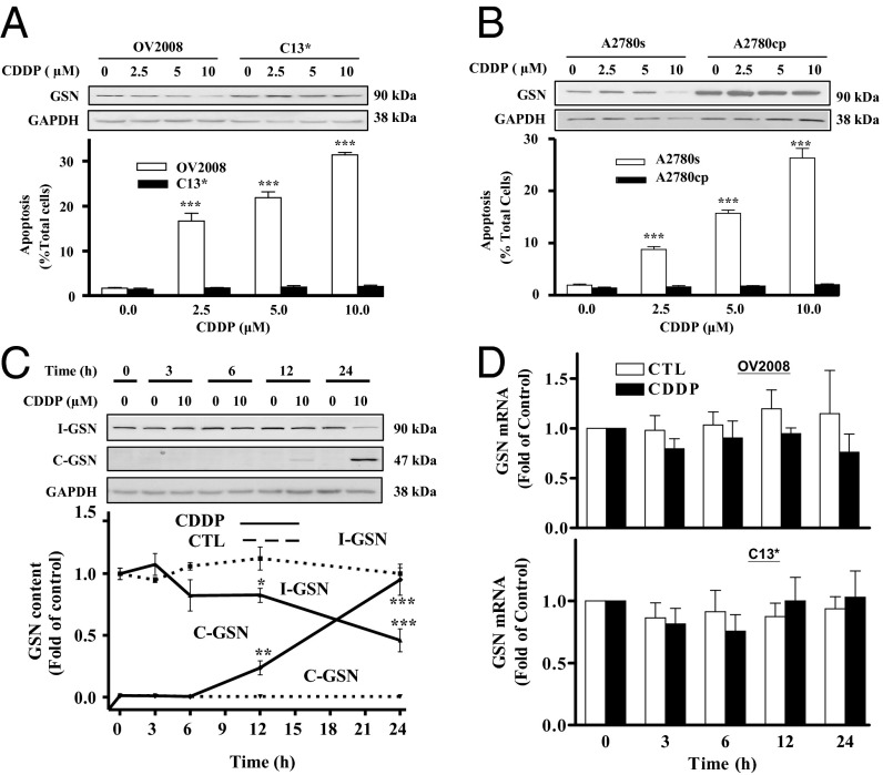
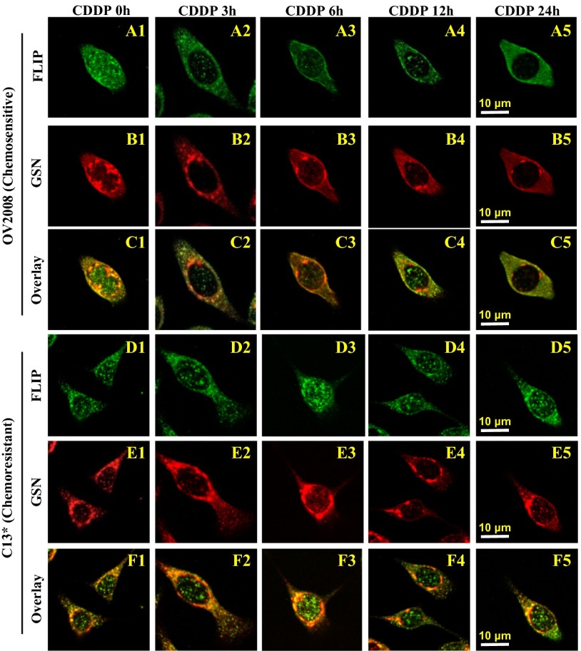
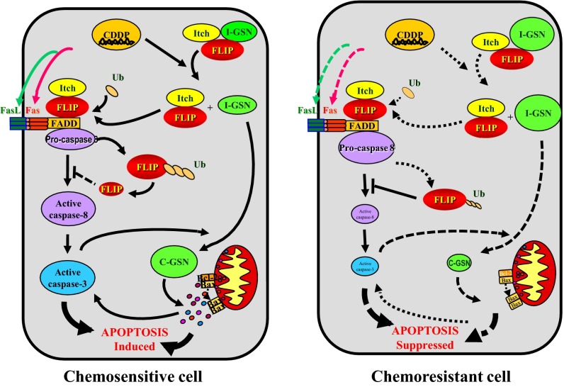
Chemoresistance is a major hurdle in cancer treatment. Down-regulation of apoptosis pathways is one of the key determinants for chemoresistance. Here, we report higher gelsolin (GSN) levels in chemoresistant gynecological cancer cells compared with their sensitive counterparts. cis-Diammine dichloroplatinium (II) (CDDP)-induced GSN down-regulation is associated with its cleavage and apoptosis. Although the C-terminal GSN fragment (C-GSN) sensitized chemoresistant cells to CDDP, intact GSN and its N-terminal fragment (N-GSN) attenuated this response. GSN silencing also facilitated CDDP-induced apoptosis in chemoresistant cells. In contrast, intact GSN (I-GSN) was prosurvival in the presence of CDDP through a FLICE-like inhibitory protein (FLIP)-Itch interaction. This interaction was colocalized in the perinuclear region that could be dissociated by CDDP in sensitive cells, thereby inducing FLIP ubiquitination and degradation, followed by apoptosis. In resistant cells, GSN was highly expressed and CDDP failed to abolish the I-GSN-FLIP-Itch interaction, resulting in the dysregulation of the downstream responses. In addition, we investigated the association between GSN expression in ovarian serous adenocarcinoma and progression free survival and overall survival, as well as clinical prognosis. GSN overexpression was significantly associated with more aggressive behavior and more cancer deaths and supported our hypothesis that high GSN expression confers chemoresistance in cancer cells by altering the GSN-FLIP-Itch interaction. These findings are in agreement with the notion that GSN plays an important role in the regulation of gynecological cell fate as reflected in dysregulation in chemosensitivity.

Keywords: cervical cancer; ovarian cancer.


Conflict of interest statement

The authors declare no conflict of interest.

Figures

Fig. 1.
Fig. 1.
Overall survival (OS) and progression-free survival (PFS) curves of all-stage patients and subgroups with serous ovarian cancer, stratified according to GSN expression. High GSN expression significantly correlated with the long-term OS and PFS in all patients (A and B), subgroup patients with PFI ≤ 12 mo (n = 50) (C and D), and late-stage subgroup patients (n = 84) (E and F). Among late-stage subgroup patients with PFI > 12 mo, the OS (G) was significantly poorer in those with GSN-positive tumors than those with GSN-negative tumors. Although the negative impact of GSN overexpression on PFS (H) was found in this subgroup, it was not statistically significant.
Fig. 2.
Fig. 2.
CDDP-induced apoptosis in cancer cells is associated with decreased intact GSN (I-GSN) protein content. (A and B) CDDP induced GSN down-regulation and apoptosis. CECA cells (A) (OV2008 and C13*) and OVCA cells (B) (A2780s and A2780cp) were cultured with CDDP (0-10 μM; 24 h) and analyzed for I-GSN and GAPDH contents by Western blotting (Upper). Apoptosis was determined morphologically by Hoechst 33258 staining (Lower; ***P < 0.001 vs. control). (C) CDDP induced I-GSN down-regulation and enhanced GSN protein cleavage into C-GSN in a time-dependent manner. OV2008 was cultured with CDDP (0-10 µM; 0–24 h) and analyzed for I-GSN, C-GSN, and GAPDH contents by Western blotting. (D) CDDP failed to alter GSN mRNA abundance in sensitive CECA cells. OV2008 was cultured with CDDP (0-10 µM; 0–24 h) and analyzed for GSN mRNA contents by RT-PCR. CTL, control.
Fig. 3.
Fig. 3.
GSN regulates CDDP-induced apoptosis in cancer cells. GSN silencing facilitated CDDP-induced apoptosis in C13* cells (A) and A2780cp cells (B). Cells were transfected with GSN or control siRNA (50-200 nM; 24 h) and then cultured with CDDP (0-10 μM; 24 h). I-GSN, C-GSN, and GAPDH contents were assessed by Western blotting (Upper), and apoptosis was determined morphologically by Hoechst 33258 staining (Lower; ***P < 0.001 vs. control; n = 3). Expression of C-terminal GSN (C-GSN; 0–2 µg, 24 h) facilitated CDDP-induced apoptosis [0 and 10 µM; 24 h; Lower; ***P < 0.001 (vs. control); n = 3] in both OV2008 (C and E) and C13* (D and F) whereas full-length (I-GSN; 0–2 µg, 24 h), N-terminal GSN (N-GSN; 0–2 µg, 24 h) and caspase cleavage mutant (M-GSN, 0–2 µg, 24 h) attenuated this response in OV2008 cells (C).
Fig. 4.
Fig. 4.
CDDP attenuates the GSN-FLIP-Itch interaction in chemosensitive but not resistant counterparts. The FLIP-GSN-Itch interaction was attenuated by CDDP in chemosensitive (OV2008) but not resistant (C13*) cells as detected by immunocoprecipitation. Cells were transfected with I-GSN (2 μg; 24 h), infected (MOI = 25; 24 h) with either adenoviral V5-FLIPL, (A) or V5-FLIPS (B) and cultured with CDDP (0-10 μM; 0–24 h). Cell lysates were immunoprecipitated with IgG (control; lanes 1 and 5) or corresponding antibody, as indicated. Protein–protein interaction was determined by immunoprecipitation–Western blots (IP-Western). GSN and V5-FLIP immunoprecipitates were immunoblotted [IP: GSN, WB: V5 and Itch; IP: V5-tagged FLIPL or FLIPS, WB: GSN and Itch (A and B, Upper)].
Fig. 5.
Fig. 5.
CDDP attenuates FLIP-GSN colocalization at OV2008 but not C13* cells. Confocal imaging of double-stained FLIP and GSN in OV2008 and C13* cells. FLIP (A1) distributed throughout the cytoplasm and nucleus in OV2008 treated with DMSO (control). CDDP treatment (0–2.5 μM) induced relocation of FLIP to the cytoplasm from 3 h to 24 h (A2A5). GSN was detected in the perinuclear region (B1) and decreased over time in the presence of CDDP (B2B5). FLIP and GSN nuclear colocalization was not detected in OV2008 cells in the absence (C1) or presence (C2C5) of CDDP, but they were colocalized in the perinuclear area and decreased over time with CDDP (C2C5). In resistant C13* cells, although perinuclear and nuclear FLIP (D1) and perinuclear of GSN (E1) were extensively detected, they were not affected by CDDP (D2D5 and E2E5, respectively). FLIP-GSN was found to be colocalized in the perinuclear area (F1) independent of the CDDP treatment (F2F5). Scale bars in A5 and D5 apply to A1C5 and D1F5, respectively (representative of total of 65 cells for each treatment group, n = 3).
Fig. 6.
Fig. 6.
A hypothetical model illustrating the role and regulation of GSN in the control of chemosensitivity in cancer cells. In nonstress state, GSN forms a complex with FLIP and Itch. CDDP leads to the dissociation of GSN from the GSN-FLIP-Itch complex in sensitive cells, thereby inducing FLIP ubiquitination and degradation, caspase-8 and -3 activation, caspase-3-mediated GSN cleavage, and apoptosis. In resistant cells, CDDP fails to alter the GSN-FLIP-Itch interaction, resulting in the attenuation of the downstream responses to CDDP.

References

    1. Kartalou M, Essigmann JM. Mechanisms of resistance to cisplatin. Mutat Res. 2001;478(1-2):23–43. - PubMed
    1. Markman M, Bookman MA. Second-line treatment of ovarian cancer. Oncologist. 2000;5(1):26–35. - PubMed
    1. Ledermann JA, Kristeleit RS. Optimal treatment for relapsing ovarian cancer. Ann Oncol. 2010;21(Suppl 7):vii218–vii222. - PubMed
    1. Sehouli J, Alfaro V, Gonzalez-Martin A. Trabectedin plus pegylated liposomal doxorubicin in the treatment of patients with partially platinum-sensitive ovarian cancer: Current evidence and future perspectives. Ann Oncol. 2012;23(3):556–562. - PubMed
    1. Cheng JQ, et al. Role of X-linked inhibitor of apoptosis protein in chemoresistance in ovarian cancer: Possible involvement of the phosphoinositide-3 kinase/Akt pathway. Drug Resist Updat. 2002;5(3-4):131–146. - PubMed

Publication types

MeSH terms