. 2014 Nov;3(11):1363-74.
doi: 10.5966/sctm.2014-0017. Epub 2014 Sep 22.

Remotely Activated Mechanotransduction via Magnetic Nanoparticles Promotes Mineralization Synergistically With Bone Morphogenetic Protein 2: Applications for Injectable Cell Therapy

Affiliations

Remotely Activated Mechanotransduction via Magnetic Nanoparticles Promotes Mineralization Synergistically With Bone Morphogenetic Protein 2: Applications for Injectable Cell Therapy

James R Henstock et al. Stem Cells Transl Med. 2014 Nov.

Abstract

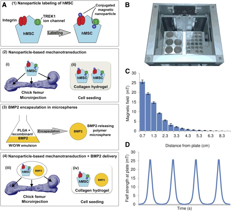
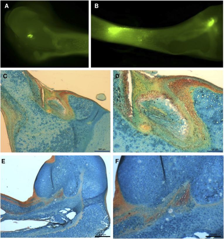
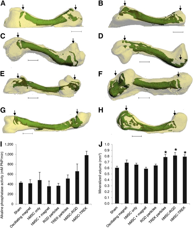
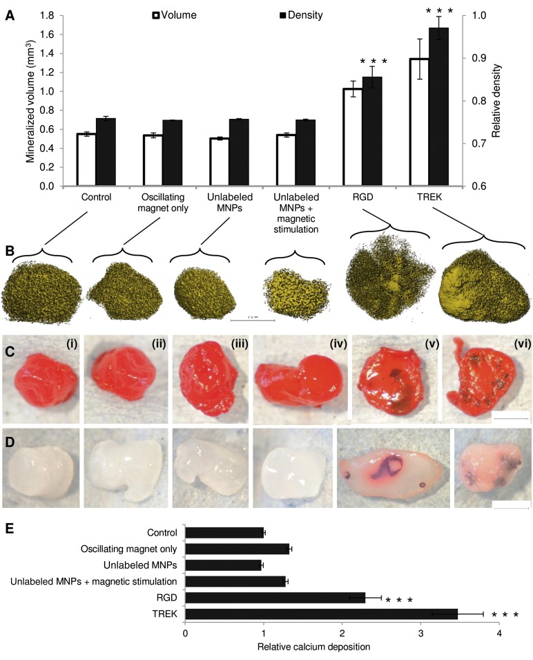
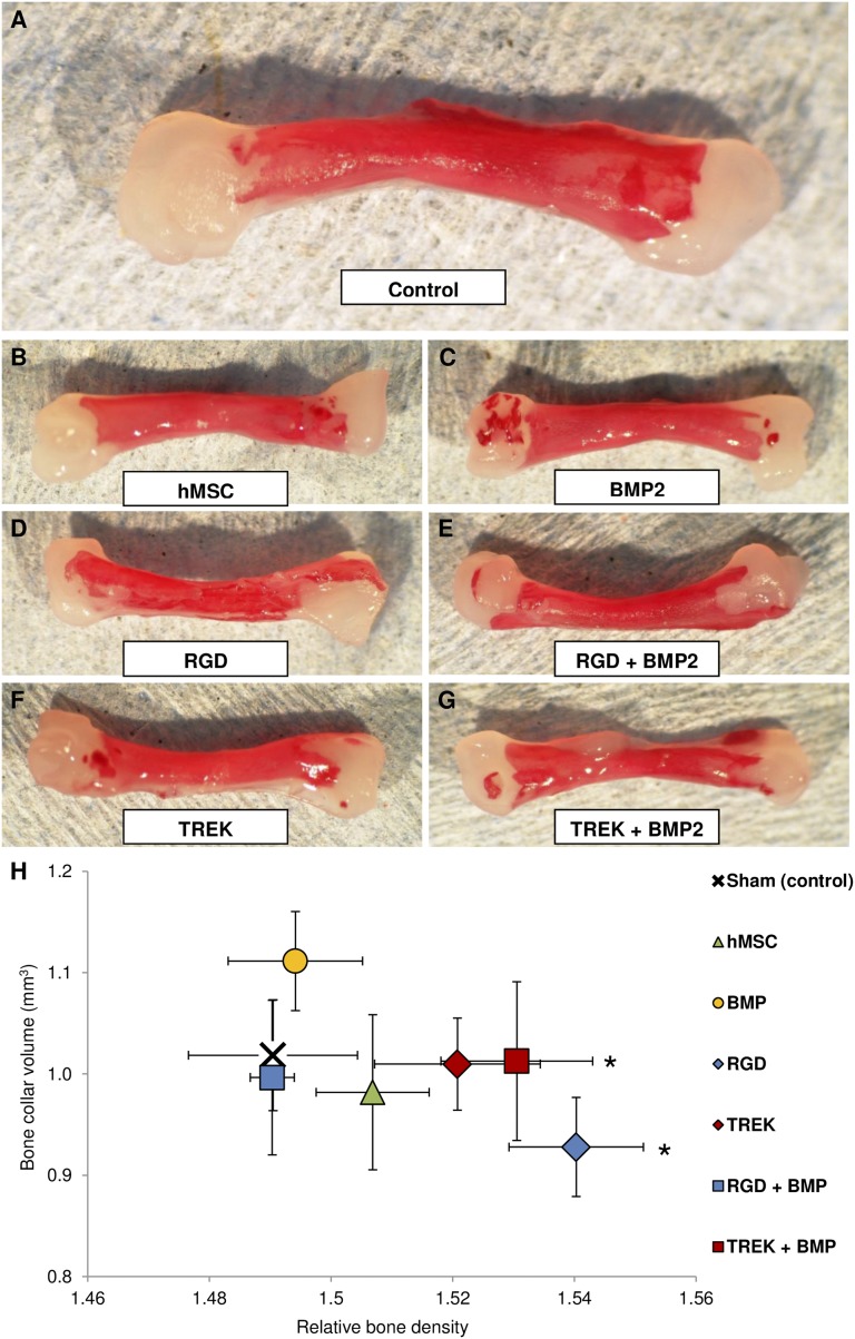
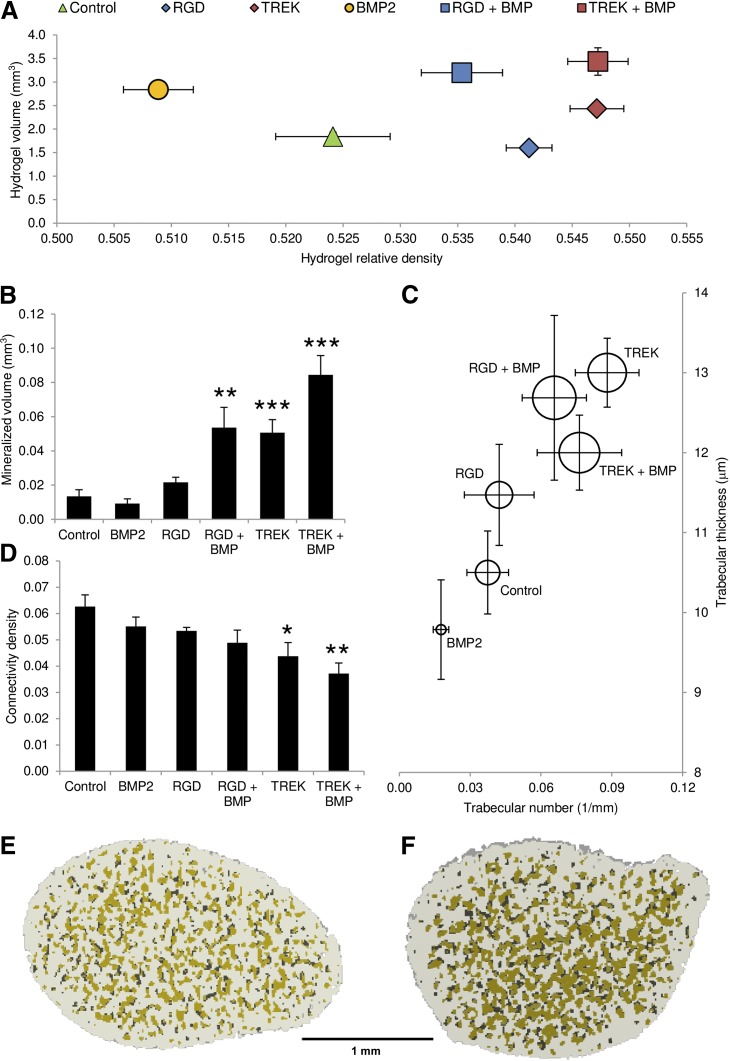
Bone requires dynamic mechanical stimulation to form and maintain functional tissue, yet mechanical stimuli are often lacking in many therapeutic approaches for bone regeneration. Magnetic nanoparticles provide a method for delivering these stimuli by directly targeting cell-surface mechanosensors and transducing forces from an external magnetic field, resulting in remotely controllable mechanotransduction. In this investigation, functionalized magnetic nanoparticles were attached to either the mechanically gated TREK1 K+ channel or the (integrin) RGD-binding domains of human mesenchymal stem cells. These cells were microinjected into an ex vivo chick fetal femur (embryonic day 11) that was cultured organotypically in vitro as a model for endochondral bone formation. An oscillating 25-mT magnetic field delivering a force of 4 pN per nanoparticle directly against the mechanoreceptor induced mechanotransduction in the injected mesenchymal stem cells. It was found that cells that received mechanical stimuli via the nanoparticles mineralized the epiphyseal injection site more extensively than unlabeled control cells. The nanoparticle-tagged cells were also seeded into collagen hydrogels to evaluate osteogenesis in tissue-engineered constructs: in this case, inducing mechanotransduction by targeting TREK1 resulted in a 2.4-fold increase in mineralization and significant increases in matrix density. In both models, the combination of mechanical stimulation and sustained release of bone morphogenetic protein 2 (BMP2) from polymer microspheres showed a significant additive effect on mineralization, increasing the effectiveness of BMP2 delivery and demonstrating that nanoparticle-mediated mechanotransduction can be used synergistically with pharmacological approaches for orthopedic tissue engineering to maximize bone formation.

Keywords: Bone marrow stromal cells; Cellular therapy; Clinical translation; Differentiation; Mesenchymal stem cells; Tissue regeneration; Transduction.


Figures

Figure 1.
Figure 1.
Experimental overview. (A): Schematic of the experimental design. (A1): hMSCs were labeled with magnetic nanoparticles targeting either the Arg-Gly-Asp-binding domains of cell-surface molecules such as integrins or the TREK1 mechanosensitive ion channel. (A2): The cells were then either injected into a cultured ex vivo chick femur (A2i) or seeded into a collagen hydrogel scaffold (A2ii). (A3): In subsequent experiments, the combined effects of directed mechanotransduction and BMP2 delivery were studied using BMP2-releasing poly(d,l-lactide-coglycolide) microspheres formed by an emulsion method. (A4): Labeled cells and BMP2-releasing microspheres were codelivered into either the chick femur or collagen hydrogels. (B): The nanoparticle-receptor complex was stimulated using a vertically oscillating external field delivered by a moving magnetic array situated beneath the culture plates for 1 hour per day (Mica Biosystems bioreactor). (C): The maximum magnetic field strength experienced at the plate was ∼25 mT, which attenuated rapidly as the array was withdrawn. (D): The cycle was repeated at 1 Hz. Abbreviations: BMP2, bone morphogenetic protein 2; hMSC, human mesenchymal stem cells; W/O/W, water-in-oil-in-water emulsion.
Figure 2.
Figure 2.
Microinjection into the chick fetal femur. (A, B): Human mesenchymal stem cells (hMSCs) labeled with the live cell tracker membrane dye DiL and injected into both cartilaginous epiphyses (A) and the middiaphysis (B), imaged immediately after microinjection. (C–F): After 2 weeks of ex vivo organotypic culture, femur epiphyses from sham control groups (C, D) and femurs injected with TREK1 magnetic nanoparticle-labeled hMSCs (E, F) were sectioned and histologically stained for glycosaminoglycans (blue) and calcium (red). Damage to the cell layers at the injection site appears to stimulate mineralization in the sham-injected control groups, whereas the nanoparticle-injected epiphyses show more widespread mineralization distal to the injection site. Scale bars = 300 μm (C, E) and 120 μm (D, F).
Figure 3.
Figure 3.
Mineralization in the chick fetal femur at the sites of microinjection. (A–D): Bone collars and secondary mineralization sites (green) within the cartilaginous chick fetal femur (white) showing the location and extent of mineralization following sham injection (A), exposure to magnetic bioreactor alone (B), injection of hMSCs (C), and injection of hMSCs followed by exposure to the magnetic bioreactor (D). (E, F): The addition of magnetic nanoparticles coated with RGD tripeptide (E) or TREK-Ab (F) resulted in mineralization at the epiphyseal injection sites. (G, H): Femurs injected with hMSCs prelabeled with either RGD-coated (G) or TREK-Ab-coated (H) magnetic nanoparticles showed the greatest extent of mineralization. Most femurs (including sham-injected femurs) displayed a secondary mineralization site in the epiphysis at the site of injection, whereas the diaphyseal injection site was not visible in any femur after 2 weeks of organotypic ex vivo culture. (I, J): After 14 days of in vitro culture, femurs injected with either phosphate-buffered saline (sham) or exposed to the oscillating magnetic bioreactor alone showed similar alkaline phosphatase activity (I) and mineralization (J) to injections of hMSCs and injections of RGD-coated magnetic nanoparticles alone. Injecting TREK-Ab-coated magnetic nanoparticles or hMSCs pretagged with either RGD or TREK nanoparticles caused significant increases in the extent of mineralization in the femur (J), and tagged cells exhibited more alkaline phosphatase activity (I). Arrows show the locations of the epiphyseal injections. Bars show standard error of the mean (n = 3 for alkaline phosphatase; n = 9 for x-ray microtomography). ∗, p < .05. Scale bars = 1 mm. Abbreviations: hMSC, human mesenchymal stem cells; PNP, p-nitrophenyl phosphate; RGD, Arg-Gly-Asp tripeptide.
Figure 4.
Figure 4.
Effect of magnetic nanoparticles on human mesenchymal stem cells (hMSCs) cultured in vitro in collagen hydrogels. Controls included MSCs alone (Ci), MSCs cultured with the oscillating field but no nanoparticles (Cii), and RGD-conjugated nanoparticles in the hydrogel (but not attached to cells) both alone (Ciii) and with the oscillating magnetic field (Civ). The experimental conditions included hydrogels seeded with MSCs prelabeled with RGD-coated nanoparticles (Cv) and TREK antibody-coated nanoparticles (Cvi). (A): X-ray microtomography revealed that mineralization in the gels similar under all the control conditions. Both RGD- and TREK-Ab-conjugated magnetic nanoparticles had significant effects on both the volume and density of the mineralized material in the gel when the hMSCs were labeled prior to seeding into the hydrogel. (B): X-ray microtomography reconstructions showing the mineralizing higher density material within the seeded collagen hydrogels. (C): Sirius red staining for collagen. (D): Alizarin red staining for calcium following partial destaining with 1% cetylpyridinium chloride. All controls containing no MNPs or MNPs unattached to cells showed similarly amorphous and nonlocalized mineralization (microtomography [μCT]) with no concentrations of collagen or calcification remaining after partial destaining. Hydrogels containing cells prelabeled with either RGD- or TREK-Ab-conjugated magnetic nanoparticles formed large regions of high-density material (μCT), which stained intensely for collagen, showing up against the background 2% collagen hydrogel. Calcium deposition was similarly localized into nodules and ridges and was sufficiently bound to dense matrix as to resist being destained by 1% cetylpyridinium chloride solution. (E): Destaining allowed quantification of calcium deposition, which was equivalent for all controls and significantly greater in hydrogels seeded with RGD- and TREK-Ab-tagged cells. Scale bars = 1 mm. Error bars in (A) and (E) show standard error of the mean (n = 9). ∗∗∗, p < .001. Abbreviations: MNPs, magnetic nanoparticles; RGD, Arg-Gly-Asp tripeptide.
Figure 5.
Figure 5.
Mineralization in the chick fetal femur in response to nanoparticle-directed mechanotransduction and BMP2 release. (A): Calcium deposition (alizarin red S staining) of whole femurs after 2 weeks of in vitro organotypic culture shows that the bone collar is the only mineralization site in control (sham-injected) femurs. (B, C): When epiphyses were injected with hMSCs (B) or BMP2-releasing microparticles (C), mineralization occurred at the injection sites. (D, E): Injection of cells prelabeled with either RGD-coated (D) or TREK-Ab-coated magnetic nanoparticles (E) showed increased bone formation, generally through extension of the bone collar into the epiphysis. (F, G): Combination injections of BMP2-releasing microparticles with RGD-labeled (F) or TREK-Ab-labeled (G) hMSCs showed both extension of the bone collar and areas of de novo mineralization at the injection site. (H): Quantification of the x-ray microtomography data reveals that injection of BMP2-releasing microparticles into the chick fetal femur caused an increase in the volume of bone formed (y-axis) compared with the sham-injected control but did not affect bone density (x-axis). Injections of unlabeled hMSCs caused a slight increase in the density of the bone (x-axis). Targeting TREK1 with magnetic nanoparticles and coinjecting BMP2-releasing microparticles resulted in a significant increase in bone density. Error bars show standard error of the mean (n = 9). ∗, p < .05. Abbreviations: BMP2, bone morphogenetic protein 2; hMSC, human mesenchymal stem cells; RGD, Arg-Gly-Asp tripeptide.
Figure 6.
Figure 6.
Combinations of magnetic nanoparticle-labeled human mesenchymal stem cells (hMSCs) and BMP2-releasing microparticles in 2.0% collagen hydrogels compared with either nanoparticles or BMP2 alone and controls, which were hMSCs alone. All hydrogels initially contracted within 72 hours of seeding; subsequent matrix formation and proliferation of MSCs resulted in an increase in hydrogel size by day 28 of the experiment. Significant differences were seen in the total size and density of the gels, which approximately match the positions of the equivalent groups from femur-injection studies (Fig. 4) as determined by x-ray microtomography data. (A): BMP2 alone resulted in an increase in hydrogel size, whereas prelabeling hMSCs with either magnetic nanoparticle caused an increase in gel density. Combinations of both BMP2-releasing microparticles and magnetic nanoparticles resulted in an increase in both volume and density, with the greatest effect seen from the combination of BMP2 and TREK-Ab labeled hMSCs. (B): Increasing the analysis threshold for the microtomography to quantify only the mineralized portion of the hydrogel revealed that the volume of the mineralizing high-density material was similar for the control and BMP2 alone, whereas cells treated with either nanoparticle in combination with BMP2 were significantly more mineralized. Nanoparticle-only and nanoparticle + BMP combinations resulted in the formation of more numerous and thicker mineralized regions within the hydrogel than either controls or BMP2 alone when analyzed as “trabeculae.” (C): The diameter of each circle surrounding the data point reflects the average density of these regions. (D–F): These mineralized nodular regions were significantly less interconnected in the TREK-nanoparticle-containing groups compared with the control group (D) and are highlighted in green in the microtomography reconstructions, shown as cross-sections through the center of MSC-seeded hydrogels containing BMP2 particles alone (E), compared with hydrogels containing BMP2 particles plus TREK-nanoparticle-labeled hMSC (F). Error bars show standard error of the mean (n = 7). Scale bar = 1 mm. ∗, p < .05; ∗∗, p < 0.01; ∗∗∗, p < .001. Abbreviations: BMP2, bone morphogenetic protein 2; RGD, Arg-Gly-Asp tripeptide.

References

    1. Simon JA, Ricci JL, Di Cesare PE. Bioresorbable fracture fixation in orthopedics: A comprehensive review. Part I. Basic science and preclinical studies. Am J Orthop. 1997;26:665–671. - PubMed
    1. Kim IS, Song YM, Cho TH, et al. Synergistic action of static stretching and BMP-2 stimulation in the osteoblast differentiation of C2C12 myoblasts. J Biomech. 2009;42:2721–2727. - PubMed
    1. Kopf J, Petersen A, Duda GN, et al. BMP2 and mechanical loading cooperatively regulate immediate early signalling events in the BMP pathway. BMC Biol. 2012;10:37. - PMC - PubMed
    1. Griffin XL, Costello I, Costa ML. The role of low intensity pulsed ultrasound therapy in the management of acute fractures: A systematic review. J Trauma. 2008;65:1446–1452. - PubMed
    1. Frias C, Reis J, Capela e Silva F, et al. Polymeric piezoelectric actuator substrate for osteoblast mechanical stimulation. J Biomech. 2010;43:1061–1066. - PubMed

Publication types

Substances

LinkOut - more resources