. 2014 Sep 9:9:4293-304.
doi: 10.2147/IJN.S68012. eCollection 2014.

Improving effects of chitosan nanofiber scaffolds on osteoblast proliferation and maturation

Affiliations

Improving effects of chitosan nanofiber scaffolds on osteoblast proliferation and maturation

Ming-Hua Ho et al. Int J Nanomedicine. .

Abstract

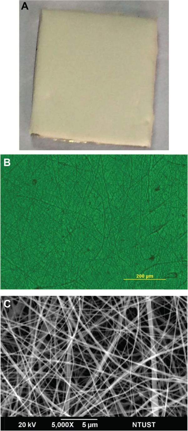
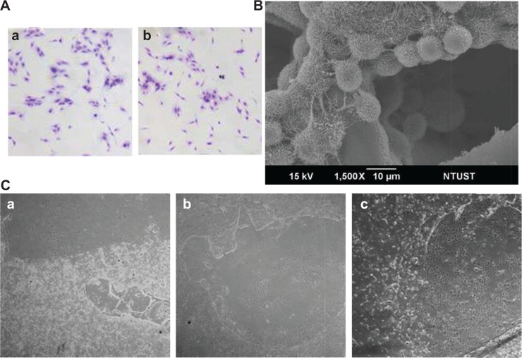
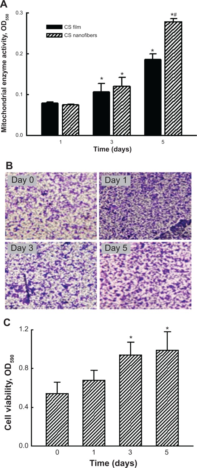
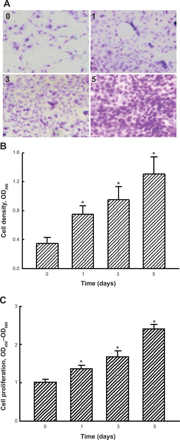
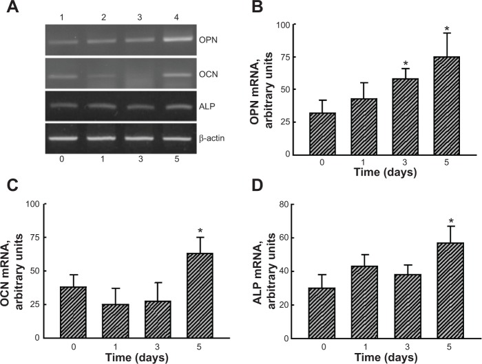
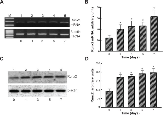
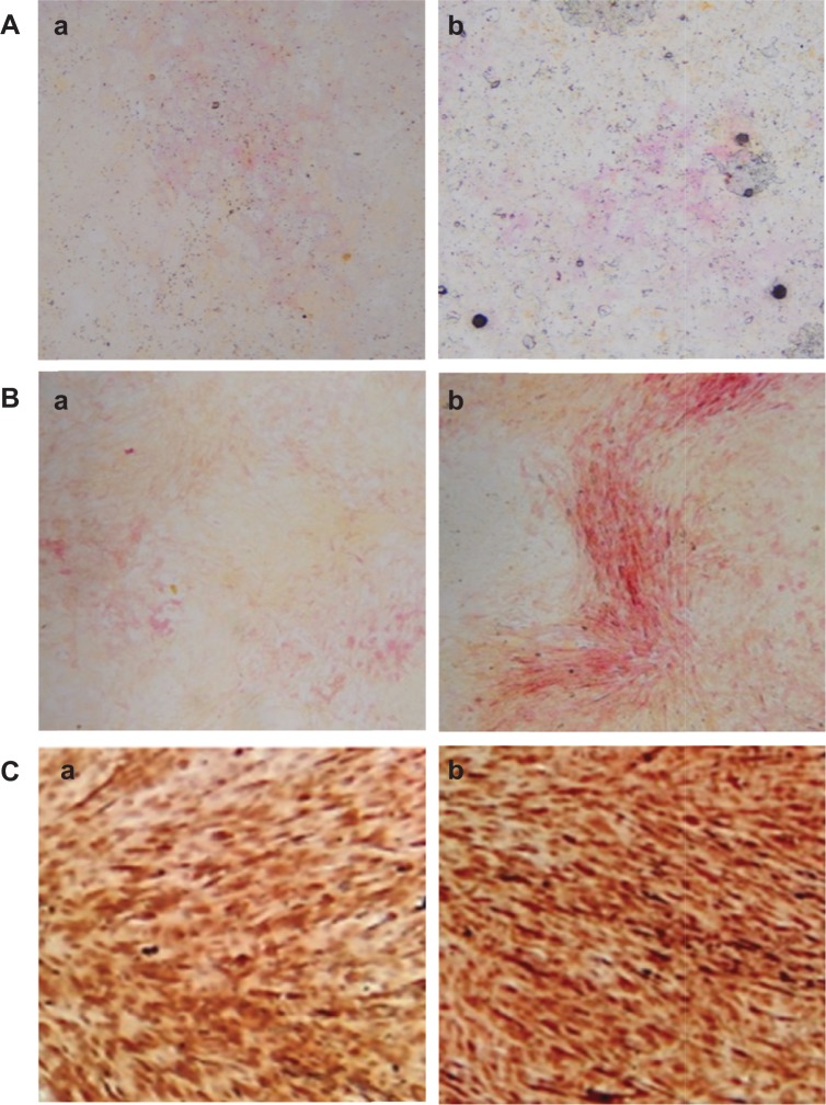
Osteoblast maturation plays a key role in regulating osteogenesis. Electrospun nanofibrous products were reported to possess a high surface area and porosity. In this study, we developed chitosan nanofibers and examined the effects of nanofibrous scaffolds on osteoblast maturation and the possible mechanisms. Macro- and micro observations of the chitosan nanofibers revealed that these nanoproducts had a flat surface and well-distributed fibers with nanoscale diameters. Mouse osteoblasts were able to attach onto the chitosan nanofiber scaffolds, and the scaffolds degraded in a time-dependent manner. Analysis by scanning electron microscopy further showed mouse osteoblasts adhered onto the scaffolds along the nanofibers, and cell-cell communication was also detected. Mouse osteoblasts grew much better on chitosan nanofiber scaffolds than on chitosan films. In addition, human osteoblasts were able to adhere and grow on the chitosan nanofiber scaffolds. Interestingly, culturing human osteoblasts on chitosan nanofiber scaffolds time-dependently increased DNA replication and cell proliferation. In parallel, administration of human osteoblasts onto chitosan nanofibers significantly induced osteopontin, osteocalcin, and alkaline phosphatase (ALP) messenger (m)RNA expression. As to the mechanism, chitosan nanofibers triggered runt-related transcription factor 2 mRNA and protein syntheses. Consequently, results of ALP-, alizarin red-, and von Kossa-staining analyses showed that chitosan nanofibers improved osteoblast mineralization. Taken together, results of this study demonstrate that chitosan nanofibers can stimulate osteoblast proliferation and maturation via runt-related transcription factor 2-mediated regulation of osteoblast-associated osteopontin, osteocalcin, and ALP gene expression.

Keywords: Runx2; chitosan nanofibers; osteoblast maturation; osteoblast-associated gene expression.


Figures

Figure 1
Figure 1
Macro- and microstructures of chitosan nanofibers. Notes: Chitosan nanofiber scaffolds were prepared using an electrospinning setup. The macro- (A) and microstructures (B) of chitosan nanofibers were observed and photographed using a camera and light microscopy, respectively. The surface morphology of chitosan nanofiber scaffolds was further scanned and photographed using scanning electron microscopy (C). Abbreviation: NTUST, National Taiwan University of Science and Technology.
Figure 2
Figure 2
Osteoblast adhesion and biocompatibility of chitosan nanofibers. Notes: Mouse osteoblasts were seeded overnight in 6 cm tissue culture plates coated with chitosan nanofiber scaffolds. Cell adhesion was assayed using a crystal violet staining protocol (A): (a) CS film; (b) CS nanofibers. Also, adhesion of mouse osteoblasts onto chitosan nanofiber scaffolds was further analyzed using scanning electron microscopy (B). (C) After culturing mouse osteoblasts on chitosan nanofiber scaffolds for (a) 1, (b) 3, and (c) 5 days, the biocompatibility of the electrospun scaffolds was observed and photographed using light microscopy. 40× magnification. Abbreviations: CS, chitosan; NTUST, National Taiwan University of Science and Technology.
Figure 3
Figure 3
Effects of chitosan nanofibers on stimulating the growth of mouse osteoblasts. Notes: Mouse osteosarcoma UMR-106 cells were seeded on chitosan films and chitosan nanofiber scaffolds for 1, 3, and 5 days. Cell growth was assayed by detecting mitochondrial complex I enzyme activity (A). Analysis of crystal violet staining was carried out to further verify the effects of chitosan nanofibers on the growth of mouse calvarial MC3T3-E1 cells (B). Stained cells were dissolved, and the signals were quantified and statistically analyzed (C). Each value represents the mean ± standard error of the mean from four independent experiments. * and # indicate that values significantly (P<0.05) differed from the control and chitosan film-treated groups, respectively. 40× magnification. Abbreviations: CS, chitosan; OD550, optical density at 550 nM wavelength; OD590, optical density at 590 nM wavelength.
Figure 4
Figure 4
Effects of chitosan nanofibers on stimulating the growth and proliferation of human osteoblasts. Notes: Human osteoblast-like MG63 cells were seeded on chitosan nanofiber scaffolds for 1, 3, and 5 days. Growth of human osteoblasts was determined by crystal violet staining (A). Stained cells were dissolved, and the signals were quantified and statistically analyzed (B). Cell proliferation was assayed using an enzyme-linked immunosorbent assay bromodeoxyuridine kit (C). Each value represents the mean ± standard error of the mean from four independent experiments. * indicates that values significantly differed from the respective control, P<0.05. 40× magnification. Abbreviations: OD450, optical density at 450 nM wavelength; OD590, optical density at 590 nM wavelength; OD690, optical density at 690 nM wavelength.
Figure 5
Figure 5
Effects of chitosan nanofibers on the induction of osteoblast differentiation-related OPN, OCN, and ALP gene expression. Notes: Human osteoblast-like MG63 cells were seeded on chitosan nanofiber scaffolds for 1, 3, and 5 days. (A) Analyses of OPN, OCN, and ALP mRNAs were conducted using RT-PCR; β-actin mRNA was analyzed as an internal control. The DNA bands were quantified and statistically analyzed (BD). Each value represents the mean ± standard error of the mean from four independent experiments. * indicates that values significantly differed from the respective control, P<0.05. Abbreviations: ALP, alkaline phosphatase; mRNA, messenger RNA; OCN, osteocalcin; OPN, osteopontin; RT-PCR, reverse transcription polymerase chain reaction.
Figure 6
Figure 6
Effects of chitosan nanofibers on the induction of Runx2 mRNA and protein expressions Notes: Human osteoblast-like MG63 cells were seeded on chitosan nanofiber scaffolds for 1, 3, 5, and 7 days. (A) Runx2 mRNA was analyzed using RT-PCR; β-actin mRNA was analyzed as an internal control. The DNA bands were quantified and statistically analyzed (B). (C) Runx2 was detected using a mouse monoclonal antibody; β-actin was immunodetected as an internal control. The immunoreactive protein bands were quantified and statistically analyzed (D). Each value represents the mean ± standard error of the mean from four independent experiments. * indicates that values significantly differed from the respective control, P<0.05. Abbreviations: mRNA, messenger RNA; Runx2, runt-related transcription factor 2; RT-PCR, reverse transcription polymerase chain reaction.
Figure 7
Figure 7
Effects of chitosan nanofibers on the mineralization of human osteoblasts. Notes: Human osteoblast-like MG63 cells were seeded on chitosan films and chitosan nanofiber scaffolds, and then exposed to a differentiation reagent (10 nM dexamethasone, 100 μg/mL ascorbic acid, and 10 mM β-glycerophosphate) for 21 days. The differentiation reagent was renewed every 2 days. Mineralization of human osteoblasts was determined using ALP-, Alizarin red S dye-, and von Kossa-staining protocols. (A) ALP staining: (a) chitosan film and (b) chitosan nanofibers. (B) Alizarin red staining: (a) chitosan film and (b) chitosan nanofibers. (C) von-Kossa staining: (a) chitosan film and (b) chitosan nanofibers. 40× magnification. Abbreviation: ALP, alkaline phosphatase.

References

    1. Seeman E, Delmas PD. Bone quality – the material and structural basis of bone strength and fragility. New Engl J Med. 2006;354:2250–2261. - PubMed
    1. Ducy P, Schinke T, Karsenty G. The osteoblast: a sophisticated fibroblast under central surveillance. Science. 2000;289:1501–1504. - PubMed
    1. Chen RM, Lin YL, Chou CW. GATA-3 transduces survival signals in osteoblasts through upregulation of bcl-xL gene expression. J Bone Min Res. 2010;25:2193–2204. - PubMed
    1. Aubin JE, Liu F, Malaval L, Gupta AK. Osteoblast and chondroblast differentiation. Bone. 1995;17:77S–83S. - PubMed
    1. Giustina A, Mazziotti G, Canalis E. Growth hormone, insulin-like growth factors, and the skeleton. Endocrine Rev. 2008;29:535–559. - PMC - PubMed

Publication types

MeSH terms