. 2014 Sep 16:5:1553-68.
doi: 10.3762/bjnano.5.167. eCollection 2014.

Ionic liquid-assisted formation of cellulose/calcium phosphate hybrid materials

Affiliations

Ionic liquid-assisted formation of cellulose/calcium phosphate hybrid materials

Ahmed Salama et al. Beilstein J Nanotechnol. .

Abstract

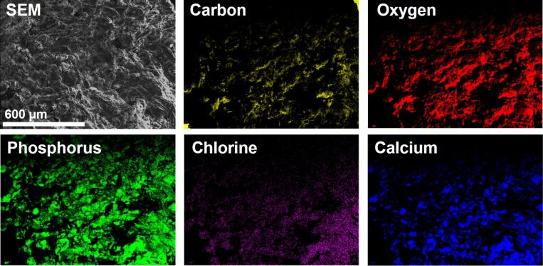
Cellulose/calcium phosphate hybrid materials were synthesized via an ionic liquid-assisted route. Scanning electron microscopy, transmission electron microscopy, energy-dispersive X-ray spectroscopy, X-ray diffraction, infrared spectroscopy, and thermogravimetric analysis/differential thermal analysis show that, depending on the reaction conditions, cellulose/hydroxyapatite, cellulose/chlorapatite, or cellulose/monetite composites form. Preliminary studies with MC3T3-E1 pre-osteoblasts show that the cells proliferate on the hybrid materials suggesting that the ionic liquid-based process yields materials that are potentially useful as scaffolds for regenerative therapies.

Keywords: biomineralization; calcium phosphate; carbohydrates; cellulose; hybrid materials; ionic liquid.


Figures

Figure 1
Figure 1
(A) ATR-FTIR spectra and (B) XRD patterns of calcium phosphates obtained from [Bmim][Cl]. The reflection at 14.2 degrees 2θ is from the sample holder (Si).
Figure 2
Figure 2
Low magnification (top row) and higher magnification (bottom row) SEM images of the precipitates. High magnification imaging of CPNaOH24 led to rapid sample charging and very poor imaging conditions even after sputtering; no image is thus shown.
Figure 3
Figure 3
SEM images of as-received microcrystalline and regenerated cellulose.
Figure 4
Figure 4
Low (top row) and high magnification (bottom row) SEM images of the hybrid materials prepared in the presence of GAA. Note the different scale bar in the lower row for CCPH3.
Figure 5
Figure 5
Low (top row) and high magnification (bottom row) SEM images of the hybrid materials prepared in the presence of NaOH.
Figure 6
Figure 6
TEM images of thin sections of CCPH2 and CCPH6. Top row are low magnification and bottom row are high magnification images of the same samples.
Figure 7
Figure 7
SEM image and elemental map of CCPH6.
Figure 8
Figure 8
XRD patterns of cellulose and mineralized samples. Panel A shows effects of acid or base addition, panel B shows effects of cellulose concentration in the case of the samples grown with NaOH.
Figure 9
Figure 9
ATR-FTIR spectra of neat cellulose, for sample nomenclature see Table 3. Panels B and D are higher magnification views of the region showing the calcium phosphate vibration bands. Spectra are shifted vertically for better visibility.
Figure 10
Figure 10
Representative TGA and DTA data of select samples. For full data see Table 3.

References

    1. Ravi Kumar M N V. React Funct Polym. 2000;46:1. doi: 10.1016/S1381-5148(00)00038-9. - DOI
    1. Mohanty A K, Misra M, Hinrichsen G. Macromol Mater Eng. 2000;276-277:1–24. doi: 10.1002/(SICI)1439-2054(20000301)276:1<1::AID-MAME1>3.0.CO;2-W. - DOI
    1. Klemm D, Heublein B, Fink H, Bohn A. Angew Chem, Int Ed. 2005;44:3358. doi: 10.1002/anie.200460587. - DOI - PubMed
    1. Eichhorn S J, Dufresne A, Aranguren M, Marcovich N E, Capadona J R, Rowan S J, Weder C, Thielemans W, Roman M, Renneckar S, et al. J Mater Sci. 2009;45:1. doi: 10.1007/s10853-009-3874-0. - DOI
    1. Epple M, editor. Biomaterialien und Biomineralisation. Vol. 1. Suttgart-Leipzig-Wiesbaden: Teubner; 2003.

LinkOut - more resources