. 2014 Sep 23;9(9):e108576.
doi: 10.1371/journal.pone.0108576. eCollection 2014.

APP is cleaved by Bace1 in pre-synaptic vesicles and establishes a pre-synaptic interactome, via its intracellular domain, with molecular complexes that regulate pre-synaptic vesicles functions

Affiliations

APP is cleaved by Bace1 in pre-synaptic vesicles and establishes a pre-synaptic interactome, via its intracellular domain, with molecular complexes that regulate pre-synaptic vesicles functions

Dolores Del Prete et al. PLoS One. .

Abstract

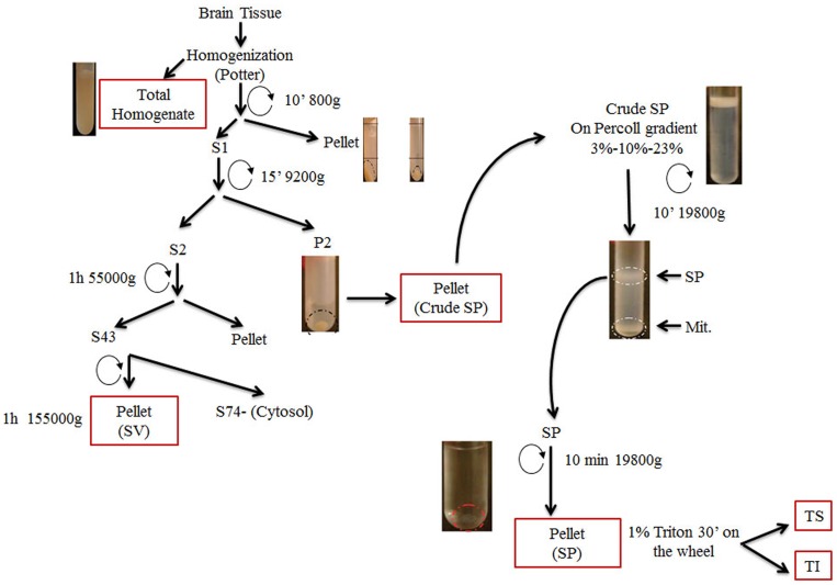
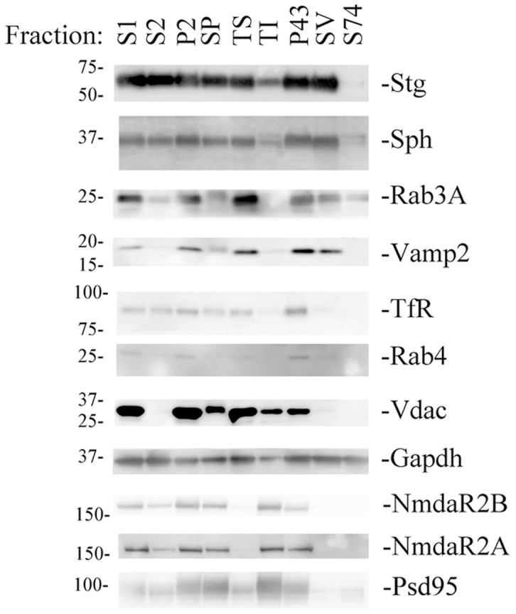
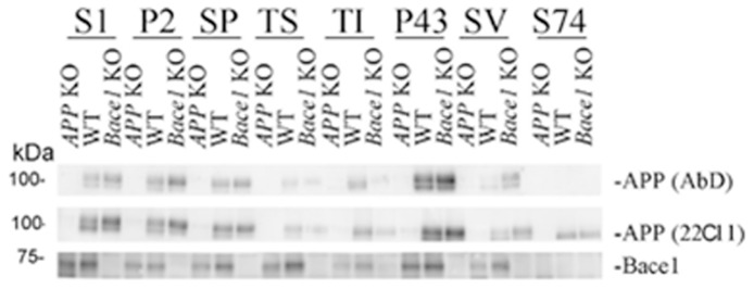
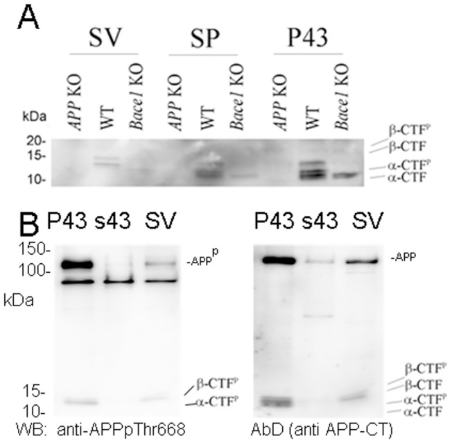
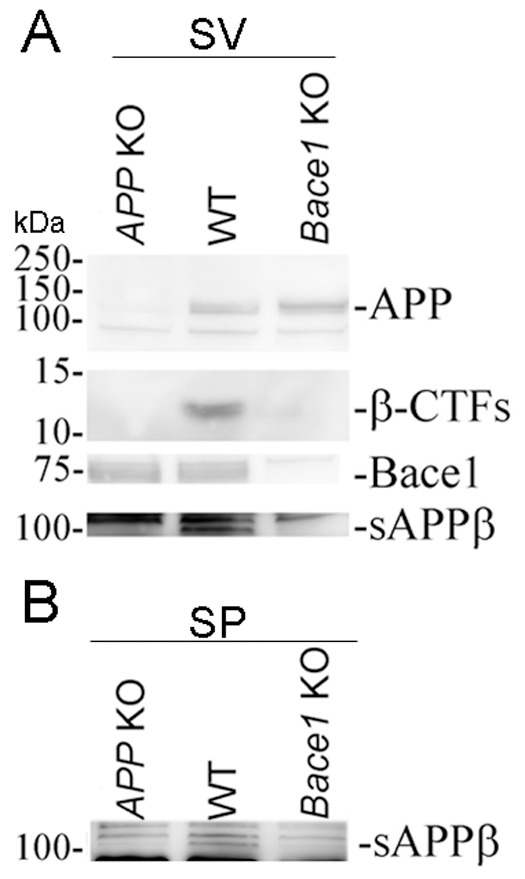
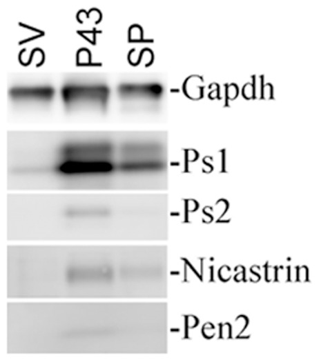
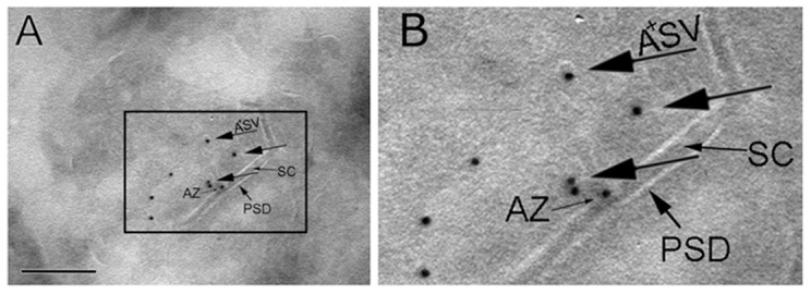
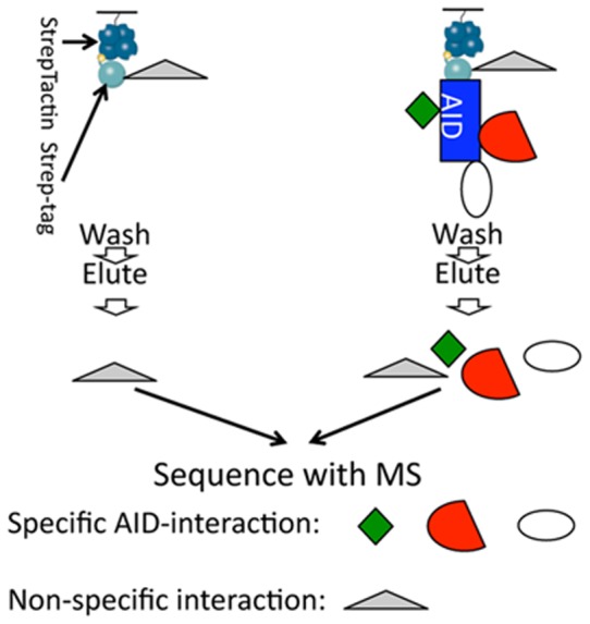
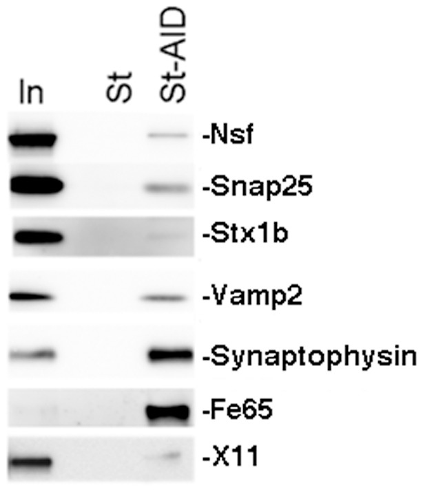
Amyloid Precursor Protein (APP) is a type I membrane protein that undergoes extensive processing by secretases, including BACE1. Although mutations in APP and genes that regulate processing of APP, such as PSENs and BRI2/ITM2B, cause dementias, the normal function of APP in synaptic transmission, synaptic plasticity and memory formation is poorly understood. To grasp the biochemical mechanisms underlying the function of APP in the central nervous system, it is important to first define the sub-cellular localization of APP in synapses and the synaptic interactome of APP. Using biochemical and electron microscopy approaches, we have found that APP is localized in pre-synaptic vesicles, where it is processed by Bace1. By means of a proteomic approach, we have characterized the synaptic interactome of the APP intracellular domain. We focused on this region of APP because in vivo data underline the central functional and pathological role of the intracellular domain of APP. Consistent with the expression of APP in pre-synaptic vesicles, the synaptic APP intracellular domain interactome is predominantly constituted by pre-synaptic, rather than post-synaptic, proteins. This pre-synaptic interactome of the APP intracellular domain includes proteins expressed on pre-synaptic vesicles such as the vesicular SNARE Vamp2/Vamp1 and the Ca2+ sensors Synaptotagmin-1/Synaptotagmin-2, and non-vesicular pre-synaptic proteins that regulate exocytosis, endocytosis and recycling of pre-synaptic vesicles, such as target-membrane-SNAREs (Syntaxin-1b, Syntaxin-1a, Snap25 and Snap47), Munc-18, Nsf, α/β/γ-Snaps and complexin. These data are consistent with a functional role for APP, via its carboxyl-terminal domain, in exocytosis, endocytosis and/or recycling of pre-synaptic vesicles.


Conflict of interest statement

Competing Interests: The authors have declared that no competing interests exist.

Figures

Figure 1
Figure 1. Schematic illustration of the fractionation protocol.
Mouse brain tissues were homogenized in homogenization buffer (HB) and centrifuged. The S1 supernatant fraction was collected and further centrifuged obtaining supernatant (S2) and pellet (P2) fractions. The P2 fraction was diluted in HB and directly applied to a discontinuous Percoll gradient to obtain the synaptosome fraction (SP) which was then lysed using 1% TritonX-100 to yield soluble (TS) and insoluble fractions (TI). The S2 fraction was ultra-centrifuged in two different steps to obtain two pellet fractions (P43 and SV) and supernatant (S74).
Figure 2
Figure 2. Analysis of the efficacy of the fractionation method.
The same amount of protein from each fraction was subjected to SDS-PAGE, followed by immuno-blotting for various marker proteins. Antibodies for Stg, Sph, Rab3A and Vamp2 were used as marker for pre-synaptic vesicles; the antibody for Vdac was used as markers for mitochondria; the antibodies for Rab4 and TfR were used as markers for endosomes: the antibodies for NmdaR2A, NmdaR2B and PSD95 were used as marker post-synaptic membrane. Anti-Gapdh was used to normalize the loading.
Figure 3
Figure 3. APP and Bace1 are found in SV fractions.
Western blot analysis was used to determine the levels of APP and Bace1 in the different brain fractions. APP KO and Bace1 KO mice fractions were used as controls for the specificity of the APP and Bace1 signals observed in the corresponding fractions isolated from WT brains.
Figure 4
Figure 4. The APP metabolite β-CTF, but not α-CTF, is found in SV fractions.
A) Analysis of APP-CTFs in membranous fractions of WT, APP KO and Bace1 KO mouse brains. Only β-CTF fragments are detected in the SV fraction of WT mice. Traces of α-CTF are seen in the Bace1 KO SV fraction. Since we did not detect APP-CTF in other SV preparations from Bace1 KO mice (see Fig. 5 for example), we conclude that this signal probably indicates a minute contamination of this SV preparation with other membranous fractions. B) Western blot analysis of P43, the soluble fraction s43 and SV with an anti-APPpThr668 antibody confirms the presence of APP and CTFs phosphorylated on Thr688. Of note, the membrane bound APP and APP-CTFs are absent, as expected, in the soluble fraction. Again, while α-CTF, α-CTF phosphorylated on Thr668 (α-CTFp), β-CTF and β-CTF phosphorylated on Thr668 (β-CTFp) are all present in the P43 fraction, only β-CTF and β-CTFp are detected in the SV fraction.
Figure 5
Figure 5. Evidence that Bace1 cleaves APP in pre-synaptic vesicles.
A) Equal amounts of proteins from SV fractions of WT, APP KO and Bace1 KO mice were subjected to SDS-PAGE, followed by immuno-blotting for APP (using AbD), Bace1 and sAPPβ. sAPPβ was detected in the SV fraction of WT mice supporting the hypothesis that Bace1 cleaves APP in pre-synaptic vesicles. B) sAPPβ was also detected in the SP fraction of WT mice.
Figure 6
Figure 6. Minimal levels of γ-secretase components are found in SV fractions.
Analysis of γ-secretase subunits distribution in brain fractions isolated from WT mice. SV, P43 and SP fractions were prepared using the protocol described in Fig. 1 and analyzed for the presence of the γ-secretase components Ps-1, Ps-2, Nct and Pen2 using Western blots. While all four γ-secretase components were readily detected in the P43 and, albeit at lower levels, SP fractions, only Ps-1 was detected, at very minimal level, in the SV fraction. Gapdh was used to normalize the loading.
Figure 7
Figure 7. APP localizes in pre-synaptic vesicles.
A) and B) Immuno-EM with the anti-APP-C-terminal antibody Y188 in hippocampus of wild type mouse brain shows APP in SV. We have used Y188 because this anti-APP antibody has demonstrated specificity for APP in Immuno-fluorescence experiments. The experiment was performed using cryosectioning, and immunogold labeling technique. Synapses were identified by morphology, i.e. SV, clefts (SC), the active zone (AZ) and the post-synaptic density (PSD). Larger arrows pointing gold particles (10 nm) indicate distribution of APP predominately on SV (A+SV). Scale bar in A is 250 nm. B) Enlarged version of a part of A.
Figure 8
Figure 8. Schematic explanation of the proteomic method used to determine the synaptic interactome of the intracellular domain of APP.
St and St-AID peptides were immobilized on StrepTactin resin. Mouse brain fractions were applied on columns; proteins were eluted, digested with trypsin and analyzed by nano LC/MS/MS.
Figure 9
Figure 9. Pre-synaptic proteins that regulate pre-synaptic vesicles endocytosis bind to the intracellular domain of APP.
Western blot analysis of pull-downs shows that Nsf, Snap25, Stx1b, Vamp2 and Synaptophysin specifically bind St-AID but not St peptides. The evidence that two previously known APP-interactors, Fe65 and X11, bind St-AID validate the proteomic approach used. In indicates the input.

References

    1. Tanzi RE (2012) The genetics of Alzheimer disease. Cold Spring Harb Perspect Med 2. - PMC - PubMed
    1. Muller UC, Zheng H (2012) Physiological Functions of APP Family Proteins. Cold Spring Harb Perspect Med 2: a006288. - PMC - PubMed
    1. Scheinfeld MH, Ghersi E, Laky K, Fowlkes BJ, D'Adamio L (2002) Processing of beta-amyloid precursor-like protein-1 and -2 by gamma-secretase regulates transcription. J Biol Chem 277: 44195–44201. - PubMed
    1. Heber S, Herms J, Gajic V, Hainfellner J, Aguzzi A, et al. (2000) Mice with combined gene knock-outs reveal essential and partially redundant functions of amyloid precursor protein family members. J Neurosci 20: 7951–7963. - PMC - PubMed
    1. Herms J, Anliker B, Heber S, Ring S, Fuhrmann M, et al. (2004) Cortical dysplasia resembling human type 2 lissencephaly in mice lacking all three APP family members. EMBO J 23: 4106–4115. - PMC - PubMed

Publication types

MeSH terms

Substances

LinkOut - more resources