

Impact of computer-aided detection systems on radiologist accuracy with digital mammography
- PMID: 25247960
- PMCID: PMC4286296
- DOI: 10.2214/AJR.12.10187
Impact of computer-aided detection systems on radiologist accuracy with digital mammography
Abstract
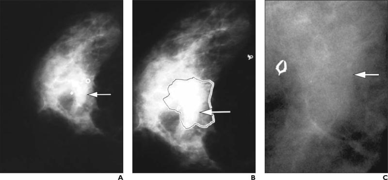
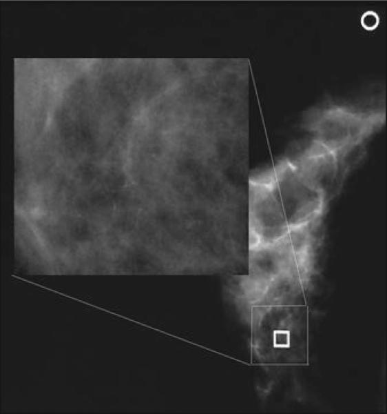
Objective: The purpose of this study was to assess the impact of computer-aided detection (CAD) systems on the performance of radiologists with digital mammograms acquired during the Digital Mammographic Imaging Screening Trial (DMIST).
Materials and methods: Only those DMIST cases with proven cancer status by biopsy or 1-year follow-up that had available digital images were included in this multireader, multicase ROC study. Two commercially available CAD systems for digital mammography were used: iCAD SecondLook, version 1.4; and R2 ImageChecker Cenova, version 1.0. Fourteen radiologists interpreted, without and with CAD, a set of 300 cases (150 cancer, 150 benign or normal) on the iCAD SecondLook system, and 15 radiologists interpreted a different set of 300 cases (150 cancer, 150 benign or normal) on the R2 ImageChecker Cenova system.
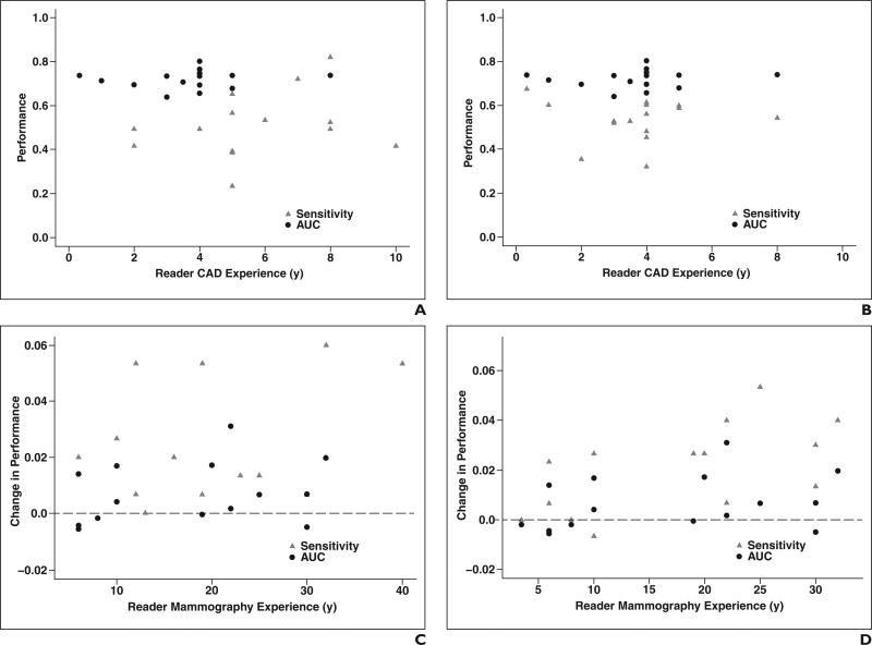
Results: The average AUC was 0.71 (95% CI, 0.66-0.76) without and 0.72 (95% CI, 0.67-0.77) with the iCAD system (p = 0.07). Similarly, the average AUC was 0.71 (95% CI, 0.66-0.76) without and 0.72 (95% CI 0.67-0.77) with the R2 system (p = 0.08). Sensitivity and specificity differences without and with CAD for both systems also were not significant.
Conclusion: Radiologists in our studies rarely changed their diagnostic decisions after the addition of CAD. The application of CAD had no statistically significant effect on radiologist AUC, sensitivity, or specificity performance with digital mammograms from DMIST.
Keywords: AUC; computer-aided detection; mammography; sensitivity; specificity.
Figures
References
-
- Pisano ED, Gatsonis CA, Yaffe MJ, et al. American College of Radiology Imaging Network Digital Mammographic Imaging Screening Trial: objectives and methodology. Radiology. 2005;236:404–412. - PubMed
-
- Pisano ED, Gatsonis C, Hendrick E, et al. Diagnostic performance of digital versus film mammography for breast-cancer screening. N Engl J Med. 2005;353:1773–1783. - PubMed
-
- Rao VM, Levin DC, Parker L, Cavanaugh B, Fran-gos AJ, Sunshine JH. How widely is computer-aided detection used in screening and diagnostic mammography? J Am Coll Radiol. 2010;7:802–805. - PubMed
-
- Nishikawa RM, Schmidt RA, Linver MN, Edwards AV, Papaioannou J, Stull MA. Clinically missed cancer: how effectively can radiologists use computer-aided detection? AJR. 2012;198:708–716. - PubMed
Publication types
MeSH terms
Grants and funding
LinkOut - more resources
Full Text Sources
Other Literature Sources
Medical
Miscellaneous
