. 2014 Sep 23;9(9):e108245.
doi: 10.1371/journal.pone.0108245. eCollection 2014.

Crystal structure of patatin-17 in complex with aged and non-aged organophosphorus compounds

Affiliations

Crystal structure of patatin-17 in complex with aged and non-aged organophosphorus compounds

Sanjeeva J Wijeyesakere et al. PLoS One. .

Abstract

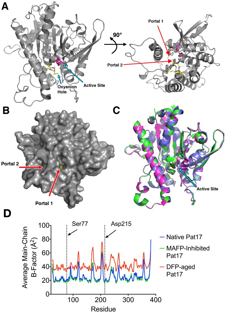
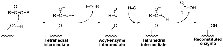
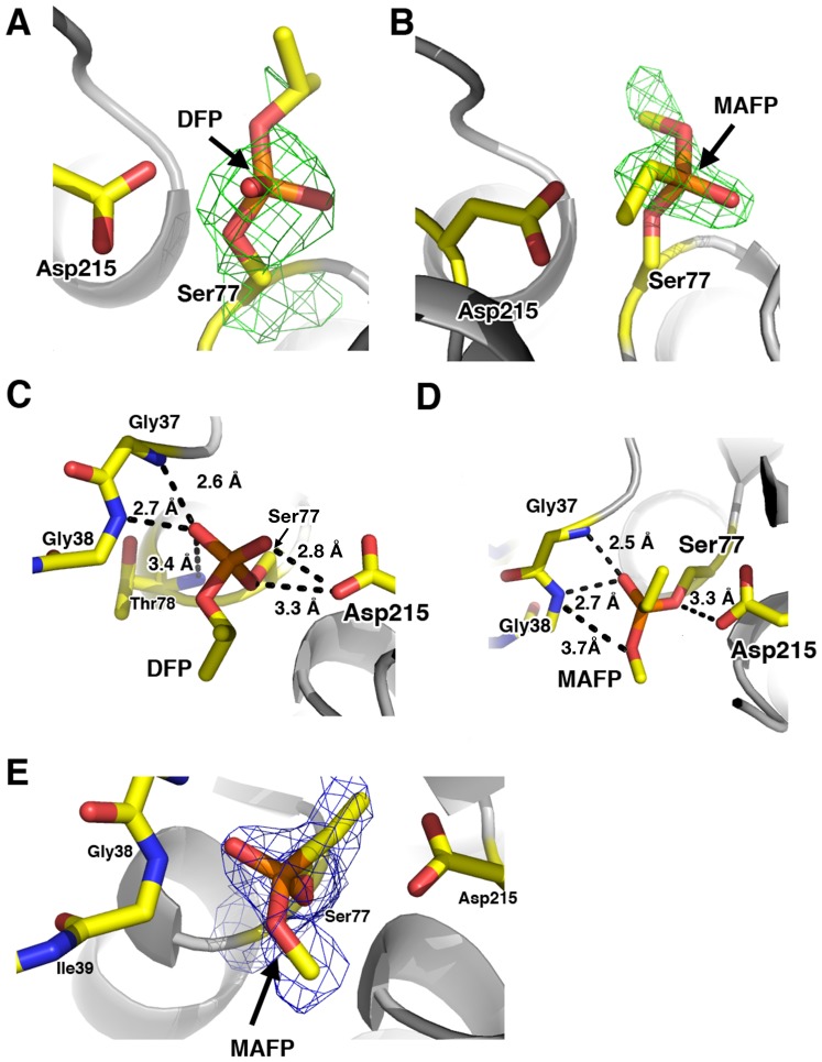
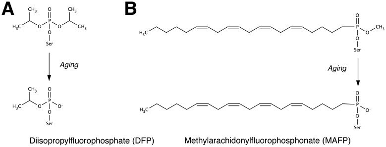
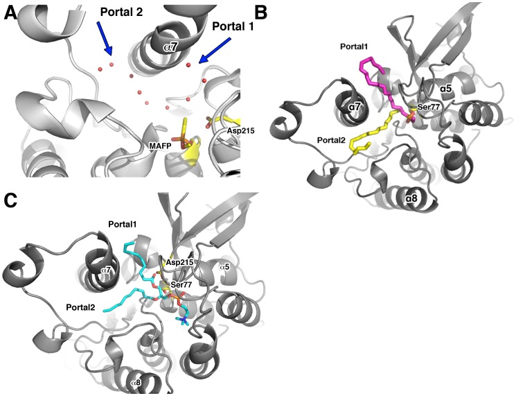
Patatin is a non-specific plant lipase and the eponymous member of a broad class of serine hydrolases termed the patatin-like phospholipase domain containing proteins (PNPLAs). Certain PNPLA family members can be inhibited by organophosphorus (OP) compounds. Currently, no structural data are available on the modes of interaction between the PNPLAs and OP compounds or their native substrates. To this end, we present the crystal structure of patatin-17 (pat17) in its native state as well as following inhibition with methyl arachidonyl fluorophosphonate (MAFP) and inhibition/aging with diisopropylphosphorofluoridate (DFP). The native pat17 structure revealed the existence of two portals (portal1 and portal2) that lead to its active-site chamber. The DFP-inhibited enzyme underwent the aging process with the negatively charged phosphoryl oxygen, resulting from the loss of an isopropyl group, being within hydrogen-binding distance to the oxyanion hole. The MAFP-inhibited pat17 structure showed that MAFP did not age following its interaction with the nucleophilic serine residue (Ser77) of pat17 since its O-methyl group was intact. The MAFP moiety is oriented with its phosphoryl oxygen in close proximity to the oxyanion hole of pat17 and its O-methyl group located farther away from the oxyanion hole of pat17 relative to the DFP-bound state. The orientation of the alkoxy oxygens within the two OP compounds suggests a role for the oxyanion hole in stabilizing the emerging negative charge on the oxygen during the aging reaction. The arachidonic acid side chain of MAFP could be contained within portals 1 or 2. Comparisons of pat17 in the native, inhibited, and aged states showed no significant global conformational changes with respect to their Cα backbones, consistent with observations from other α/β hydrolases such as group VIIA phospholipase A2.


Conflict of interest statement

Competing Interests: The authors did not receive direct funding from the Michigan Economic Development Corporation for this work. The Michigan Economic Development Corporation/Michigan Technology Tri-Corridor provided funding for the development of the LS-CAT beamlines at APS. The beamline operations request all users to print the prescribed acknowledgement statement in all publications describing structures whose data were collected at LS-CAT. The LS-CAT beam lines were used to collect diffraction data for the structures described in this manuscript. The use of the beamlines in no way restricts our ability to publish or share information, therefore this does not alter the authors' adherence to PLOS ONE policies on sharing data and materials.

Figures

Figure 1
Figure 1. Pat17 structure.
(A) The left panel is the overall view of pat17. The protein consists of 8 β-strands and 9 α-helices. The patatin fold is a modified α/β hydrolase fold with a central β-sheet sandwiched by α-helices. The residues that form the oxyanion hole (Gly37, Gly38, Ile39, Arg40 and Gly41) are colored yellow while residues in the active site dyad (Ser77 and Asp215) are shown in magenta. The right panel is a 90° rotation of the left panel about the x-axis depicting the location of the two portals (shown as red arrows) flanking helix 7 (α7) that lead into the active site chamber of the enzyme. (B) Solvent-accessible surface of native pat17 depicting the location of the two portals (denoted via red arrows). Pat17 is shown in the same orientation as the left panel in (A) with Ser77 rendered as yellow spheres. (C) Overlay of pat17 in its native (green), MAFP-inhibited (magenta) and DFP-aged (blue) states showing that there is no significant global conformational change associated with inhibition and aging of an OP compound on the active site serine (Ser77) of the enzyme (denoted as sticks with carbons colored green (native), magenta (MAFP-inhibited) or blue (DFP-aged)). An arrow denotes the active site of pat17. (D) Overlay of average main-chain B-factors for the residues in native, inhibited and aged pat17.
Figure 2
Figure 2. Proposed catalytic mechanism for the active site of Pat17.
Asp215 (not shown) acts as a general base and activates the Ser77 nucleophile by abstracting its terminal hydrogen. The activated Ser77 (shown as –OH) attacks the acyl carbon of the substrate forming a tetrahedral intermediate whose negative charge is shielded by the oxyanion hole of Pat17 (not shown). Loss of R-OH yields an acyl-enzyme intermediate that is hydrolyzed rapidly (passing through another tetrahedral intermediate) to release the acyl moiety and regenerate the enzyme.
Figure 3
Figure 3. Pat17 bound to OPs.
Initial difference (Fo-Fc) maps contoured at 3σ (depicted as a green mesh) showing the positive electron densities around (A) the aged DFP and (B) the non-aged MAFP adducts on the catalytic Ser77 residue of pat17. (C and D) The active site of patatin with (C) the aged DFP and (D) non-aged MAFP adducts showing contact distances between the adducts and the oxyanion hole of pat17 as well as the contact distance between the negatively charged oxygen atoms of the aged DFP and the backbone amide nitrogen of Thr78. For clarity, the orientation of the molecule is a 180° rotation about the X- and Y-axes in panel A. (E) 2Fo-Fc electron density (contoured at 1 σ; depicted as a blue mesh) around the catalytic Ser77 residue in complex with MAFP. The active site Ser77 and Asp215 residues as well as the residues that comprise the oxyanion hole of pat17 are rendered as sticks with the following color scheme: yellow  =  carbon, blue  =  nitrogen and red  =  oxygen.
Figure 4
Figure 4. Structures of (A) DFP and (B) MAFP in their native state as well as the putative structures of these inhibitors in their aged forms.
Chemical structures were generated in MarvinSketch 5.5 (ChemAxon, http://www.chemaxon.com).
Figure 5
Figure 5. Substrate entry and exit points in Pat17.
(A) Pat17 in complex with MAFP (with crystallographic water molecules shown as red spheres) depicting the trail of water molecules leading from the active site chamber through portals 1 and 2. (B) Model depicting the non-aged MAFP adduct with its arachidonic acid chain modeled as going through portal1 (with carbons colored magenta) or portal2 (with carbons colored yellow). (C) Model showing the phospholipid POPC (PDB ligand ID POV; depicted with carbons colored cyan) accommodated within the active site of pat17 with the lipid side-chains modeled within both, portal1 and portal2. The active site Ser77 and Asp215 residues as well as the MAFP and POPC moieties are rendered as sticks with the following color scheme: yellow/magenta/cyan  =  carbon, blue  =  nitrogen and red  =  oxygen.

References

    1. Andrews DL, Beames B, Summers MD, Park WD (1988) Characterization of the lipid acyl hydrolase activity of the major potato (Solanum tuberosum) tuber protein, patatin, by cloning and abundant expression in a baculovirus vector. Biochem J 252: 199–206. - PMC - PubMed
    1. Hirayama O, Matsuda H, Takeda H, Maenaka K, Takatsuka H (1975) Purification and properties of a lipid acyl-hydrolase from potato tubers. Biochim Biophys Acta 384: 127–137. - PubMed
    1. Ganal MW, Bonierbale MW, Roeder MS, Park WD, Tanksley SD (1991) Genetic and physical mapping of the patatin genes in potato and tomato. Mol Gen Genet 225: 501–509. - PubMed
    1. Pots AM, Gruppen H, Hessing M, van Boekel MA, Voragen AG (1999) Isolation and characterization of patatin isoforms. J Agric Food Chem 47: 4587–4592. - PubMed
    1. Strickland JA, Orr GL, Walsh TA (1995) Inhibition of Diabrotica Larval Growth by Patatin, the Lipid Acyl Hydrolase from Potato Tubers. Plant Physiol 109: 667–674. - PMC - PubMed

Publication types

LinkOut - more resources