
Smoking accelerates aging of the small airway epithelium

Matthew S Walters et al. Respir Res. .

Abstract

Background: Aging involves multiple biologically complex processes characterized by a decline in cellular homeostasis over time leading to a loss and impairment of physiological integrity and function. Specific cellular hallmarks of aging include abnormal gene expression patterns, shortened telomeres and associated biological dysfunction. Like all organs, the lung demonstrates both physiological and structural changes with age that result in a progressive decrease in lung function in healthy individuals. Cigarette smoking accelerates lung function decline over time, suggesting smoking accelerates aging of the lung. Based on this data, we hypothesized that cigarette smoking accelerates the aging of the small airway epithelium, the cells that take the initial brunt of inhaled toxins from the cigarette smoke and one of the primary sites of pathology associated with cigarette smoking.

Methods: Using the sensitive molecular parameters of aging-related gene expression and telomere length, the aging process of the small airway epithelium was assessed in age matched healthy nonsmokers and healthy smokers with no physical manifestation of lung disease or abnormalities in lung function.

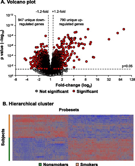
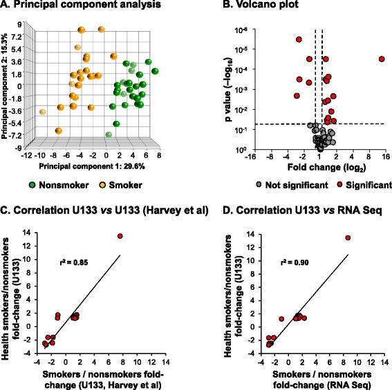
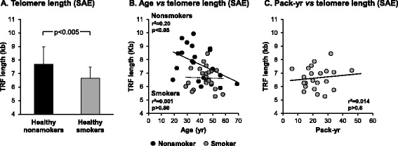
Results: Analysis of a 73 gene aging signature demonstrated that smoking significantly dysregulates 18 aging-related genes in the small airway epithelium. In an independent cohort of male subjects, smoking significantly reduced telomere length in the small airway epithelium of smokers by 14% compared to nonsmokers.

Conclusion: These data provide biologic evidence that smoking accelerates aging of the small airway epithelium.


Figures

Figure 1
Figure 1
Smoking dysregulated genes in the small airway epithelium. A. Volcano plot, smoker vs nonsmoker small airway epithelium (n = 29 nonsmokers and n = 29 smokers) using all expressed (P call ≥10) probesets as input dataset. Ordinate – p value; abscissa – fold-change (log2). Data demonstrates 2488 smoking-dysregulated probesets [fold-change ≥1.2, p < 0.05 with false discovery rate (FDR) correction] representing a total of 1737 unique genes. B. Hierarchical cluster analysis of smoker vs nonsmoker small airway epithelium based on expression of 2488 smoking-dysregulated probesets [fold-change ≥1.2, p < 0.05 with false discovery rate (FDR) correction]. Probesets expressed above the average are represented in red, below average in blue and average in grey. The probesets are represented horizontally and individual samples vertically.
Figure 2
Figure 2
Expression of an aging gene signature in small airway epithelium of age matched healthy smokers vs healthy nonsmokers. A. Principal component analysis of gene expression of small airway epithelium of smokers (n = 29, orange circles) and nonsmokers (n = 29, green circles) using the de Magalhaes et al. [26] 67 gene aging signature as an input dataset. Data is corrected for all covariates except smoking status. B. Volcano plot, smoker vs nonsmoker small airway epithelium of the 67 aging gene signature. Ordinate – p value (log10); abscissa – fold-change (log2). Red circles represent genes significantly differentially expressed in smoker vs nonsmoker small airway epithelium (≥1.2 fold-change up- or down-regulated; p < 0.05 using a Benjamini-Hochberg correction of false discovery rate). C. Correlation analysis of the 18 aging genes differentially expressed in the small airway epithelium of healthy smokers vs healthy nonsmokers, comparing smoker vs nonsmoker fold-changes in U133 gene expression with U133 gene expression from an independent dataset (n = 12 nonsmokers and n = 10 smokers, GSE4498) [25]. D. Correlation analysis of the 18 aging genes differentially expressed in the small airway epithelium of healthy smokers vs healthy nonsmokers, comparing smoker vs nonsmoker fold-changes in U133 gene expression with RNA Seq gene expression from an independent dataset (n = 5 nonsmokers and n = 6 smokers) [27].
Figure 3
Figure 3
Immunohistochemical staining analysis of smoking dysregulated aging genes in the small airway epithelium. Normal nonsmoker human bronchus sections from 3 independent donors were analyzed for expression of C3, clusterin (CLU), CX3CL1, LAPTM5, MGST1 and SPP1 using gene specific antibodies. Isotype specific antibody was used as negative control. Scale bar 20 μm.
Figure 4
Figure 4
Telomere length in the small airway epithelium of nonsmokers and smokers. DNA was isolated from the small airway epithelium of male healthy nonsmokers (n = 21) and male healthy smokers (n = 22) and telomere length [terminal restriction fragment (TRF)] quantified by Southern analysis. A. Data shown is represented as the average ± standard deviation of the TRF length (kb). The difference in mean TRF length between phenotypes was calculated by 2-tailed Students t test. B. Correlation of telomere length with age. Telomere length was quantified by Southern analysis and correlated with the age of each individual subject. Correlations between mean telomere length and age were performed using linear regression. Black circles represent healthy nonsmokers and grey circles represent healthy smokers. C. Correlation of telomere length with smoking history (pack-yr). Telomere length from healthy smokers was quantified by Southern analysis and correlated with the smoking history for each individual subject. Correlations between mean telomere length and smoking history were performed using linear regression.

References

    1. Kirkwood TB. Understanding the odd science of aging. Cell. 2005;120:437–447. doi: 10.1016/j.cell.2005.01.027. - DOI - PubMed
    1. Lopez-Otin C, Blasco MA, Partridge L, Serrano M, Kroemer G. The hallmarks of aging. Cell. 2013;153:1194–1217. doi: 10.1016/j.cell.2013.05.039. - DOI - PMC - PubMed
    1. Kerstjens HA, Rijcken B, Schouten JP, Postma DS. Decline of FEV1 by age and smoking status: facts, figures, and fallacies. Thorax. 1997;52:820–827. doi: 10.1136/thx.52.9.820. - DOI - PMC - PubMed
    1. Janssens JP, Pache JC, Nicod LP. Physiological changes in respiratory function associated with ageing. Eur Respir J. 1999;13:197–205. doi: 10.1183/09031936.99.14614549. - DOI - PubMed
    1. Fletcher C, Peto R. The natural history of chronic airflow obstruction. Br Med J. 1977;1:1645–1648. doi: 10.1136/bmj.1.6077.1645. - DOI - PMC - PubMed

Publication types

MeSH terms