Case Reports
. 2014 Sep 23;2014(9):rju094.
doi: 10.1093/jscr/rju094.

Primary thyroid schwannoma masquerading as a thyroid nodule

Affiliations
Case Reports

Primary thyroid schwannoma masquerading as a thyroid nodule

Harsh Dhar et al. J Surg Case Rep. .

Abstract

The thyroid gland is a very rare site for head and neck schwannomas. Till date there have been only 19 reported cases in English literature. Only 25% of schwannomas occur in the head and neck region, most of them arising in relation to the peripheral nerves and cervical sympathetic chain. We report a similar case, with clinical and sonological features of a benign thyroid nodule. The diagnosis of schwannoma was established on the final histopathology report and a review of the slides and the imaging was done to confirm the site of origin. A thorough review of earlier reported cases was done. We summarize the existing knowledge on this entity, emphasizing the challenge of diagnosing it pre-operatively.


Figures

Figure 1:
Figure 1:
USG of the neck: hyperechoic cystic lesion abutting the postero-lateral aspect of the right lobe of thyroid.
Figure 2:
Figure 2:
Contrast enhanced CT neck (axial section). Heterogeneously enhancing lesion with multiple cystic areas within abutting the postero-lateral aspect of the right lobe of thyroid, pushing it anteriorly and medially.
Figure 3:
Figure 3:
Contrast enhanced CT neck (coronal section). Coronal section showing good plane of delineation between the lesion and carotid sheath, along with mass effect on the thyroid gland and the right common carotid.
Figure 4:
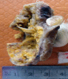
Figure 4:
Cross section of hemithyroidectomy specimen: a well-circumscribed cystic lesion with yellowish colour of the inner cyst wall; a rim of normal thyroid is seen at the periphery.
Figure 5:
Figure 5:
Microscopic section (H&E slide, 40×) of well-encapsulated lesion with hyper- (Antoni A) and hypocellular (Antoni B) areas. Multiple cystic areas are also seen.
Figure 6:
Figure 6:
Microscopic section (H&E slide, 40×) of Verocay bodies within Antoni A pattern.
Figure 7:
Figure 7:
Microscopic section (H&E slide, 100×) of magnified view of a region in Fig. 6 showing Verocay bodies.

References

    1. De Paoli F, Giugliano G, Casadio C, Tredici P, Bruschini R, De Fiori E. Schwannoma of thyroid bed. A case report and considerations on interdisciplinary collaboration. Acta Otorhinolaryngol Ital. 2005;25:250–4. - PMC - PubMed
    1. Delaney WE, Fry KE. Neurilemoma of the thyroid gland. Ann Surg. 1964;160:1014–7. - PMC - PubMed
    1. Cashman E, Skinner LJ, Timon C. Thyroid swelling: an unusual presentation of a cervical sympathetic chain schwannoma. Medscape. 2008;10:201. - PMC - PubMed
    1. An J, Oh YL, Shin JS, Jeong HS. Primary schwannoma of thyroid gland. Acta Cytol. 2010;54:857–62. - PubMed
    1. Kandil E, Khalek MA, Abdullah O, Dali D, Faruqui S, Khan A, Friedlander P, Jaffe BM, Crawford B. Primary peripheral nerve sheath tumours of the thyroid gland. Thyroid. 2010;20:583–6. - PubMed

Publication types

LinkOut - more resources