

doi: 10.1038/leu.2014.285.
Epub 2014 Sep 24.
MARIMO cells harbor a CALR mutation but are not dependent on JAK2/STAT5 signaling
Affiliations
- PMID: 25249012
- PMCID: PMC4320290
- DOI: 10.1038/leu.2014.285
Item in Clipboard
MARIMO cells harbor a CALR mutation but are not dependent on JAK2/STAT5 signaling
Leukemia.
2015 Feb.
Free PMC article
No abstract available
Figures
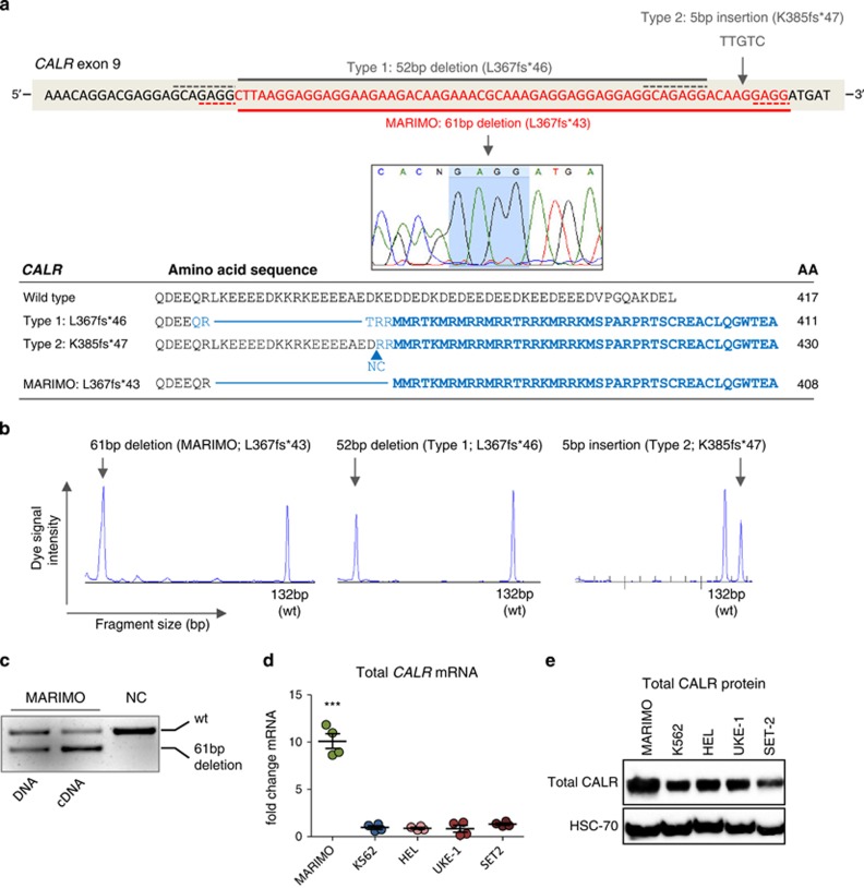
Identification of a CALR-mutated human cell line. (a) Top panel shows the mutated region in CALR exon 9 (red bases). The commonest CALR mutations are shown above the DNA sequence. Solid gray line shows type 1 (52- bp deletion; c.1099_1150del; L367fs*46) and gray arrow shows type 2 (5- bp insertion; c.1154_1155_ins; K385fs*47 mutations). The CALR mutation in human cell line MARIMO is shown below the DNA sequence. Solid red line and capillary sequencing image show a heterozygous 61-bp deletion (c.1099_1159del; L367fs*43) in MARIMO. Dashed gray and red lines represent the homologous sequence flanking the deleted regions in type 1 and MARIMO mutations, respectively, also highlighted in the capillary sequencing image (pale blue) for MARIMO. Lower panel shows the predicted protein sequence of the commonest CALR mutations and of MARIMO with total protein sizes. Amino acids (AA) in the new reading frame are shaded blue and the common novel peptide sequence shared by the different CALR variants are in bold blue. (b) PCR amplification of CALR exon 9 followed by fragment size analysis, as used for diagnostic testing for CALR mutations. Vertical heights of peaks represent dye signal intensity and horizontal position of peaks reflect the fragment size of the PCR amplicon. Wild type (wt) peak occurs at 132- bp. Left panel shows wt and mutated alleles of MARIMO (61-bp separation in peaks), middle panel shows Type 1/L367fs*46 with peak separation of 52 bp and right panel shows Type 2/K385fs*47 peaks separated by 5 bp. (c) Agarose gel image showing wt (upper band) and mutated-CALR (lower band) in MARIMO DNA and cDNA. (d) Quantitative real-time PCR of total CALR mRNA levels expressed as a fold change relative to house-keeping RPLP0 levels, for the cell lines MARIMO, the BCR-ABL1+ CML cell line K562, and the JAK2V617F+ cell lines HEL, UKE-1 and SET-2. Graph depicts all data points generated in two independent experiments performed in duplicate. ***P<0.001 (e) Western blot showing total CALR protein levels of MARIMO and four other myeloid cell lines.
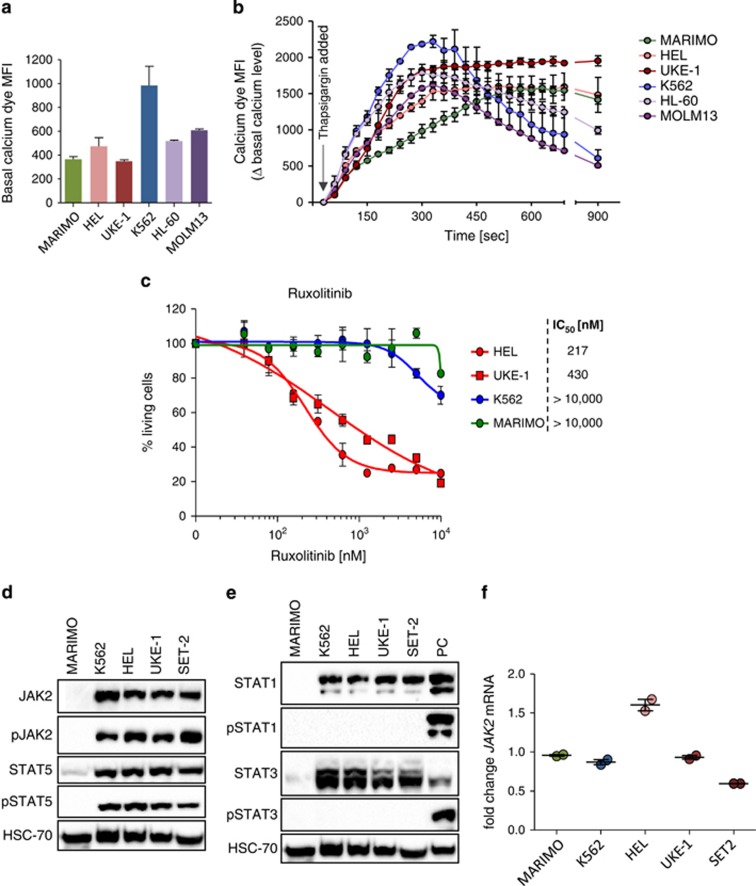
Characterization of the cell line MARIMO. (a and b) Basal cytoplasmic calcium level (a) and changes in cytoplasmic calcium levels over time upon addition of thapsigargin (b) in MARIMO and five other leukemic cell lines. (c) Dose response curves for the JAK2-inhibitor Ruxolitinib in the cell lines MARIMO, K562, HEL and UKE-1. (d and e) Western blots showing protein levels of the inactive and phosphorylated forms of JAK2 and STAT5 (d) and STAT1 and STAT3 (e). PC, positive control. (f) JAK2 mRNA levels expressed as fold changes relative to RPLP0 in MARIMO and four myeloid cell lines.
References
-
- Klampfl T, Gisslinger H, Harutyunyan AS, Nivarthi H, Rumi E, Milosevic JD, et al. Somatic mutations of calreticulin in myeloproliferative neoplasms. N Engl J Med. 2013;369:2379–2390. - PubMed
-
- Tefferi A, Thiele J, Vannucchi AM, Barbui T. An overview on CALR and CSF3R mutations and a proposal for revision of WHO diagnostic criteria for myeloproliferative neoplasms. Leukemia. 2014;28:1407–1413. - PubMed
-
- Reilly JT, McMullin MF, Beer PA, Butt N, Conneally E, Duncombe AS, et al. Use of JAK inhibitors in the management of myelofibrosis: a revision of the British Committee for Standards in Haematology Guidelines for Investigation and Management of Myelofibrosis 2012 Br J Haematol 2014. e-pub ahead of print 25 June 2014; doi:10.1111/bjh.12985 - DOI - PubMed
Publication types
MeSH terms
Substances
Grants and funding
LinkOut - more resources
Full Text Sources
Other Literature Sources
Research Materials
Miscellaneous
