. 2015 May;8(3):533-44.
doi: 10.1038/mi.2014.86. Epub 2014 Sep 24.

Human gastric epithelial cells contribute to gastric immune regulation by providing retinoic acid to dendritic cells

Affiliations

Human gastric epithelial cells contribute to gastric immune regulation by providing retinoic acid to dendritic cells

D Bimczok et al. Mucosal Immunol. 2015 May.

Abstract

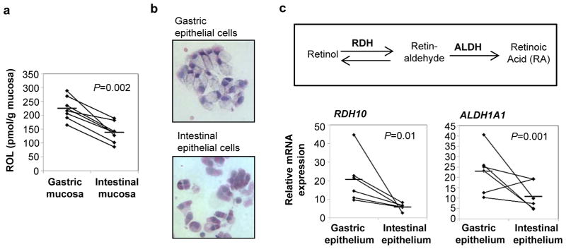
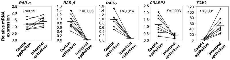
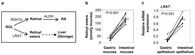
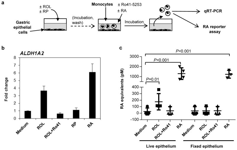
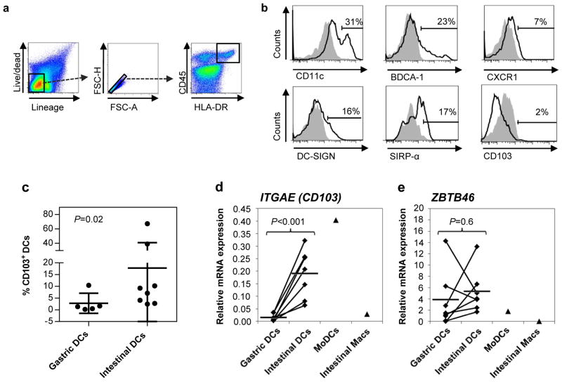
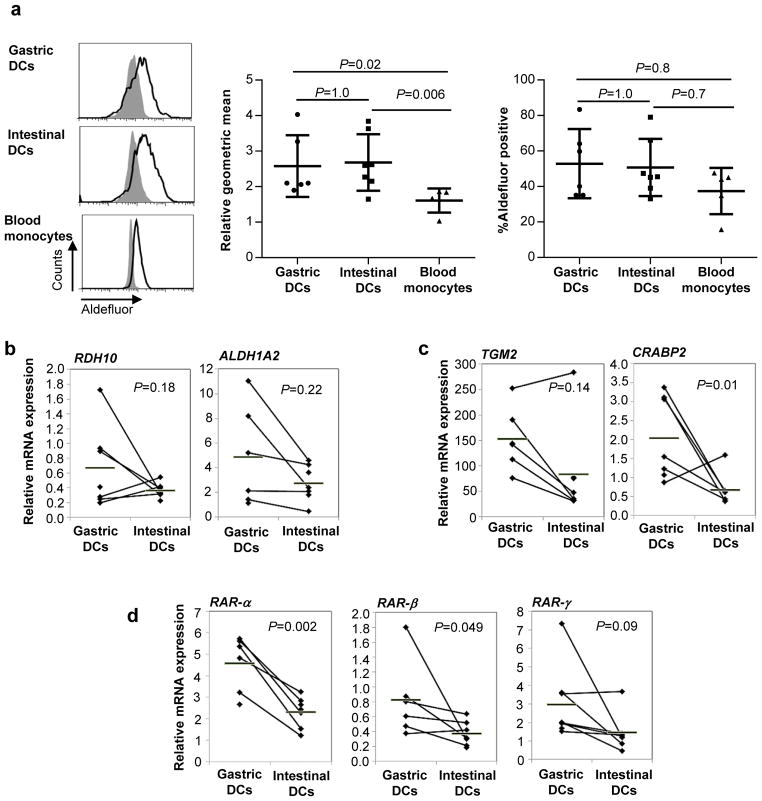
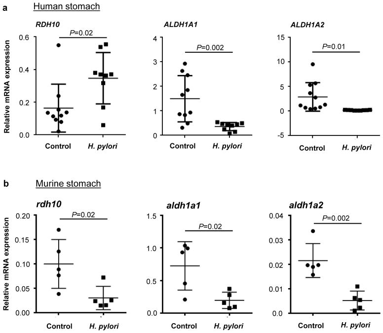
Despite the high prevalence of chronic gastritis caused by Helicobacter pylori, the gastric mucosa has received little investigative attention as a unique immune environment. Here, we analyzed whether retinoic acid (RA), an important homeostatic factor in the small intestinal mucosa, also contributes to gastric immune regulation. We report that human gastric tissue contains high levels of the RA precursor molecule retinol (ROL), and that gastric epithelial cells express both RA biosynthesis genes and RA response genes, indicative of active RA biosynthesis. Moreover, primary gastric epithelial cells cultured in the presence of ROL synthesized RA in vitro and induced RA biosynthesis in co-cultured monocytes through an RA-dependent mechanism, suggesting that gastric epithelial cells may also confer the ability to generate RA on gastric dendritic cells (DCs). Indeed, DCs purified from gastric mucosa had similar levels of aldehyde dehydrogenase activity and RA biosynthesis gene expression as small intestinal DCs, although gastric DCs lacked CD103. In H. pylori-infected gastric mucosa, gastric RA biosynthesis gene expression was severely disrupted, which may lead to reduced RA signaling and thus contribute to disease progression. Collectively, our results support a critical role for RA in human gastric immune regulation.


Conflict of interest statement

Disclosure The authors declared no conflicts of interest.

Figures

Figure 1
Figure 1. RA biosynthesis by human gastric epithelial cells
(a) Tissue levels of retinol in human gastric and small intestinal mucosa were measured by normal phase HPLC after lipid extraction (n=7). (b, c) Epithelial cells were recovered from fresh human gastric and intestinal tissue specimens by incubation with EDTA, and expression of genes involved in RA biosynthesis was analyzed by quantitative (q)RT-PCR using the random standard curve method. (b) Cytospins of H&E stained gastric and intestinal epithelial cells, original magnification 20x. (c) Relative gene expression for RDH10 and ALDH1A1. Diamonds: individual samples; bars: mean; lines connect donor-matched samples; n=6. Statistical significance was determined using the Student’s t- test.
Figure 2
Figure 2. Increased expression of RA response genes in primary human gastric compared with intestinal epithelial cells
Epithelial cells were recovered from fresh human gastric and intestinal tissue specimens by incubation with EDTA, and gene expression of RAR-α, RAR-β, RAR-γ, CRABP2 and TGM2 was analyzed by qRT-PCR using the random standard curve method. Diamonds: individual samples; bars: mean; lines connect donor-matched samples; n=6–8. Statistical significance was determined using the Student’s t-test.
Figure 3
Figure 3. Small intestinal, but not gastric, epithelial cells are involved in biosynthesis of retinyl esters
(a) Alternative metabolic pathways for retinol usage by epithelial cells. (b) Tissue levels of retinyl esters in human gastric and small intestinal mucosa were measured by reverse phase HPLC after lipid extraction (n=7). (c) Freshly isolated human gastric and intestinal epithelial cells were analyzed for their expression of LRAT by qRT-PCR using the standard curve method. Diamonds: individual samples; bars: mean; lines connect paired samples; n=7. Statistical significance was determined using the Student’s t-test.
Figure 4
Figure 4. ROL-treated gastric epithelial cells induce RA biosynthesis in co-cultured antigen-presenting cells
(a) Experiment overview. (b) Monolayers of primary gastric epithelial cells were treated with ROL (2 μM) or retinyl palmitate (RP; 2 μM) for 4 – 6 h and washed. Blood monocytes were then added at 1 x10⁶/well, together with medium alone, the RAR inhibitor Ro-41-5253 (Ro-41; 1 μM) or RA (50 nM). Monocytes were harvested after 16 h, and gene expression of ALDH1A2 in recovered monocytes was analyzed by qRT-PCR. ITGAX (CD11c) was used as a housekeeping gene. Data are from one representative experiment of four similar experiments. Mean±SEM of duplicates. (c) Monolayers of primary gastric epithelial cells were kept in medium (n=4) or fixed with 70% Ethanol (n=3) and then treated with ROL, ROL and Ro-41-5253, or RA. Blood monocytes were added at 1 x10⁶/well for 6 – 8 h and then were harvested for analysis of RA production using the Sil-15 reporter cell line. Symbols: individual samples; bars: mean and SD, n=3–4; Kruskal-Wallis test with Bonferroni correction.
Figure 5
Figure 5. Gastric DCs lack expression of CD103
(a) Gating strategy for isolation of gastric and intestinal DCs. (b) Phenotype of isolated gastric DCs. Data are representative of n=6–8. (c) Intestinal DCs express higher levels of CD103 than gastric DCs. Diamonds: individual samples; bars: mean; P=0.02, Kruskal-Wallis test with Bonferroni correction; n=6–8. (d, e) Gastric DCs lack ITGAE (CD103) gene expression but express similar levels of ZBTB46. RNA was isolated from FACS-sorted gastric and intestinal dendritic cells (live HLA-DR+/CD45+/lineage cells), and expression of (d) ITGAE and (e) ZBTB46 was analyzed by qRT-PCR. Monocyte-derived DCs (MoDCs; n=1) and elutriated intestinal macrophages (Macs, n=1) were analyzed for comparison. Diamonds: individual samples; bars: mean; lines connect paired samples; n=7. Statistical significance was determined using the Student’s t-test.
Figure 6
Figure 6. Gastric DC synthesize RA as efficiently as intestinal DCs
(a) ALDH activity of isolated gastric and intestinal DCs and blood monocytes was analyzed with the Aldefluor® assay. Left panels: representative FACS data; grey histograms: DEAB-treated control cells, black outline: untreated samples. Middel panel: Combined data from 5–7 experiments shown as geometric mean fluorescence (normalized to geomean of the DEAB-treated control); Right panel: Combined data from 5–7 experiments shown as % positive cells. (b, c) RNA was isolated from FACS-sorted gastric and intestinal dendritic cells (live HLA-DRhigh/CD45+/lineage cells), and expression of (b) RA biosynthesis genes RDH10 and ALDH1A2, (c) RA response genes and TGM2 and CRABP2 and (d) RA receptors RAR-α, RAR-β and RAR-γ was analyzed by qRT-PCR using the random standard curve method. Diamonds: individual samples; bars: mean; lines connect paired samples; n=5–7. Statistical significance was determined using the Student’s t-test.
Figure 7
Figure 7. Gastric H. pylori infection interferes with retinoid metabolism
(a) RNA was isolated from human gastric biopsy specimens obtained from healthy or H. pylori-infected subjects. Gene expression of RDH10, ALDH1A1 and ALDH1A2 was determined by quantitative RT-PCR using the random standard curve method, with data normalized to GAPDH expression (n=9–11). (b) 6 – 8 week old female C57/BL6 mice (n=5) were infected with H. pylori (SS1) for 2 months or were mock-infected, stomachs were harvested, and gene expression of rdh10, aldh1a1 and aldh1a2 was determined by qRT-PCR. Data were normalized to hprt expression and analyzed using the ΔΔcT method. (a, b) Dots and squares: individual samples; bars: mean and SD; Statistical significance was determined using the Student’s t-test.

References

    1. Atherton JC, Blaser MJ. Coadaptation of Helicobacter pylori and humans: ancient history, modern implications. J Clin Invest. Sep;119(9):2475–87. Epub 2009/09/05. - PMC - PubMed
    1. Bimczok D, Grams JM, Stahl RD, Waites KB, Smythies LE, Smith PD. Stromal regulation of human gastric dendritic cells restricts the Th1 response to Helicobacter pylori. Gastroenterology. 2011;141(3):929–38. Epub 2011/06/28. - PMC - PubMed
    1. Yang I, Nell S, Suerbaum S. Survival in hostile territory: the microbiota of the stomach. FEMS Microbiol Rev. 2013;37(5):736–61. Epub 2013/06/25. - PubMed
    1. Bik EM, et al. Molecular analysis of the bacterial microbiota in the human stomach. Proc Natl Acad Sci U S A. 2006;103(3):732–7. Epub 2006/01/13. - PMC - PubMed
    1. Iwata M. Retinoic acid production by intestinal dendritic cells and its role in T-cell trafficking. Semin Immunol. 2009;21(1):8–13. - PubMed

Publication types

MeSH terms