. 2015 May;8(3):582-95.
doi: 10.1038/mi.2014.90. Epub 2014 Sep 24.

Reciprocal regulation of lymphoid tissue development in the large intestine by IL-25 and IL-23

Affiliations

Reciprocal regulation of lymphoid tissue development in the large intestine by IL-25 and IL-23

D S Donaldson et al. Mucosal Immunol. 2015 May.

Abstract

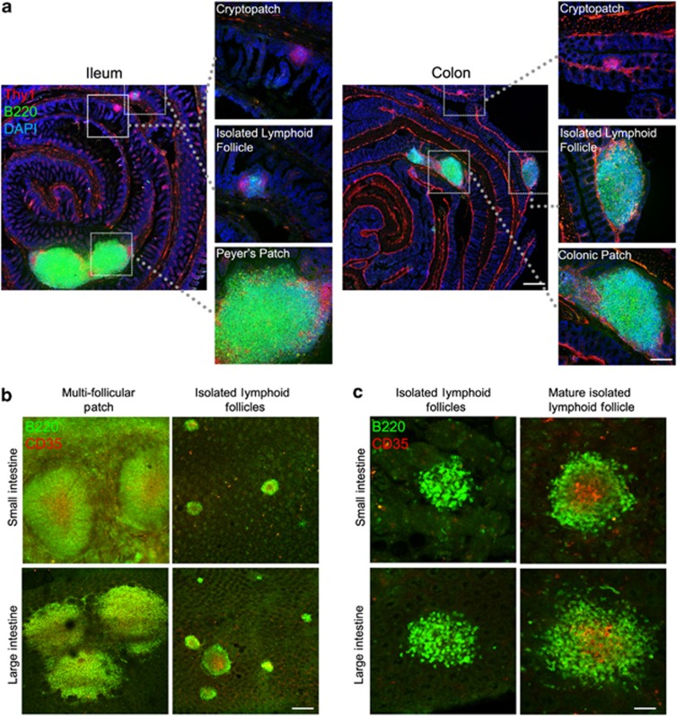
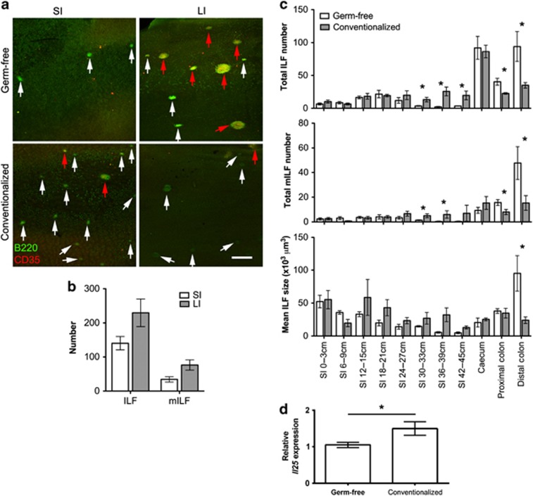
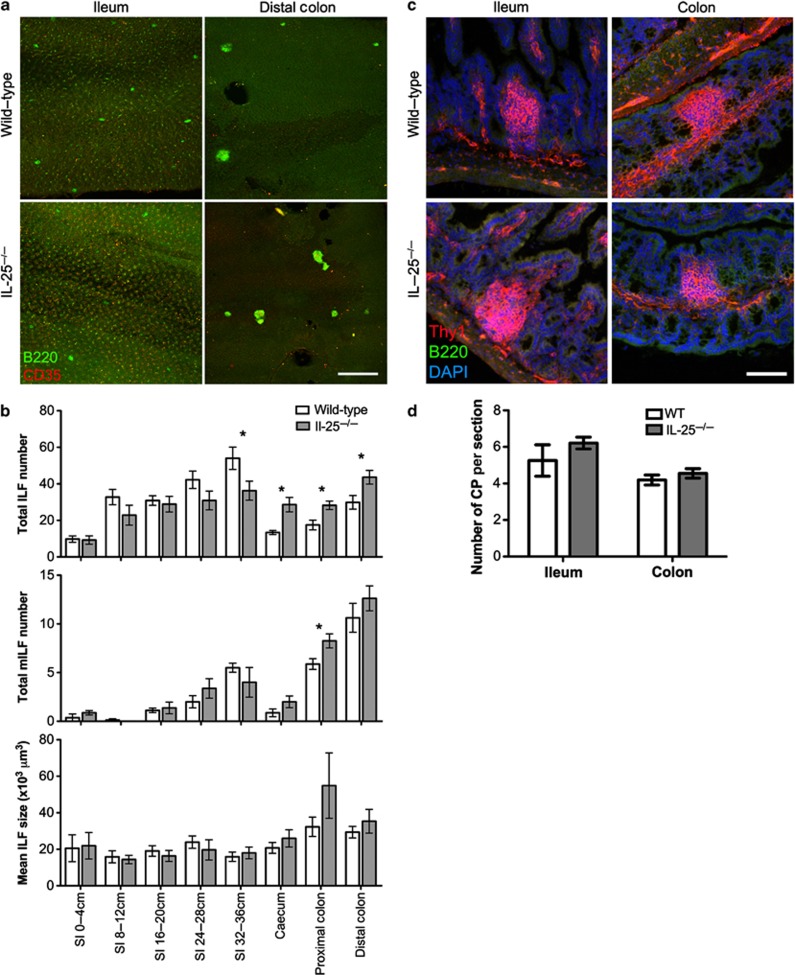
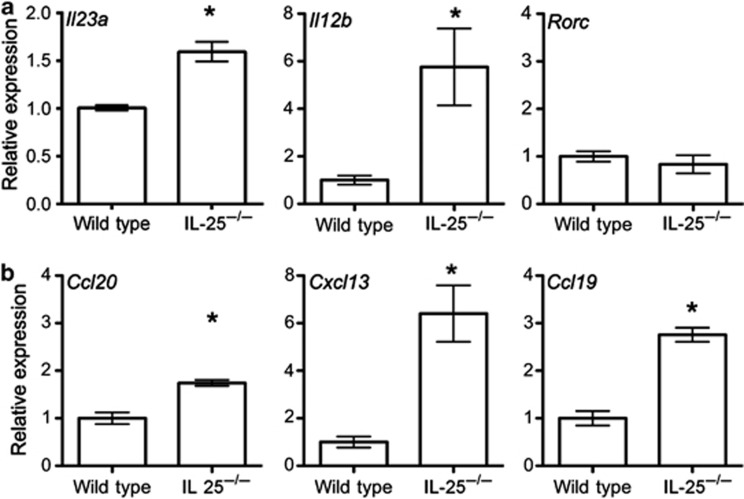
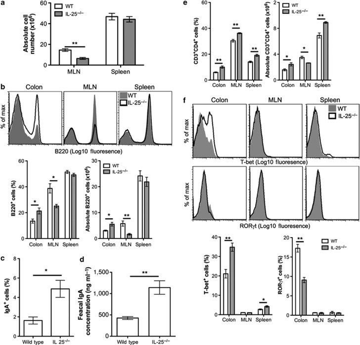
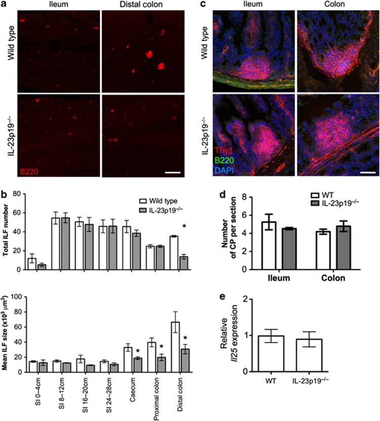
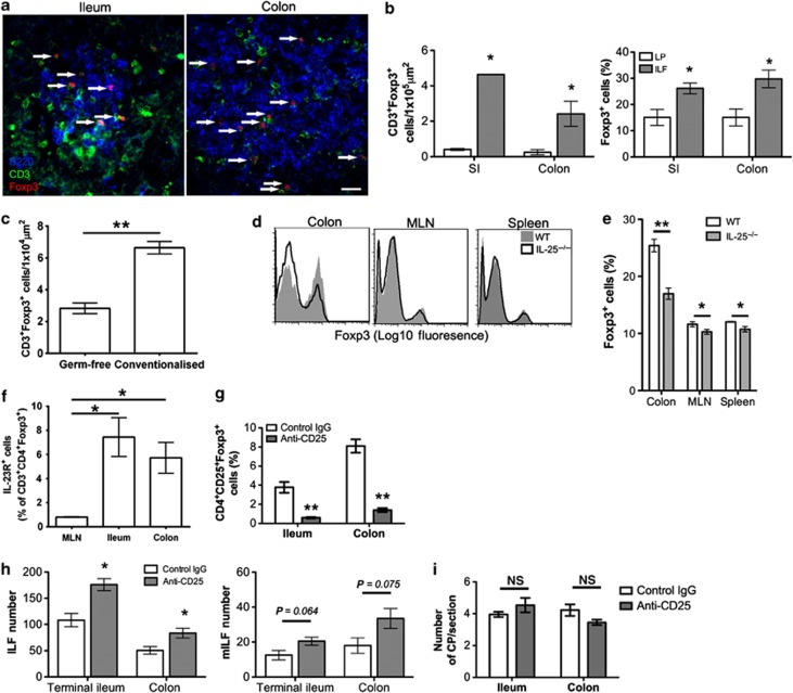
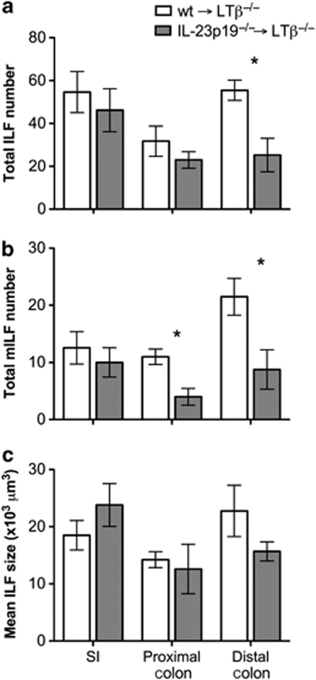
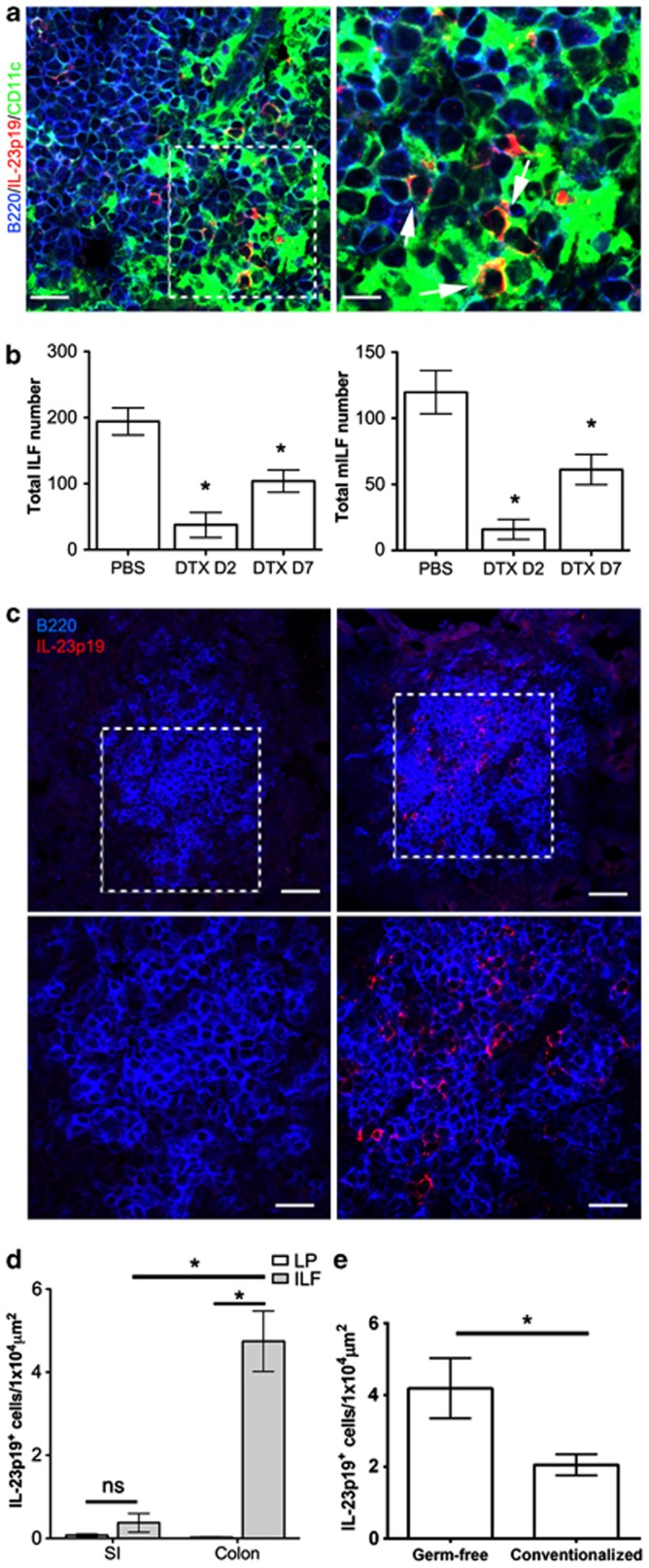
Isolated lymphoid follicles (ILFs) develop after birth in the small and large intestines (SI and LI) and represent a dynamic response of the gut immune system to the microbiota. Despite their similarities, ILF development in the SI and LI differs on a number of levels. We show that unlike ILF in the SI, the microbiota inhibits ILF development in the colon as conventionalization of germ-free mice reduced colonic ILFs. From this, we identified a novel mechanism regulating colonic ILF development through the action of interleukin (IL)-25 on IL-23 and its ability to modulate T regulatory cell (Treg) differentiation. Colonic ILF develop in the absence of a number of factors required for the development of their SI counterparts and can be specifically suppressed by factors other than IL-25. However, IL-23 is the only factor identified that specifically promotes colonic ILFs without affecting SI-ILF development. Both IL-23 and ILFs are associated with inflammatory bowel disease, suggesting that disruption to this pathway may have an important role in the breakdown of microbiota-immune homeostasis.


Figures

Figure 1
Figure 1
Gastrointestinal lymphoid tissue of the small and large intestines. (a) Sections of small and large intestine were stained for Thy1 (red), B220 (green), and nuclei (blue) to detect cryptopatches, isolated lymphoid follicles, and multifollicular patches (Peyer's patches in the small intestine and colonic patches in the large intestine). Bar=500 μm in low power panels and 200 μm in higher power panels. (b) Intestinal segments were whole-mount stained for B220 (green) and CD35 (red) to detect multifollicular patches and isolated lymphoid follicles. Bar=200 μm. (c) Mature isolated lymphoid follicles can be differentiated by CD35 staining (red), visualizing follicular dendritic cell networks. Bar=50 μm.
Figure 2
Figure 2
Isolated lymphoid follicle (ILF) development and maturation in the large intestine (LI) of germ-free mice is suppressed by the microbiota. (a) Alternate 3 cm sections of small intestine (SI) and the whole LI were whole-mount immunostained for B220 (green) to detect ILFs and CD35 (red) to detect mature ILFs (mILFs). Representative images of SI and LI are shown. White arrowheads highlight immature ILFs and red arrowheads highlight mILFs. Bar=500 μm. (b) The total number of ILFs and mILFs in the SI and LI of germ-free mice was determined by microscopy (n=3). SI numbers were estimated from representative sections. (c) The number of ILFs and mILFs and the mean ILF size were determined in the intestines of germ-free and conventionalized germ-free mice by microscopy (n=3). (d) The relative level of Il25 mRNA expression in the colon of germ-free and conventionalized mice was determined by quantitative real-time reverse-transcriptase–PCR (qRT–PCR; n=3). Statistical differences were determined by Student's t-test (*P<0.05).
Figure 3
Figure 3
Interleukin (IL)-25−/− mice have increased large intestinal (LI) isolated lymphoid follicles (ILFs). (a) Alternate 4 cm sections of small intestine (SI) and the whole LI of wild-type (WT) and IL-25−/− mice were whole-mount immunostained for B220 (green) to detect ILFs and CD35 (red) to detect mature ILFs (mILFs). Representative images of ileum and distal colon are shown. Bar=500 μm. (b) Total ILF and mILF numbers and mean ILF size were determined by microscopy (n=8). Results are representative of two independent experiments. (c) Sections of ileum and colon were stained for Thy1 (red), B220 (green), and nuclei (blue) to detect cryptopatches (CPs). Bar=50 μm. (d) CP numbers in 20 sections (100 μm apart) of 10 cm of terminal ileum or the whole colon were determined by microscopy and converted to CP/section (n=4). Statistical differences were determined by Student's t-test (*P<0.05).
Figure 4
Figure 4
The expression of Il23a, Il12b, Ccl20, Cxcl13, and Ccl19 mRNA is increased in the colon of interleukin (IL)-25−/− mice. (a) The relative levels of Il23a, Il12b, and Rorc mRNA expression in the colon of wild-type and IL-25−/− mice were determined by quantitative real-time reverse-transcriptase–PCR (qRT–PCR; n=3). (b) The relative levels of Ccl20, Cxcl13, and Ccl19 mRNA expression in the colon of wild-type and IL-25−/− mice were determined by qRT–PCR (n=3). Results are representative of two independent experiments. Statistical differences were determined by Student's t-test (*P<0.05).
Figure 5
Figure 5
Interleukin (IL)-25−/− mice have increased colonic B cells, immunoglobulin A (IgA) responses, and T helper type 1 (Th1) cells. (a) Mesenteric lymph node (MLN) and spleen cells from wild-type (WT) and IL-25−/− mice were isolated and counted (n=4). (b) The relative frequency of B220+ B cells in the colonic lamina propria (LP), MLN, and spleen of WT and IL-25−/− mice was determined by flow cytometry and the absolute cell number calculated (n=4). Representative histograms are shown. (c) The proportion of B220+ cells that express IgA in the colonic LP was determined by flow cytometry (n=3). (d) Homogenates of fecal pellets (10%) from wild-type and IL-25−/− mice were prepared in phosphate-buffered saline (PBS) and IgA levels determined by enzyme-linked immunosorbent assay (ELISA; n=4). Absolute values were calculated from a standard curve of control mouse IgA. (e) The relative frequency of CD3+CD4+ T cells in the colonic LP, MLN, and spleen of WT and IL-25−/− mice was determined by flow cytometry and the absolute cell number calculated (n=4). (f) The relative frequency of T-bet+ and RORγt+ cells in CD3+CD4+ T-cell population from the colonic LP, MLN, and spleen of WT and IL-25−/− mice. Representative histograms are shown. Results are representative of three independent experiments. Statistical differences were determined by Student's t-test (*P<0.05, **P<0.01).
Figure 6
Figure 6
Interleukin (IL)-23p19−/− mice have a colon-specific reduction in isolated lymphoid follicles (ILFs). (a) Alternate 4 cm sections of small intestine (SI) and the whole large intestine from wild-type (WT) or IL-23p19−/− mice were whole-mount immunostained for B220 (red) to detect ILFs. Bar=500 μm. (b) ILF numbers and mean ILF size in WT or IL-23p19−/− mice was determined by microscopy (n=4). (c) Sections of ileum and colon were stained for Thy1 (red), B220 (green), and nuclei (blue) to detect cryptopatches (CPs). Bar=50 μm. (d) CP numbers in 20 sections (100 μm apart) of 10 cm of terminal ileum or the whole colon were determined by microscopy and converted to CP/section (n=4). (e) The relative level of Il25 mRNA expression in the colon of WT and IL-23p19−/− mice was determined by quantitative real-time reverse-transcriptase–PCR (qRT–PCR; n=4). Statistical differences were determined by Student's t-test (*P<0.05).
Figure 7
Figure 7
Anti-CD25 treatment enhances isolated lymphoid follicle (ILF) numbers in the ileum and colon. (a) Sections of small intestine (SI) and large intestine (LI) were immunostained for B220 (blue), CD3 (green), and Foxp3 (red). Representative images of ILFs from the ileum and colon are shown. Bar=20 μm. (b) The number of CD3+Foxp3+ cells and the proportion of CD3+ cells expressing Foxp3 within ILFs and adjacent areas of lamina propria (LP) were determined by microscopy (n=3). (c) The number of CD3+Foxp3+ within ILFs of germ-free and conventionalized mice (n=3) (d, e) The relative frequency of Foxp3+ cells in the CD3+CD4+ T-cell population of the colonic LP, mesenteric lymph node (MLN), and spleen of wild-type (WT) and interleukin (IL)-25−/− mice was determined by flow cytometry. Representative histograms are shown (n=4). (f) The relative frequency of IL-23R+ cells within the CD4+Foxp3+ T cell population of the MLN, ileum, and colon was determined by flow cytometry (n=3). (g) Mice were treated with anti-CD25 or control Ig. At 7 days after final treatment, mice were killed and the proportion of CD25+Foxp3+ cells within the CD4+ population of the ileal and colonic LP were determined by flow cytometry (n=4). (h) Portions of terminal ileum (8 cm) and the whole colon of anti-CD25 or control Ig-treated mice were whole-mount immunostained for B220 and CD35 to detect ILFs and mature ILFs (mILFs), respectively. ILF and mILF numbers were determined by microscopy (n=4). (i) Sections of ileum and colon were stained for Thy1 (red) and B220 (green) to detect cryptopatches (CPs). CP numbers in 12 sections (100 μm apart) of 10 cm of terminal ileum or the whole colon were determined by microscopy and converted to CP/section (n=4). Statistical differences were determined by Student's t-test (*P<0.05, **P<0.01, NS, not significant).
Figure 8
Figure 8
LTβ−/− mice reconstituted with interleukin (IL)-23p19−/− bone marrow have a specific reduction in colonic isolated lymphoid follicles (ILFs). LTβ−/− were lethally irradiated and reconstituted with wild-type (wt) or IL-23p19−/− bone marrow. After 10 weeks, alternate 4 cm sections of small intestine (SI) and colon were whole-mount immunostained for B220 and CD35 to detect ILFs and mature ILFs (mILFs), respectively. (a) Total ILF and (b) mILF numbers were determined by microscopy (n=4). SI values are the mean ILF or mILF numbers present in a 4-cm section of SI (5 sections/mouse). (c) Mean ILF size was also determined. Statistical differences were determined by Student's t-test (*P<0.05).
Figure 9
Figure 9
Interleukin (IL)-23p19+ cells are enriched in colonic isolated lymphoid follicles (ILFs). (a) Sections of colonic ILFs were also immunostained for B220 (blue), IL-23p19 (red), and CD11c (green). Bar=20 μm in the left panel and 5 μm in the right panel. (b) LTβ−/− were irradiated and reconstituted with bone marrow from CD11c-DTR mice. Mice were treated with diphtheria toxin (DTX) and culled 2 or 7 days after treatment. Phosphate-buffered saline (PBS)-treated mice culled 7 days after treatment were included as controls. The colon of each mouse was whole-mount immunostained for B220 to detect ILFs and CD35 to detect mature ILFs (mILFs). ILF and mILF numbers were determined by microscopy (n=3–4). (c) Sections of small intestine (SI) and colon from wild-type mice were immunostained for B220 (blue) and IL-23p19 (red). Representative images of ILF are shown. Bar=40 μm in the upper panel and 20 μm in the lower panel. (d) IL-23p19+ cells within ILFs or adjacent areas of lamina propria (LP) were enumerated and the density of IL-23p19+ cells determined (n=3). (e) IL-23p19+ cells within ILFs of germ-free and conventionalized mice were enumerated and the density of IL-23p19+ cells determined (n=3). Statistical differences were determined by Student's t-test (*P<0.05).

Similar articles

Cited by

References

    1. Blumberg R., Powrie F. Microbiota, disease, and back to health: a metastable journey. Sci. Transl. Med. 2012;4:137rv7. - PMC - PubMed
    1. Round J.L., Mazmanian S.K. The gut microbiota shapes intestinal immune responses during health and disease. Nat. Rev. Immunol. 2009;9:313–323. - PMC - PubMed
    1. Fujimura Y., Kamoi R., Iida M. Pathogenesis of aphthoid ulcers in Crohn's disease: correlative findings by magnifying colonoscopy, electron microscopy, and immunohistochemistry. Gut. 1996;38:724–732. - PMC - PubMed
    1. Leithauser F., et al. Foxp3-expressing CD103+ regulatory T cells accumulate in dendritic cell aggregates of the colonic mucosa in murine transfer colitis. Am. J. Pathol. 2006;168:1898–1909. - PMC - PubMed
    1. Leithauser F., Trobonjaca Z., Moller P., Reimann J. Clustering of colonic lamina propria CD4(+) T cells to subepithelial dendritic cell aggregates precedes the development of colitis in a murine adoptive transfer model. Lab. Invest. 2001;81:1339–1349. - PubMed

Publication types

MeSH terms