Review
. 2014 Dec 1;307(11):E955-68.
doi: 10.1152/ajpendo.00130.2014. Epub 2014 Sep 23.

Physiological effects and therapeutic potential of proinsulin C-peptide

Affiliations
Review

Physiological effects and therapeutic potential of proinsulin C-peptide

Gina L C Yosten et al. Am J Physiol Endocrinol Metab. .

Abstract

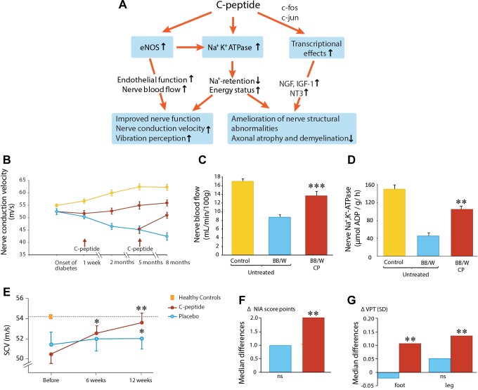
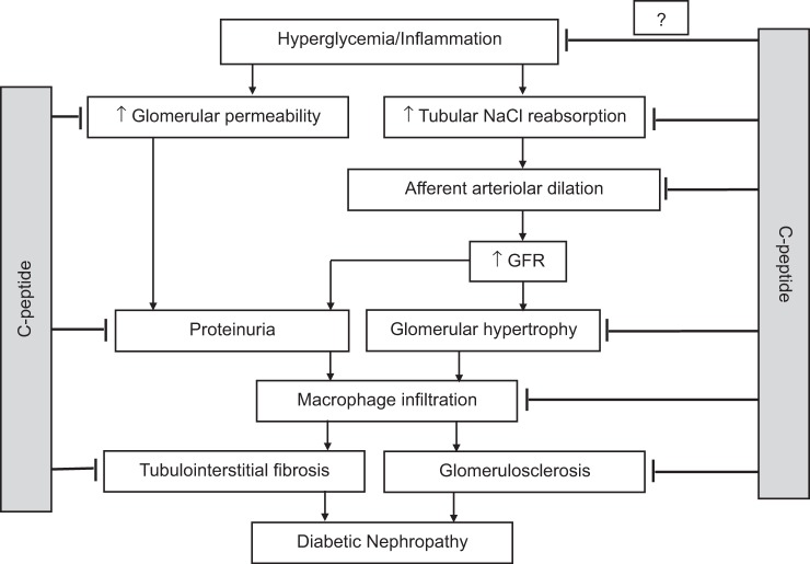
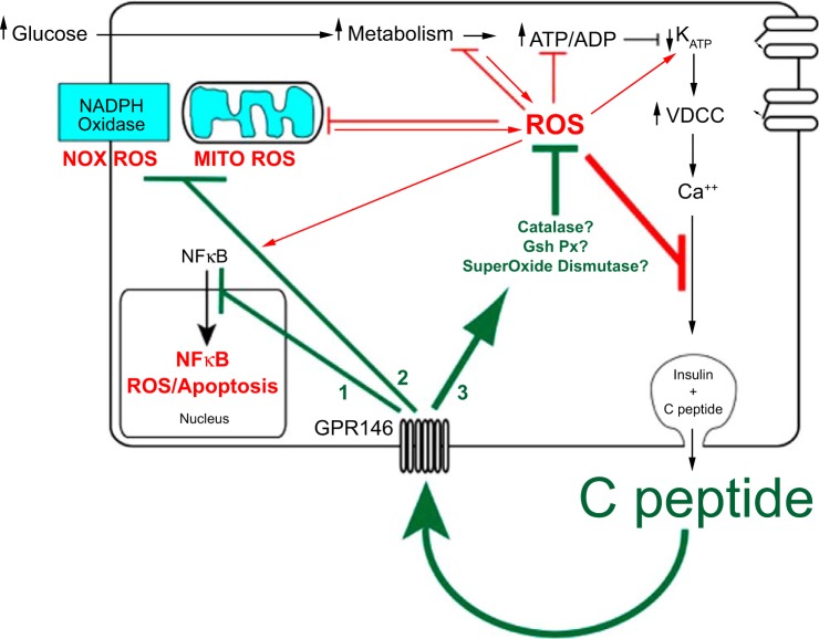
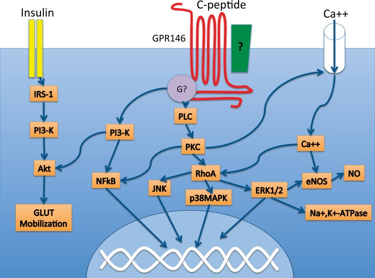
Connecting Peptide, or C-peptide, is a product of the insulin prohormone, and is released with and in amounts equimolar to those of insulin. While it was once thought that C-peptide was biologically inert and had little biological significance beyond its role in the proper folding of insulin, it is now known that C-peptide binds specifically to the cell membranes of a variety of tissues and initiates specific intracellular signaling cascades that are pertussis toxin sensitive. Although it is now clear that C-peptide is a biologically active molecule, controversy still remains as to the physiological significance of the peptide. Interestingly, C-peptide appears to reverse the deleterious effects of high glucose in some tissues, including the kidney, the peripheral nerves, and the vasculature. C-peptide is thus a potential therapeutic agent for the treatment of diabetes-associated long-term complications. This review addresses the possible physiologically relevant roles of C-peptide in both normal and disease states and discusses the effects of the peptide on sensory nerve, renal, and vascular function. Furthermore, we highlight the intracellular effects of the peptide and present novel strategies for the determination of the C-peptide receptor(s). Finally, a hypothesis is offered concerning the relationship between C-peptide and the development of microvascular complications of diabetes.

Keywords: C-peptide; diabetes; diabetes-associated complications.


Figures

Fig. 1.
Fig. 1.
C-peptide and diabetic neuropathy. A: schematic overview of C-peptide's effects on nerve function and structure in diabetes. eNOS, endothelial NO synthase. B: effects of C-peptide (red) or placebo (blue) administration for up to 8 mo on nerve conduction velocity (NCV) in diabetic rats. Yellow symbols indicate NCV development in healthy nondiabetic animals; data from Ref. . C: sciatic nerve blood flow in healty (yellow), untreated diabetic (blue), and C-peptide-treated (red) diabetic BB/W rats; data from Ref. . D: Na+,K+-ATPase activity of the sciatic nerve in healthy (yellow), untreated diabetic (blue), and C-peptide-treated diabetic rats; data from Ref. . E: effects of C-peptide in replacement dose (red) or placebo (blue) for 3 mo on sensory nerve conduction velocity (SCV) in type 1 diabetes subjects with early-stage nerve impairment (Ref. 19). F: influence of C-peptide (red) or placebo (blue) administration for 6 mo on neuropathy impairment score (NIA) (Ref. 20). G: vibration perception in subjects with type 1 diabetes and manifest neuropathy; data from Ref. .
Fig. 2.
Fig. 2.
Simplified model of the pathogenesis of diabetic nephropathy. C-peptide is renoprotective in type 1 diabetes via inhibiting tubular sodium reabsorption, reducing afferent arteriolar diameter and glomerular permeability, preventing and attenuating the progression of glomerular hyperfiltration, hypertrophy and microalbuminuria, renal inflammation, glomerulosclerosis, and tubulointerstitial fibrosis. GFR, glomerular filtration rate. Studies from our laboratory have also shown that C-peptide may also have blood glucose-lowering effects; however, it appears that C-peptide exerts its renoprotective effects independently of blood glucose regulation.
Fig. 3.
Fig. 3.
Autocrine C-peptide model underlying β-cell adaptation to oxidative stress. The centerpiece of functional β-cell mass is insulin secretion, which is cosecreted in equimolar amounts with C-peptide. Autocrine action of C-peptide, which protects functional β-cell mass, should therefore provide a buffer of protection for its own secretion as well as that of insulin. In addition to binding to its receptor GPR146 for such autocrine actions, C-peptide is predicted to regulate at least 3 distinct pathways in the β-cell: 1) deactivation of the NF-κB pathway, which protects against apoptosis; 2) inhibition of pathways that generate ROS including the plasmalemma NADPH oxidase and the mitochondrial electron transport chain; 3) activation of pathways that catalyze the degradation of ROS by activating antioxidant enzymes superoxide dismutase, catalase, and glutathione peroxidase. In the absence of these autocrine actions of C-peptide, more prolonged and higher accumulation of ROS and apoptosis would accelerate loss of β-cell functional mass. This suggests a tipping point in loss of functional β-cell mass where the loss in C-peptide secretion and its autocrine protection results in a downward spiral of both secretion and protection, ultimately leading to few if any β-cells.
Fig. 4.
Fig. 4.
C-peptide-initiated signaling cascades. C-peptide is associated with the regulation of several signaling cascades, including phospholipase C (PLC) and the NF-κB pathway. These intracellular signaling events are likely mediated by a G protein-coupled receptor such as GPR146, which has been shown to be necessary for C-peptide signaling in KATOIII cells. GPR146 interacts with an as yet unknown G protein, which could be either Gαi or Gαo, since several of the cellular actions of C-peptide were shown to be pertussis toxin sensitive. GPR146 may interact physically with additional proteins on the cell membrane, such as an integrin (green box). C-peptide and insulin appear to functionally interact, particularly at the level of Akt. Akt, protein kinase B; Ca++, calcium ion; eNOS, endothelial NO synthase; ERK1/2, extracellular signal-regulated kinase; G?, G protein; GLUT, glucose transporter; IRS-1, insulin receptor substrate 1; JNK, c-Jun NH2-terminal kinase; Na+,K+-ATPase, sodium/potassium ATPase; NO, nitric oxide; NF-κB, nuclear factor κ-light-chain-enhancer of activated B cells; PKC, protein kinase C; PLC, phospholipase C; PI3-K, phosphotidylinositol 3-kinase; p38 MAPK, mitogen-activated protein kinase; RhoA, Ras homolog gene family, member A.

References

    1. Al-Rasheed NM, Meakin F, Royal EL, Lewington AJ, Brown J, Willars GB, Brunskill NJ. Potent activation of multiple signaling pathways by C-peptide in opossum kidney proximal tubular cells. Diabetologia 47: 987–997, 2004. - PubMed
    1. Al-Rasheed NM, Willars GB, Brunskill NJ. C-peptide signals via Galpha i to protect against TNF-alpha-mediated apoptosis of opossum kidney proximal tubular cells. J Am Soc Nephrol 17: 986–995, 2006. - PubMed
    1. Alpers CE, Hudkins KL. Mouse models of diabetic nephropathy. Curr Opin Nephrol Hypertens 20: 278–284, 2011. - PMC - PubMed
    1. Bathgate RA, Halls ML, van der Westhuizen ET, Callander GE, Kocan M, Summers RJ. Relaxin family peptides and their receptors. Physiol Rev 93: 405–480, 2013. - PubMed
    1. Bhatt MP, Lim YC, Hwang J, Na S, Kim YM, Ha KS. C-peptide prevents hyperglycemia-induced endothelial apoptosis through inhibition of reactive oxygen species-mediated transglutaminase 2 activation. Diabetes 62: 243–253, 2013. - PMC - PubMed

MeSH terms

LinkOut - more resources