. 2015 Mar;64(3):916-925.
doi: 10.2337/db14-0332. Epub 2014 Sep 23.

β-cell-specific CD8 T cell phenotype in type 1 diabetes reflects chronic autoantigen exposure

Affiliations

β-cell-specific CD8 T cell phenotype in type 1 diabetes reflects chronic autoantigen exposure

Ania Skowera et al. Diabetes. 2015 Mar.

Abstract

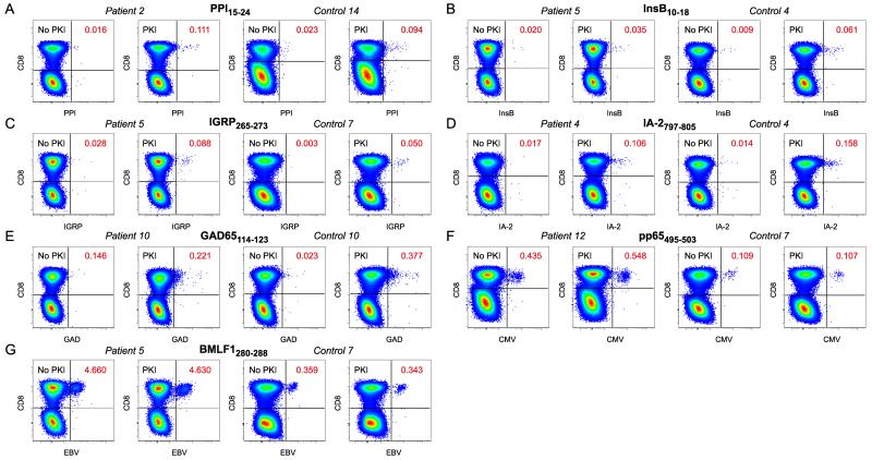
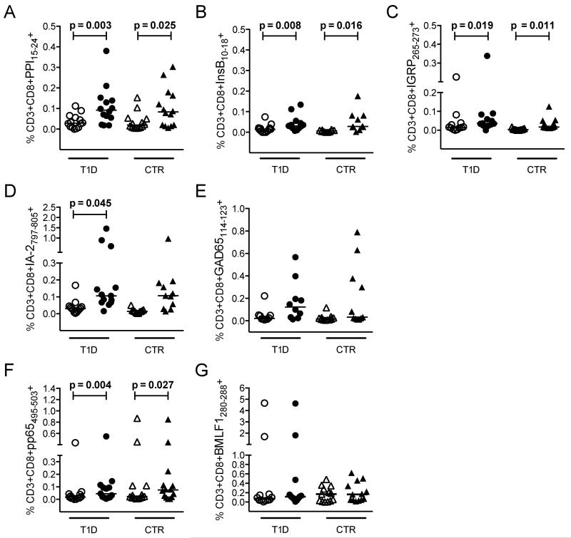
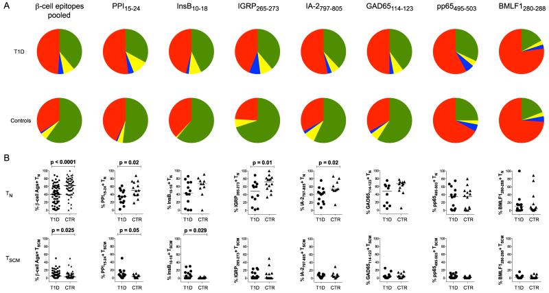
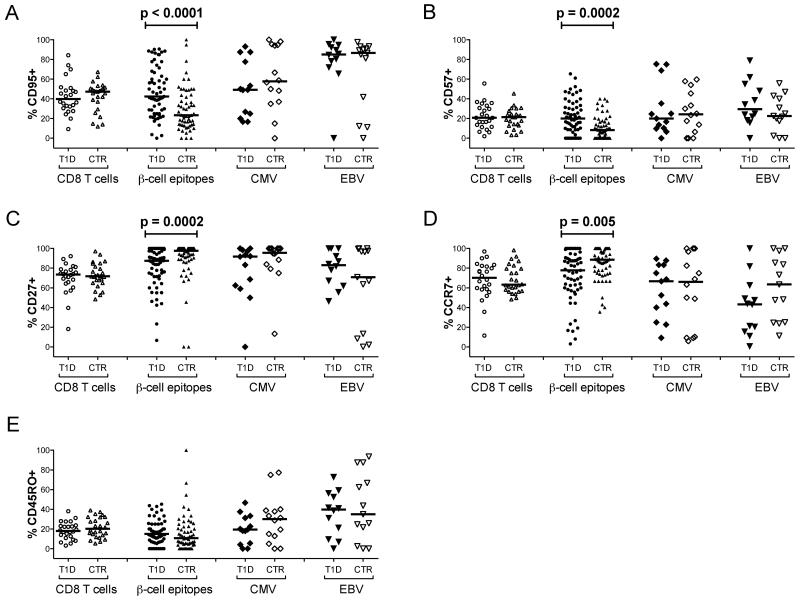
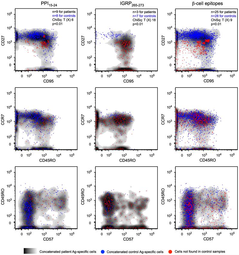
Autoreactive CD8 T cells play a central role in the destruction of pancreatic islet β-cells that leads to type 1 diabetes, yet the key features of this immune-mediated process remain poorly defined. In this study, we combined high-definition polychromatic flow cytometry with ultrasensitive peptide-human leukocyte antigen class I tetramer staining to quantify and characterize β-cell-specific CD8 T cell populations in patients with recent-onset type 1 diabetes and healthy control subjects. Remarkably, we found that β-cell-specific CD8 T cell frequencies in peripheral blood were similar between subject groups. In contrast to healthy control subjects, however, patients with newly diagnosed type 1 diabetes displayed hallmarks of antigen-driven expansion uniquely within the β-cell-specific CD8 T cell compartment. Molecular analysis of selected β-cell-specific CD8 T cell populations further revealed highly skewed oligoclonal T cell receptor repertoires comprising exclusively private clonotypes. Collectively, these data identify novel and distinctive features of disease-relevant CD8 T cells that inform the immunopathogenesis of type 1 diabetes.


Figures

Figure 1
Figure 1
Identification of antigen-specific CD8 T cell populations. A-G: Thawed PBMCs were stained with pHLA-A*0201 tetramers representing PPI15-24 (A), InsB10-18 (B), IGRP265-273 (C), IA-2797-805 (D), GAD65114-123 (E), CMV pp65495-503 (F) and EBV BMLF1280-288 (G). Gates were set serially on singlets, live CD3+CD14CD19 cells and lymphocytes prior to Boolean exclusion of dye aggregates and subsequent analysis in bivariate CD8 versus tetramer plots (Supplementary Figure 1 A). Representative paired data in the absence or presence of dasatinib are shown for type 1 diabetes patients and healthy controls. Tetramer-binding CD8 T cell frequencies are indicated.
Figure 2
Figure 2
Summary of antigen-specific CD8 T cell frequencies. A-G: Thawed PBMCs were stained with pHLA-A*0201 tetramers representing PPI15-24 (A), InsB10-18 (B), IGRP265-273 (C), IA-2797-805 (D), GAD65114-123 (E), CMV pp65495-503 (F) and EBV BMLF1280-288 (G). Graphs show tetramer-binding CD8 T cell frequencies in the absence (empty symbols) or presence (filled symbols) of dasatinib for type 1 diabetes patients (T1D) and healthy controls (CTR). No significant differences across identical comparisons were observed between subject groups. Bars represent median values. Statistical analyses were performed using the Wilcoxon signed-rank test; p values ≤ 0.05 are shown.
Figure 3
Figure 3
Phenotypic subset analysis of antigen-specific CD8 T cells. A: Pie chart representations of mean subset percentages for pooled β-cell-specific CD8 T cells and individual specificities in type 1 diabetes patients (top row) and healthy controls (bottom row). Subsets are defined and color-coded as follows: green, TN (CD27+CD45ROCD57CD95CCR7+); yellow, TSCM (CD27+CD45ROCD95+CCR7+); blue, TEFF (CD27CD45ROCD95+CCR7); red, all remaining memory cells. B: Column plots showing TN and TSCM subset percentages for pooled β-cell-specific CD8 T cells and individual specificities in type 1 diabetes patients (T1D) and healthy controls (CTR). Representative data are shown in Supplementary Figure 1 B&C. Almost identical results were obtained with the TN subset defined in the absence of CD57 (CD27+CD45ROCD95CCR7+). Bars represent median values. Statistical analyses were performed using the Mann-Whitney U-test; p values ≤ 0.05 are shown.
Figure 4
Figure 4
Single marker analysis of antigen-specific CD8 T cells. A-E: Percent expression of CD95 (A), CD57 (B), CD27 (C), CCR7 (D) and CD45RO (E) is shown for type 1 diabetes patients (T1D) and healthy controls (CTR) across all CD8 T cells and the indicated specificities. Corresponding data for individual β-cell-derived epitope-specific CD8 T cell populations are shown in Supplementary Figure 4. Bars represent median values. Statistical analyses were performed using the Mann-Whitney U-test; p values ≤ 0.05 are shown.
Figure 5
Figure 5
Frequency difference gating analysis of antigen-specific CD8 T cells. Overlays of concatenated data from type 1 diabetes patients (cloud plots) and healthy controls (blue dots) are shown for the indicated β-cell specificities across bivariate phenotypic profiles. Populations of CD27intermediateCD95+ CD8 T cells, present at significantly higher frequencies in type 1 diabetes patients compared to healthy controls, are displayed as red dots.

References

    1. Roep BO, Peakman M. Antigen targets of type 1 diabetes autoimmunity. Cold Spring Harb Perspect Med. 2012;2:a007781. - PMC - PubMed
    1. Roep BO, Peakman M. Diabetogenic T lymphocytes in human Type 1 diabetes. Curr Opin Immunol. 2011;23:746–753. - PubMed
    1. Coppieters KT, Dotta F, Amirian N, Campbell PD, Kay TW, Atkinson MA, Roep BO, von Herrath MG. Demonstration of islet-autoreactive CD8 T cells in insulitic lesions from recent onset and long-term type 1 diabetes patients. J Exp Med. 2012;209:51–60. - PMC - PubMed
    1. Willcox A, Richardson SJ, Bone AJ, Foulis AK, Morgan NG. Analysis of islet inflammation in human type 1 diabetes. Clin Exp Immunol. 2009;155:173–181. - PMC - PubMed
    1. Skowera A, Ellis RJ, Varela-Calvino R, Arif S, Huang GC, Van-Krinks C, Zaremba A, Rackham C, Allen JS, Tree TI, Zhao M, Dayan CM, Sewell AK, Unger WW, Drijfhout JW, Ossendorp F, Roep BO, Peakman M. CTLs are targeted to kill beta cells in patients with type 1 diabetes through recognition of a glucose-regulated preproinsulin epitope. J Clin Invest. 2008;118:3390–3402. - PMC - PubMed

Publication types

MeSH terms

Substances