. 2014 Sep 23;55(10):6960-7.
doi: 10.1167/iovs.14-15020.

Retinal mitochondrial DNA mismatch repair in the development of diabetic retinopathy, and its continued progression after termination of hyperglycemia

Affiliations

Retinal mitochondrial DNA mismatch repair in the development of diabetic retinopathy, and its continued progression after termination of hyperglycemia

Manish Mishra et al. Invest Ophthalmol Vis Sci. .

Abstract

Purpose: Mitochondrial DNA (mtDNA) is damaged in the retina in diabetes, and mitochondria copy numbers are decreased. The displacement-loop (D-loop) of the mtDNA, the region with transcription/replication elements, experiences more damage than other regions of mtDNA. Our aim was to examine the role of DNA mismatch repair (MMR) in mitochondria homeostasis in diabetic retinopathy, and in its continued progression after cessation of hyperglycemia.

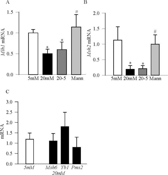
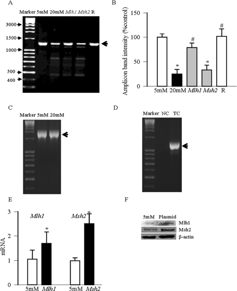
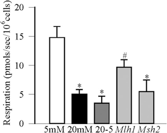
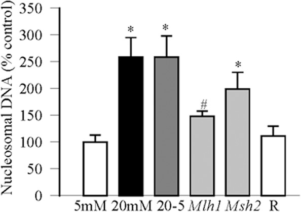
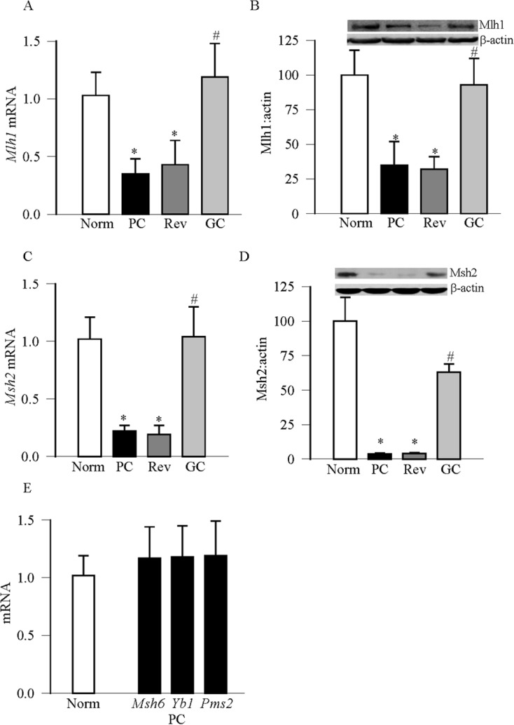
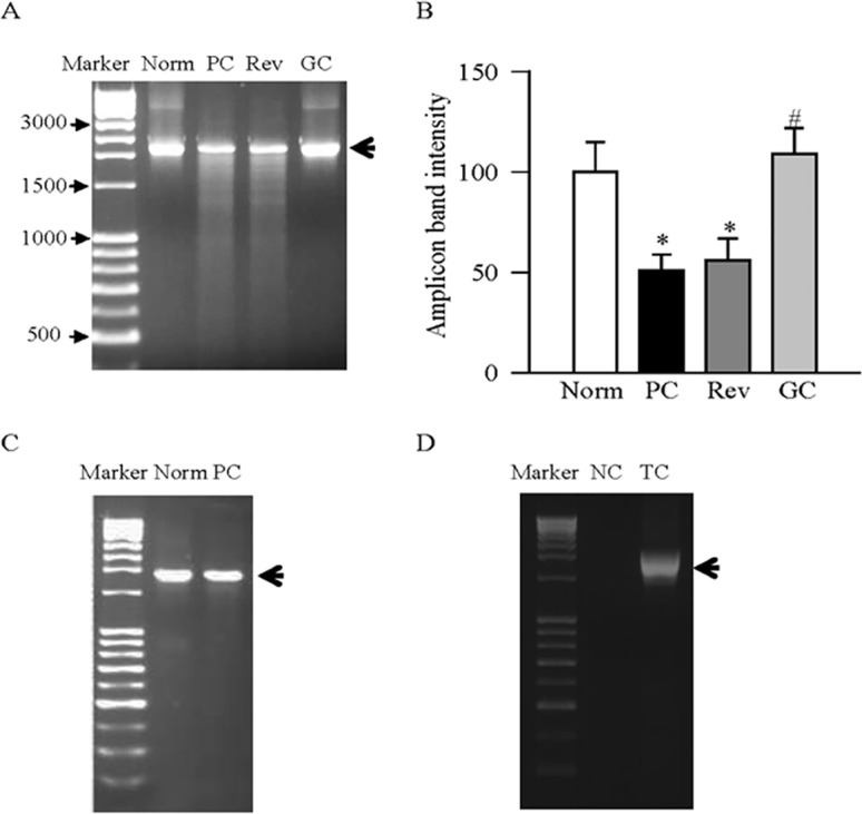
Methods: Effect of hyperglycemia on sequence variants in the D-loop region was investigated in retinal endothelial cells and in the retina from streptozotocin-induced diabetic rats using mismatch-specific surveyor nuclease. The role of MMR machinery in mtDNA damage and mitochondrial respiration was investigated in retinal endothelial cells overexpressing Mlh1, an MMR enzyme mainly associated with mtDNA polymerase gamma, or Msh2 (associated with nuclear polymerase beta).

Results: Hyperglycemia increased sequence variants in the D-loop region. While overexpression of Mlh1 in endothelial cells ameliorated glucose-induced increase in D-loop sequence variants, decrease in respiration rate and increase in apoptosis, overexpression of Msh2 did not protect the mitochondria damage. Termination of hyperglycemia failed to reverse decrease in MMR enzymes and increase in D-loop sequence variants.

Conclusions: Due to a compromised MMR system, the sequence variants in the D-loop region were not repaired, and that resulted in impaired mtDNA transcription. Mitochondria become dysfunctional, and they continued to be dysfunctional even after hyperglycemia was terminated, contributing to the development, and progression of diabetic retinopathy. Thus, strategies targeting mitochondrial MMR machinery could help maintain mitochondria homeostasis, and inhibit the development of diabetic retinopathy and its continued progression.

Keywords: DNA repair; diabetic retinopathy; metabolic memory; mitochondria damage; mtDNA mismatch.


Figures

Figure 1
Figure 1
Effect of high glucose on MMR machinery. Retinal endothelial cells were analyzed for mRNA expressions of MMR genes by qRT-PCR using β-actin as a housekeeping gene. Each measurement was made in duplicate using cells from three to four different cell preparations, and the values are represented as mean ± SD. (A) Mlh1, (B) Msh2, and (C) Msh6, Yb1, and Pms2. *P < 0.05 and #P < 0.05 compared with 5 mM glucose and 20 mM glucose, respectively. 5 mM, 5 mM glucose; 20 mM, 20 mM glucose; 20-5, 4 days of 20 mM followed by 4 days of 5mM glucose; Mann, 20 mM mannitol.
Figure 2
Figure 2
Sequence variants in the D-loop region. (A) DNA was amplified using semiquantitative PCR for D-loop regions of mtDNA, and after digesting the samples with mismatch-specific surveyor nuclease, they were analyzed on an agarose gel. The arrow indicates the amplicon product length. (B) The amplicon intensity of the D-loop region was quantified, and the intensity of the band in 5 mM glucose was considered as 100%. (C) For comparison, amplification for the Cox region (Cox1 and Cox2) of the mtDNA was analyzed after mismatch- specific surveyor nuclease digestion. (D) ‘No template' (NC), D-loop template control without surveyor nuclease digestion (TC). (E) Gene transcripts and (F) protein expression were quantified in cells transfected with Mlh1 or Msh2 by q-PCR and Western blot techniques, respectively. Values are from three to four different experiments. *P < 0.05 compared with 5 mM glucose and #P < 0.05 compared with 20 mM glucose. 5 mM and 20 mM, 5 mM and 20 mM glucose, respectively; Mlh1 and Msh2, cells after transfecting with Mlh1 or Msh2, incubated in 20 mM glucose for 4 days; R, cell incubated with transfection reagent alone during transfection, followed by incubation in 5 mM glucose for 4 days.
Figure 3
Figure 3
Effect of high glucose on mitochondrial respiration: the cellular respiration was recorded by high-resolution respirometry using 106 retinal endothelial cells. Respiration rate was calculated using the OROBOROS oxygraph software (DatLab 4.0). *P < 0.05 and #P < 0.05 compared with 5 mM glucose and 20 mM glucose, respectively.
Figure 4
Figure 4
Overexpression of mismatch proteins and cell apoptosis: apoptosis was measured by performing ELISA for cytoplasmic histone-associated DNA fragments using an assay ELISA kit. Values are presented as mean ± SD of three different experiments, each performed in duplicate. The values obtained from cells in normal glucose were considered as 100%. *P < 0.05 and #P < 0.05 compared with 5 mM glucose and 20 mM glucose, respectively.
Figure 5
Figure 5
Effect of diabetes on MMR machinery. Gene expression of the mismatch genes were quantified in the rat retina by qRT-PCR, and the Ct value of each gene was normalized to β-actin in that sample. Protein expressions were quantified by Western blot technique using β-actin as the loading proteins. Each measurement was made in duplicate, and the values are represented as mean ± SD of four to six rats/group. (A, B) Gene and protein expressions of Mlh1, and (C, D) represent those of Msh2. (E) The gene expressions of Msh6, Yb1, and Pms2. *P < 0.05 compared with normal and #P < 0.05 compared with poor control. Norm, normal rats; PC and GC, rats in continuous poor control or good control, respectively, for 6 months. Rev, rats in poor control for 3 months followed by good control for 3 additional months.
Figure 6
Figure 6
Effect of diabetes on retinal mtDNA sequence variants. (A) Retinal DNA was amplified for mtDNA region encoding D-loop using semiquantitative PCR, digested with mismatch specific surveyor nuclease, and separated on an agarose gel. The arrow indicates the D-loop amplicon product length. (B) The amplicon intensity of the D-loop region was quantified, and the intensity of the band obtained from normal rat retina was considered as 100%. (C) Amplification for the Cox1 region of the mtDNA was analyzed after mismatch specific surveyor nuclease digestion. (D) “No template” (NC) and D-loop template control without surveyor nuclease digestion (TC) were analyzed by agarose gel electrophoresis. Values are from four to six rats in each group. *P < 0.05 and #P < 0.05 compared with normal and poor control, respectively. Norm, normal rats; PC and GC, rats in continuous poor control or good control respectively, for 6 months; Rev, 3 months of poor control followed by 3 months of good control.

Similar articles

Cited by

References

    1. Kowluru RA, Abbas SN. Diabetes-induced mitochondrial dysfunction in the retina. Invest Ophthalmol Vis Sci. 2003; 44: 5327–5334 - PubMed
    1. Kanwar M, Chan PS, Kern TS, Kowluru RA. Oxidative damage in the retinal mitochondria of diabetic mice: possible protection by superoxide dismutase. Invest Ophthalmol Vis Sci. 2007; 48: 3805–3811 - PubMed
    1. Brownlee M. The pathobiology of diabetic complications: a unifying mechanism. Diabetes. 2005; 54: 1615–1625 - PubMed
    1. Stuart JA, Brown MF. Mitochondrial DNA maintenance and bioenergetics. Biochim Biophys Acta. 2006; 1757: 79–89 - PubMed
    1. Madsen-Bouterse SA, Mohammad G, Kanwar M, Kowluru RA. Role of mitochondrial DNA damage in the development of diabetic retinopathy, and the metabolic memory phenomenon associated with its progression. Antioxid Redox Signal. 2010; 13: 797–805 - PMC - PubMed

Publication types

MeSH terms