

Clinical considerations for epidermal necrolysis
- PMID: 25249808
- PMCID: PMC4171800
Clinical considerations for epidermal necrolysis
Abstract
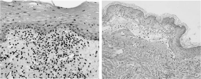
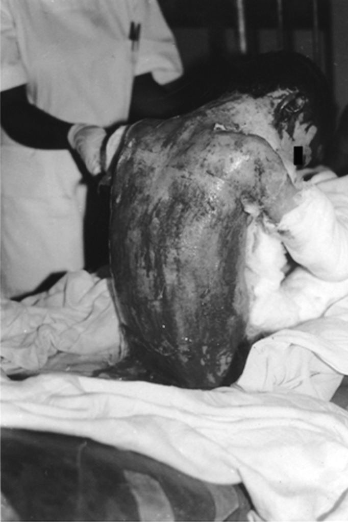
Background: Stevens-Johnson syndrome (SJS) and toxic epidermal necrolysis (TEN) are considered a spectrum of acute life-threatening mucocutaneous reactions that differ only in severity. Both diseases are characterized by mucous membrane and skin involvement, are often caused by medications, and are collectively known as epidermal necrolysis (EN).
Methods: A severity of illness score has been devised to predict prognosis in patients with EN. The scoring system addresses 7 prognostic factors.
Results: Patients with EN require supportive care. Those with extensive skin involvement should be admitted to an intensive care unit or burn unit if possible. Suspected, as well as unnecessary, medications should be discontinued. Baseline laboratory tests, imaging, cultures, and biopsies should be obtained. Intravenous access should be established and hydration and nutritional support begun. Daily oral care, wound care, pain control, and early physician consultation are also important aspects of treatment.
Conclusion: EN requires early diagnosis, appropriate workup, and appropriate treatment to minimize potential morbidity and mortality. In many clinicians' experience, EN is rare; therefore, education and improved understanding of the potential causes and appropriate treatment regimens are vital when confronted with such a patient.
Keywords: Drug eruptions; Stevens-Johnson syndrome; drug hypersensitivity.
Conflict of interest statement
Figures
References
-
- James WD, Berger TG, Elston DM, Odom RB. Andrews' Diseases of the Skin: Clinical Dermatology. 10th ed. Philadelphia, PA: Elsevier;; 2006.
-
- Valeyrie-Allanore L, Roujeau JC. Epidermal necrolysis (Stevens-Johnson syndrome and toxic epidermal necrolysis) In: Goldsmith LA, Katz SI, Gilchrest BA, Paller AS, Leffell DJ, Wolff K, editors. Fitzpatrick's Dermatology in General Medicine. Vol 1. 8th ed. New York, NY: McGraw-Hill;; 2012. pp. 439–448. In. eds.
-
- French LE, Prins C. Bolognia JL, Jorizzo JL, Rapini RP, et al, eds. Dermatology. 2nd ed. Philadelphia, PA: Elsevier;; 2008. Erythema multiforme, Stevens-Johnson syndrome and toxic epidermal necrolysis; pp. 287–300. In.
-
- Weedon D, Strutton G, Rubin AI. Weedon's Skin Pathology. 3rd ed. Philadelphia, PA: Elsevier;; 2010.
Publication types
LinkOut - more resources
Full Text Sources
Medical
Research Materials