. 2014 Sep 9:8:269.
doi: 10.3389/fncel.2014.00269. eCollection 2014.

C1-Inhibitor protects from focal brain trauma in a cortical cryolesion mice model by reducing thrombo-inflammation

Affiliations

C1-Inhibitor protects from focal brain trauma in a cortical cryolesion mice model by reducing thrombo-inflammation

Christiane Albert-Weissenberger et al. Front Cell Neurosci. .

Abstract

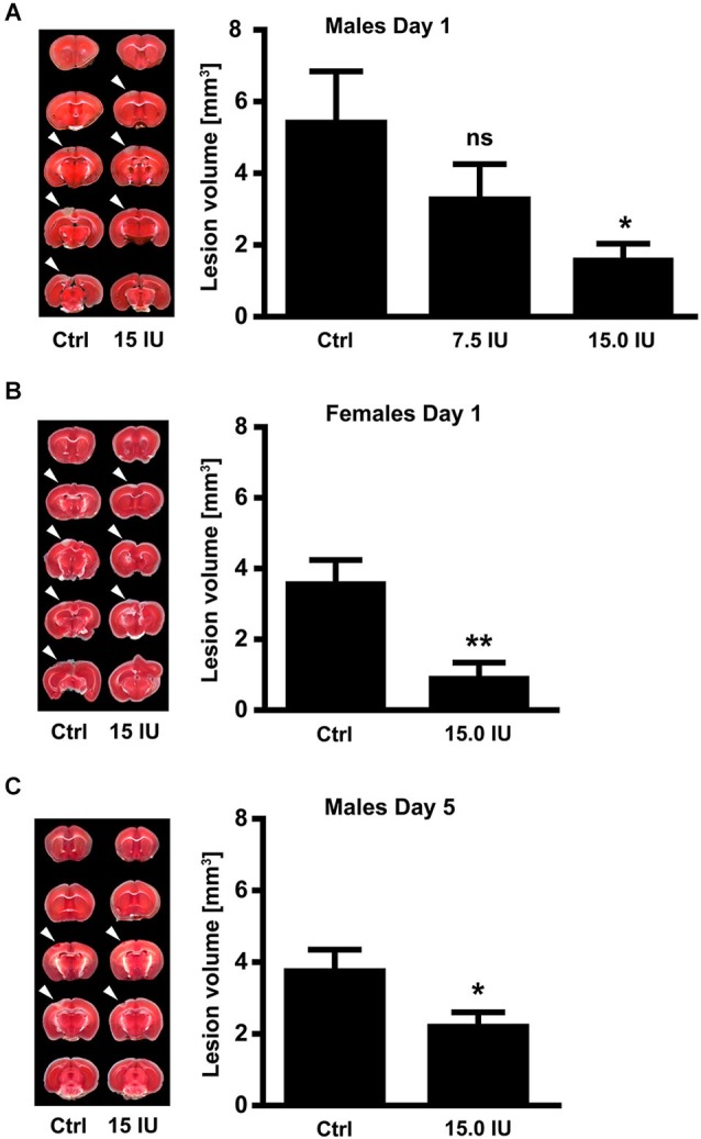
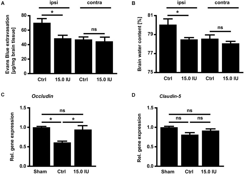
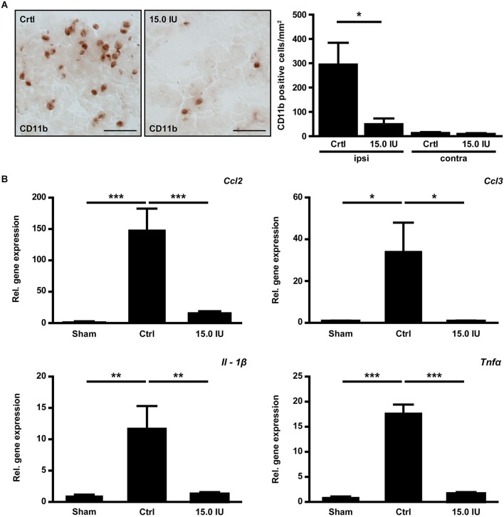
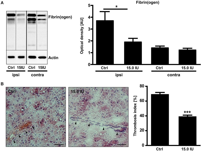
Traumatic brain injury (TBI) induces a strong inflammatory response which includes blood-brain barrier damage, edema formation and infiltration of different immune cell subsets. More recently, microvascular thrombosis has been identified as another pathophysiological feature of TBI. The contact-kinin system represents an interface between inflammatory and thrombotic circuits and is activated in different neurological diseases. C1-Inhibitor counteracts activation of the contact-kinin system at multiple levels. We investigated the therapeutic potential of C1-Inhibitor in a model of TBI. Male and female C57BL/6 mice were subjected to cortical cryolesion and treated with C1-Inhibitor after 1 h. Lesion volumes were assessed between day 1 and day 5 and blood-brain barrier damage, thrombus formation as well as the local inflammatory response were determined post TBI. Treatment of male mice with 15.0 IU C1-Inhibitor, but not 7.5 IU, 1 h after cryolesion reduced lesion volumes by ~75% on day 1. This protective effect was preserved in female mice and at later stages of trauma. Mechanistically, C1-Inhibitor stabilized the blood-brain barrier and decreased the invasion of immune cells into the brain parenchyma. Moreover, C1-Inhibitor had strong antithrombotic effects. C1-Inhibitor represents a multifaceted anti-inflammatory and antithrombotic compound that prevents traumatic neurodegeneration in clinically meaningful settings.

Keywords: C1-inhibitor; blood-brain barrier; contact-kinin system; edema; inflammation; thrombosis; traumatic brain injury.


Figures

Figure 1
Figure 1
C1-Inhibitor (C1-Inh) protects against traumatic brain injury in mice of both sexes. (A) Left panel shows representative 2,3,5-triphenyltetrazolium chloride (TTC) staining of 5 coronal brain sections of 6-week-old male control mice (Ctrl) and 6-week-old male mice treated with 7.5 IU or 15.0 IU C1-Inh. C1-Inh was always applied 1 h after cortical cryolesion and the lesion volume was assessed from TTC staining at day 1. The lesion volume was reduced in a dose-dependent manner with a significant reduction after treatment with 15.0 IU (n = 11–13, * P < 0.05, One-way analysis of variance with post hoc Bonferroni’s Multiple Comparison Test). (B) Representative TTC staining and lesion volume of 6-week-old female control and 15.0 IU C1-Inh treated mice, showing a significant reduction in lesion volume after treatment with 15.0 IU at day 1 (n = 10–11, ** P = 0.0019, Unpaired t-test). (C) A significant reduction of lesion volume was detectable up to 5 days after treatment with 15.0 IU C1-Inh in 6-week-old male mice (n = 10, * P < 0.05, Unpaired t-test).
Figure 2
Figure 2
C1-inhibitor (C1-Inh) treatment results in stabilization of the blood brain barrier. (A) Vascular leakage on day 1 after cryolesion was significantly decreased after treatment with 15.0 IU of C1-Inh as confirmed by the concentration of Evans Blue detectable in the brain parenchyma (n = 6, * P < 0.05, ns P > 0.05, One-way analysis of variance with post hoc Bonferroni’s Multiple Comparison Test, ipsi: ipsilateral hemisphere, contra: contralateral hemisphere). (B) Edema formation as reflected by the brain water content in the ipsi- and contralateral hemispheres of control and 15.0 IU treated mice on day 1 after cryolesion (n = 6, * P < 0.05, ns P > 0.05, One-way analysis of variance with post hoc Bonferroni’s Multiple Comparison Test). (C) Relative gene expression of occludin in the ipsilateral brain parenchyma of control and 15.0 IU treated mice 24 h after cryolesion or sham operation (n = 7 or 3 for sham operated, * P < 0.05, One-way analysis of variance with post hoc Bonferroni’s Multiple Comparison Test). (D) Relative gene expression of claudin-5 in the ipsilateral brain parenchyma of control and 15.0 IU treated mice 24 h after cryolesion or sham operation (n = 7 or 3 for sham operated, ns P > 0.05, One-way analysis of variance with post hoc Bonferroni’s Multiple Comparison Test).
Figure 3
Figure 3
C1-inhibitor (C1-Inh) treatment results in attenuation of the inflammatory response. (A) Representative immunohistochemical stainings of cerebral CD11b positive cells on day 1 after cryolesion. Quantification of cell number in control and C1-Inh treated mice revealed less CD11b positive cells per mm2 in the lesion site of C1-Inh treated mice (n = 5, * P < 0.05, One-way analysis of variance with post hoc Bonferroni’s Multiple Comparison Test, Scale bar 50 μm). (B) Relative gene expression of the genes encoding the proinflammatory cytokines ccl2, ccl3, interleukin (IL)-1β, and tumor necrosis factor (Tnf)α in control and C1-Inh treated mice on day 1 after cryolesion. Gene expression of all proinflammatory cytokines were significantly reduced in C1-Inh treated mice (n = 5, * P < 0.05, ** P < 0.01, *** P < 0.001, One-way analysis of variance with post hoc Bonferroni’s Multiple Comparison Test).
Figure 4
Figure 4
C1-inhibitor (C1-Inh) treatment results in inhibition of thrombus formation. (A) Accumulation of fibrin(ogen) in the ipsilateral (ipsi) and contralateral (contra) brain hemispheres of control and 15.0 IU C1-Inh treated mice was analyzed by immunoblotting 24 h after cryolesion, and bands were quantified by densitometry. The representative immunoblot and the quantification shows a significant reduction of fibrin(ogen) in the 15.0 C1-Inh treated mice (n = 5–6, * P < 0.05, One-way analysis of variance with post hoc Bonferroni’s Multiple Comparison Test; AU = arbitrary units). (B) Representative H8E staining from traumatic hemispheres of vehicle-treated and 15.0 IU C1-Inh treated mice on day 1 after cryolesion. Occluded vessels (arrowhead in the left panel) were more abundant in control mice when compared to vessels of 15.0 IU C1-Inh treated mice (arrowheads in the right panel). This finding was confirmed by the calculation of the thrombosis index showing a highly significant reduction of occluded vessels in 15.0 IU C1-Inh treated mice on day 1 after cryolesion (n = 4, *** P < 0.001, Unpaired t-test; Scale bar 50 μm).

References

    1. Albert-Weissenberger C., Sirén A. L. (2010). Experimental traumatic brain injury. Exp. Transl. Stroke Med. 2:16 10.1186/2040-7378-2-16 - DOI - PMC - PubMed
    1. Albert-Weissenberger C., Sirén A. L., Kleinschnitz C. (2013). Ischemic stroke and traumatic brain injury: the role of the kallikrein-kinin system. Prog. Neurobiol. 101–102, 65–82 10.1016/j.pneurobio.2012.11.004 - DOI - PubMed
    1. Albert-Weissenberger C., Stetter C., Meuth S. G., Gobel K., Bader M., Sirén A. L., et al. (2012). Blocking of bradykinin receptor B1 protects from focal closed head injury in mice by reducing axonal damage and astroglia activation. J. Cereb. Blood Flow Metab. 32, 1747–1756 10.1038/jcbfm.2012.62 - DOI - PMC - PubMed
    1. Auer L. M., Ott E. (1979). Disturbances of the coagulatory system in patients with severe cerebral trauma II. Platelet function. Acta Neurochir. (Wien) 49, 219–226 10.1007/bf01808961 - DOI - PubMed
    1. Banerji A. (2010). Current treatment of hereditary angioedema: an update on clinical studies. Allergy Asthma Proc. 31, 398–406 10.2500/aap.2010.31.3387 - DOI - PubMed