Review
. 2014 Sep 5:6:18.
doi: 10.3389/fnsyn.2014.00018. eCollection 2014.

Muscarinic cholinergic receptors modulate inhibitory synaptic rhythms in hippocampus and neocortex

Affiliations
Review

Muscarinic cholinergic receptors modulate inhibitory synaptic rhythms in hippocampus and neocortex

Bradley E Alger et al. Front Synaptic Neurosci. .

Abstract

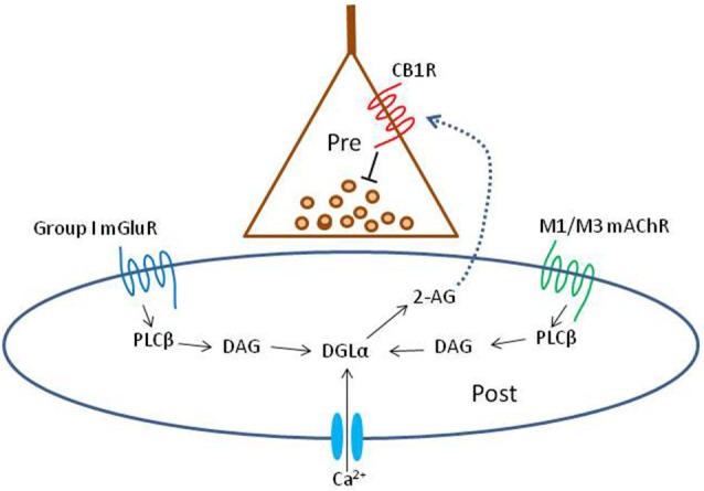
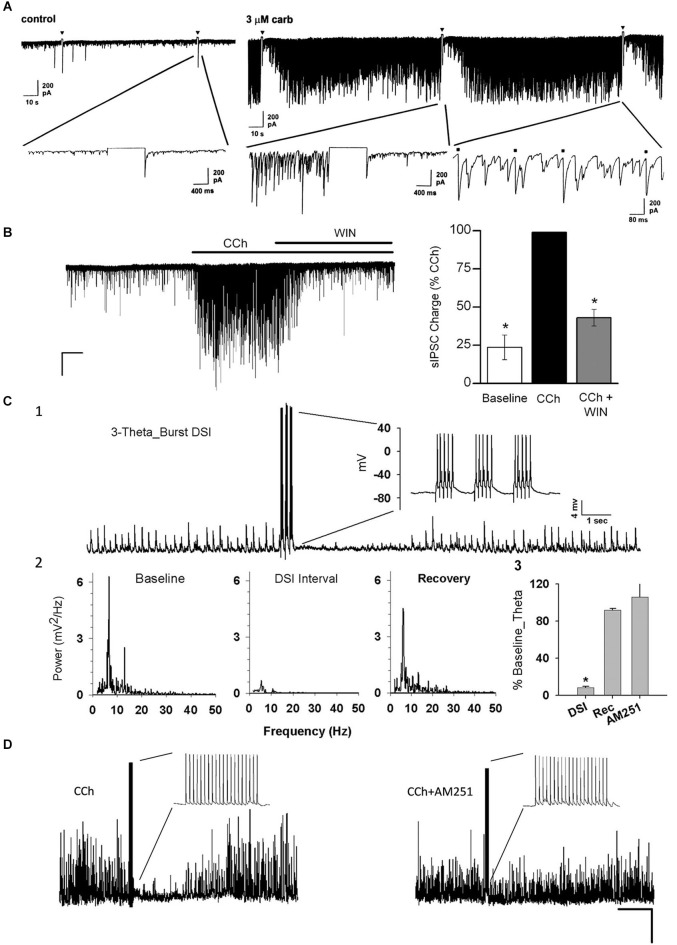
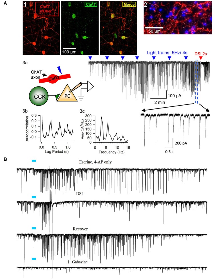
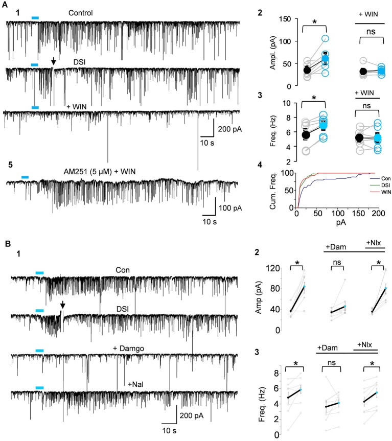
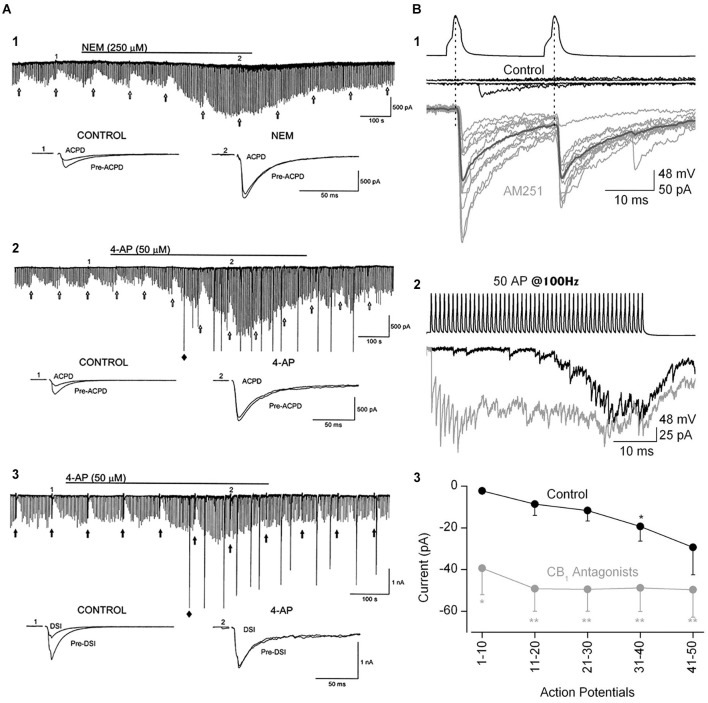
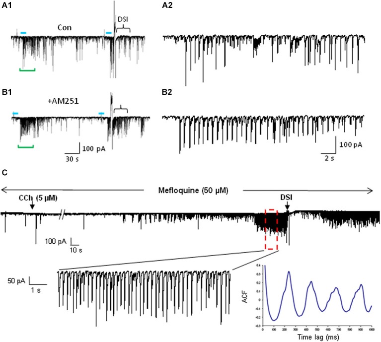
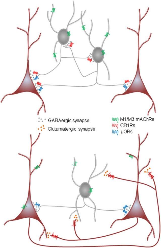
Activation of muscarinic acetylcholine (ACh) receptors (mAChRs) powerfully affects many neuronal properties as well as numerous cognitive behaviors. Small neuronal circuits constitute an intermediate level of organization between neurons and behaviors, and mAChRs affect interactions among cells that compose these circuits. Circuit activity is often assessed by extracellular recordings of the local field potentials (LFPs), which are analogous to in vivo EEGs, generated by coordinated neuronal interactions. Coherent forms of physiologically relevant circuit activity manifest themselves as rhythmic oscillations in the LFPs. Frequencies of rhythmic oscillations that are most closely associated with animal behavior are in the range of 4-80 Hz, which is subdivided into theta (4-14 Hz), beta (15-29 Hz) and gamma (30-80 Hz) bands. Activation of mAChRs triggers rhythmic oscillations in these bands in the hippocampus and neocortex. Inhibitory responses mediated by GABAergic interneurons constitute a prominent feature of these oscillations, and indeed, appear to be their major underlying factor in many cases. An important issue is which interneurons are involved in rhythm generation. Besides affecting cellular and network properties directly, mAChRs can cause the mobilization of endogenous cannabinoids (endocannabinoids, eCBs) that, by acting on the principal cannabinoid receptor of the brain, CB1R, regulate the release of certain neurotransmitters, including GABA. CB1Rs are heavily expressed on only a subset of interneurons and, at lower density, on glutamatergic neurons. Exogenous cannabinoids typically disrupt oscillations in the theta (θ) and gamma (γ) ranges, which probably contributes to the behavioral effects of these drugs. It is important to understand how neuronal circuit activity is affected by mAChR-driven eCBs, as this information will provide deeper insight into the actions of ACh itself, as well as into the effects of eCBs and exogenous cannabinoids in animal behavior. After covering some basic aspects of the mAChR system, this review will focus on recent findings concerning the mechanisms and circuitry that generate θ and γ rhythms in hippocampus and neocortex. The ability of optogenetic methods to probe the many roles of ACh in rhythm generation is highlighted.

Keywords: GABA; cholecystokinin; endocannabinoid; interneuron; opioid; optogenetics; parvalbumin; theta.


Figures

Figure 1
Figure 1
Schematic summary diagram of the endocannabinoid system. A presynaptic nerve terminal is shown synapsing on a postsynaptic cell. Agonist binding of either group I mGluRs or M1 or M3 mAChRs activates phospholipase Cβ (PLCβ) in the postsynaptic cell. The product of the reaction catalyzed by PLCβ, diacylglycerol (DAG), is metabolized by the enzyme diacylglycerol lipase α (DGLα) to form the endocannabinoid, 2-arachidonyl glycerol (2-AG). DGLα can also be activated by Ca2+ influx via a PLC-independent mechanism. 2-AG is membrane permeant and gains access through an unknown mechanism to the cannabinoid receptor, CB1R, on the presynaptic nerve terminal. Binding of CB1R by 2-AG inhibits transmitter release, mainly by inhibiting Ca2+ influx into the terminal, although other mechanisms can be involved. This is the simplest conventional model of the system and various details are controversial or have been omitted for the sake of simplicity; see a comprehensive review, e.g., Kano et al. (2009); for more information.
Figure 2
Figure 2
Carbachol (CCh) elicits persistent occurrence of large, rhythmic inhibitory synaptic responses in hippocampus and neocortex. (A) Representative whole-cell recording from a Sprague/Dawley rat CA1 pyramidal cell (PC) in control saline (iGluR antagonists present in experiments shown in all panels) in vitro and a 1-s depolarizing voltage-step (downward triangles) has no effect on the small spontaneous IPSCs visible on the baseline. Bath application of 3 μM CCh induces a persistent barrage of large IPSCs that are transiently interrupted by the periods of DSI that occur after the voltage steps (From Martin and Alger, ; with permission). (B) Representative trace from a layer II/III neocortical PC from a Swiss CD-1 mouse slice. A large barrage of IPSCs occurs after CCh (5 μM) is added to the bath, and the IPSCs are suppressed by further addition of the CB1R agonist WIN55212-2 (5 μM). From Trettel et al. (2004) with permission. (C1) Representative sharp electrode recording of large, rhythmic IPSPs induced by 5 μM CCh in a rat CA1 PC in a hippocampal slice. Brief bursts of action potentials induced by depolarizing current injections induced a period of DSI. (C2) Power spectral analyses of the IPSPs before, during and after DSI. Note peak power in the theta frequency range. (C3) Group data showing “relative theta power” (integral of power from 4–14 Hz/total power from 2–50 Hz) from experiments as in C1, C2. DSI strongly suppressed the theta power (which recovered fully following DSI). DSI was abolished by the CB1R antagonist, AM251 (3 μM). From Reich et al. (2005) with permission. (D) DSI of 5 μM, CCh-induced IPSPs produced by an action potential train in a layer II/III PC in a mouse neocortical slice. DSI was abolished by 5 μM AM251. From Trettel et al. (2004) with permission.
Figure 3
Figure 3
Release of ACh by light-stimulation of ChR2 in ChAT-expressing axons induces bursts of rhythmic IPSCs in the CA1 region of hippocampal slices. (A1) Examples of ChAT-positive cells in MS/DBB expressing ChR2+mCherry following viral injection of AAV (see text) into a ChAT-Cre mouse (from Nagode et al., , with permission), and (A2) ChR2+mCherry axons plus DAPi staining showing cholinergic axons in proximity to cells in CA1. Details of procedures are found in Nagode et al. (2011). (A3) Diagram of experimental setup; light-stimulation of ChR2-expressing axons in CA1 release ACh onto CCK+ interneurons that fire trains of action potentials and thereby induce IPSPs in CA1 PCs. Sample trace to the right shows trains of blue-light pulses (blue triangles) given at 2 min intervals gradually come to induce prolonged bursts of GABAergic IPSCs (downward deflections in the presence of iGluR blockers to prevent EPSC occurrence in experiments shown in this panel; cf. expanded portion, below) in a PC. A 2-s voltage step was given to the PC near the end of the trace (red arrow) to induce DSI, the transient interruption of the IPSCs. (A3b,c) Autocorrelation function and power spectrum of data from this cell illustrate the rhythmic nature of the ACh-induced IPSCs. Neither physostigmine nor 4-AP were used in this experiment. (B) Top trace, light pulse (blue bar) delivered to ChR2-expressing axons in a slice from a ChAT-Cre, AAV-injected mouse induced a burst of large IPSCs in a CA1 PC; second trace, the burst of IPSCs was interrupted during the period of DSI produced by a brief depolarization of the PC; third trace, recovery of the IPSC burst after the DSI trial; fourth trace, application of the GABA-A receptor antagonist, gabazine, blocks all light-induced activity, confirming their identity as IPSCs. Physostigmine, 1 μM, and 4-AP, 20 μM, were present in the bathing solution. Results are typical of numerous experiments. (A2, B) from D.A. Nagode Ph.D. thesis at http://archive.hshsl.umaryland.edu/handle/10713/2315. (A3a–c) is a typical result (c.f. Nagode et al., 2011).
Figure 4
Figure 4
IPSCs triggered by light-induced ACh release arise from CB1R+ interneurons. (A1) Top two traces as in Panel Panel 3B; the CB1R agonist, WIN55212-2 (WIN) was then applied to the bathing solution of the same cell, and prevented the ability of ACh to induce repetitive IPSCs. (A2–A4) Group data showing that the increases in IPSC amplitudes (A2) or frequency (A3) or cumulative frequency (A4) induced by light in control solution (left graphs in A2,A3), were occluded in WIN-treated slices; i.e., that they arose from CB1R+ interneurons. (A5) Shows that pretreatment with the CB1R antagonist, AM251, prevented the ability of WIN to suppress the ACh-induced IPSCs. (B1) DSI-sensitive IPSCs induced by ACh release can also be reversibly suppressed by the μOR agonist, DAMGO, and recover when the μOR antagonist, naloxone, is applied. (B2,3) Show group data for experiments such as in (A). Physostigmine, 1 μM, and 4-AP, 5–20 μM, were present in the bathing solution. Figures taken from D. Nagode Ph.D. Thesis, at http://archive.hshsl.umaryland.edu/handle/10713/2315.
Figure 5
Figure 5
The ability of eCBs to inhibit GABA release can be modulated by manipulations that increase transmitter release. (A1) Bath-application N-ethylmaleimide, an organic compound that affects G-proteins, ion channels and other biochemical processes, increases GABA release and abolishes the GPCR-dependent, eCB-mediated depression of IPSCs, as well as DSI (not shown; cf. Morishita et al., 1997). (A2,A3) The K+ channel blocker, 4-AP, increases IPSCs and abolishes mGluR-dependent, eCB-mediated IPSC suppression and DSI. From Morishita et al. (1998) with permission. (B) Paired recording from mossy-fiber associated (MFA), CCK+ interneurons and CA3 PCs. (B1) Top two sets of traces show two presynaptic action potentials in the interneuron and the absence of a response in the PC in control saline. After addition of the CB1R antagonist, AM251, the interneuron action potentials reliably elicit large unitary IPSCs. (B2) A train of 50 interneuron action potentials initially produces only a few sparse IPSCs in the PC towards the end of the train in control solution (black trace). In the presence of AM251 (gray trace) the IPSCs are detected from the first action potential and occur throughout the train. (B3) Group data showing the difference in the IPSC currents, integrated within 100-ms time windows, in control and CB1R antagonist conditions. The conclusion is that a tonic, eCB mediated suppression of GABA release can be overcome by vigorous stimulation of interneuron activity. From Losonczy et al. (2004) with permission.
Figure 6
Figure 6
mAChR-induced IPSCs are regulated by eCBs but do not depend on electrical coupling for their occurrence. (A) Bath application of the CB1R antagonist AM251 increases the occurrence of IPSCs evoked by optogenetic release of ACh (CA1 PC recording). Blue bars indicate the period of light stimulation (5 Hz). Traces in (A2) and (B2) depict an expansion of ~29 s of the traces (green brackets) in (A1) and (B1) beginning just before the onset of L-IPSC activity. The increase in number and average amplitude of the L-IPSCs caused by AM251 indicates that they had been partially suppressed by the eCBs mobilized by ACh. Bracket at the end of (A1) indicates approximate period of DSI after a voltage step given to the pyramidal cell. Comparable period in (B1) shows that AM251 prevents DSI, thus confirming that the L-IPSC activity (e.g., expanded trace in A2), despite being partially suppressed by the long-lasting ACh-induced mobilization of eCBs, could be further depressed by a sudden release of eCBs (i.e., DSI). Physostigmine and 4-AP are present. (A) and (B) modified with permission from Nagode et al. (2011). (C) Representative trace from a rat hippocampal slice pretreated and continuously perfused with the gap junction blocker, mefloquine (50 μM). Inset shows an expanded time scale of the indicated region in the top trace. The autocorrelogram of the expanded region demonstrates rhythmic, CCh-induced IPSCs despite the presence of mefloquine. Typical results (n = 5). From D.A. Nagode Ph.D. thesis at http://archive.hshsl.umaryland.edu/handle/10713/2315.
Figure 7
Figure 7
Diagrams of two models for mACh-induced inhibitory rhythmic IPSCs in hippocampus. Top, synaptically connected interneuron network is tonically activated by activation of M1/M3 mAChRs on interneurons in CA1. Interneuron firing is induced by mAChR-induced depolarization that, when integrated with intrinsic interneuron firing properties and incoming GABAergic IPSPs from other interneurons of the group, generates rhythmic synchronous interneuron firing. The target PCs receive a rhythmic barrage of IPSPs. This is analogous to the ING (“interneuron gamma”) model of gamma rhythms. Cannabinoids interrupt rhythms generated by this network by inhibiting the release of GABA from the CB1R-expressing (mainly CCK+) interneurons; opioids probably inhibit the network by acting on μORs present on a subset of the CB1R+ cells. Note evidence of Pietersen et al. (2014) for an intrinsic γ generator in CA1 that would involve a PING mechanism. Bottom, ACh drives action potential firing in an interconnected excitatory network (such as the CA3, but not the CA1, PCs) as well as in the interneurons. The glutamatergic output of the PCs excites interneurons that feed GABAergic IPSPs back onto the PCs. Interactions between the excitatory and inhibitory cells generates the rhythms. This is analogous to the PING (“pyramidal-interneuron gamma”) model. Cannabinoids inhibit rhythms generated by this network by inhibiting the release of glutamate from the PCs; opioids inhibit the rhythms by acting on the μORs on the (mainly PV+) interneurons.

References

    1. Alger B. E. (2002). Retrograde signaling in the regulation of synaptic transmission: focus on endocannabinoids. Prog. Neurobiol. 68, 247–286 10.1016/s0301-0082(02)00080-1 - DOI - PubMed
    1. Alger B. E., Kim J. (2011). Supply and demand for endocannabinoids. Trends Neurosci. 34, 304–315 10.1016/j.tins.2011.03.003 - DOI - PMC - PubMed
    1. Alger B. E., Pitler T. A., Wagner J. J., Martin L. A., Morishita W., Kirov S. A., et al. (1996). Retrograde signaling in depolarization-induced suppression of inhibition in rat hippocampal CA1 cells. J. Physiol. 496, 197–209 - PMC - PubMed
    1. Ali A. B. (2007). Presynaptic inhibition of GABAA receptor-mediated unitary IPSPs by cannabinoid receptors at synapses between CCK-positive interneurons in rat hippocampus. J. Neurophysiol. 98, 861–869 10.1152/jn.00156.2007 - DOI - PubMed
    1. Allen T. G., Abogadie F. C., Brown D. A. (2006). Simultaneous release of glutamate and acetylcholine from single magnocellular “cholinergic” basal forebrain neurons. J. Neurosci. 26, 1588–1595 10.1523/jneurosci.3979-05.2006 - DOI - PMC - PubMed

LinkOut - more resources