Review
. 2014 Aug 30:4:44.
doi: 10.4103/2156-7514.139734. eCollection 2014.

Arterial Tortuosity Syndrome: An Approach through Imaging Perspective

Affiliations
Review

Arterial Tortuosity Syndrome: An Approach through Imaging Perspective

Venkatraman Bhat. J Clin Imaging Sci. .

Abstract

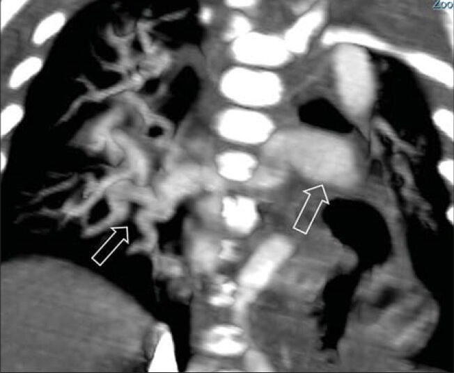
This pictorial illustration demonstrates various aspects of arterial tortuosity syndrome (ATS) obtained predominantly from a multiple detector computed tomography (MDCT) examination of a patient. In addition, a comprehensive review of typical multi-modality imaging observations in patients with ATS is presented along with a description of a few imaging signs. Non-invasively obtained, conclusive information is required in patients with ATS in view of the fragile vascular structures involved. An amazing wealth of information can be obtained by reviewing the volumetric data sets of MDCT examination. In the context of incomplete clinical information or remote reading of radiographic examination with inadequate clinical details, ability to "image data mine" the hidden, unexplored information may be vastly useful. The role of MDCT as a single modality of evaluation in ATS is highlighted.

Keywords: Aortic elongation sign; V sign; arterial tortuosity syndrome; cluster of vessels sign; image data mining; meandering vessel sign; multiple detector computed tomography.


Conflict of interest statement

Conflict of Interest: None declared.

Figures

Figure 1
Figure 1
Case 1: 7-month-old female child with poor weight gain and suspected cardiac disease was diagnosed with ATS. Frontal chest radiograph demonstrates multiple linear shadows overlying the right lung field due to excessive skin folding (black arrows). Also, there is retrocardiac lucency due to diaphragmatic hernia (empty arrow).
Figure 2
Figure 2
Case 1: 7-month-old female with poor weight gain and suspected cardiac disease was diagnosed with ATS. Surface-rendered 3-D reconstruction (a and b) of the same patient illustrates excessive skin laxity leading to multiple redundant skin folds. Changes are striking in the back, axillary regions, and lower abdomen.
Figure 3
Figure 3
Case 1: 7-month-old female with poor weight gain and suspected cardiac disease was diagnosed with ATS. Surface-rendered 3-D image in (a) frontal projection demonstrates depression in chest wall, pectus excavatum deformity (black arrow). (b) Coronal contrast-enhanced sagittal CT image shows a depression below the sternum (white arrow).
Figure 4
Figure 4
Case 1: 7-month-old female with poor weight gain and suspected cardiac disease was diagnosed with ATS. Frontal 3-D reconstructed view of spine reveals mild dorsal scoliosis with right-sided convexity. No malformation of the vertebral bodies was visualized. Ribs demonstrate a normal configuration.
Figure 5
Figure 5
Radiological signs in arterial tortuosity syndrome: Aortic elongation sign. (a and b) 4-month-old female with dysmorphic features and abnormal echo was diagnosed with ATS. Elongated tortuous aorta projects laterally (arrow). Sign is particularly useful in younger patient where the aortic knuckle is normally inconspicuous.
Figure 6
Figure 6
Case 1: 7-month-old female with poor weight gain and suspected cardiac disease was diagnosed with ATS. (a and b) Maximal intensity projection images in coronal orientation show gross elongation, tortuosity of the thoracic aorta (empty arrow). Pulmonary artery is elongated (black arrow). (b) Tortuosity and dilatation are also noted in the proximal part of great vessels (empty arrow).
Figure 7
Figure 7
Case 1: 7-month-old female with poor weight gain and suspected cardiac disease was diagnosed with ATS. Global view of 3-D rendered image of aorta in thoracic and abdominal part with extremely tortuous course of the thoracic aorta. Uneven caliber of the pulmonary artery is observed (empty arrow).
Figure 8
Figure 8
Case 2: 1-month-old female with prenatal suspicion of cardiac disease was postnatally diagnosed with ATS. a) Coronal contrast-enhanced CT shows gross dilatation and tortuosity of thoracic and abdominal aorta (arrows). Incidentally there is left lung collapse due to bronchial compression. (b) 3-D volume-rendered image shows the global sign of gross aortic dilatation and tortuosity (arrows). At the areas of kinks, there is significant luminal narrowing (triangles).
Figure 9
Figure 9
Radiological signs in arterial tortuosity syndrome: Meandering vessels sign. (a) Case 1. 7-month-female with poor weight gain and suspected cardiac disease was diagnosed with ATS. Coronal CT image shows lateral location of the tortuous aorta (empty arrows) reaching to mid-thoracic cavity. (b) 1-month-old female with prenatal suspicion of cardiac disease was postnatally diagnosed with ATS. CT image illustrates location of the meandering aorta up to the chest wall (empty arrows).
Figure 10
Figure 10
Case 1: 7-month-old female presented with poor weight gain and suspected cardiac disease was diagnosed with ATS. (a) Coronal reconstruction of contrast-enhanced CT image shows splaying of the subcarinal angle (black arrows) due to cardiomegaly and a Bochdalek's hernia (empty arrow). (b) 3-D rendered image of airways does not show significant compression or indentation by the tortuous vessels.
Figure 11
Figure 11
Case 1: 7-month-old female with poor weight gain and suspected cardiac disease was diagnosed with ATS. 3-D rendered view of the pulmonary arteries seen from the posterior aspect demonstrates an early branching of the pulmonary artery (empty arrow) with uneven caliber of right and left pulmonary arteries.
Figure 12
Figure 12
Radiological signs in arterial tortuosity syndrome: “V” sign of pulmonary artery. (a) Case 1: 7-month-old female with poor weight gain and suspected cardiac disease was diagnosed with ATS. Modified axial contrast-enhanced CT image demonstrates inverted V shape configuration of the pulmonary artery (black arrow) bifurcation and relative narrowing of proximal parts of right and left pulmonary arteries (arrows). (b) 4-month-old female with dysmorphic features and abnormal echo was diagnosed with ATS. T1-weighted MRI examination demonstrates the same configuration and narrow pulmonary arteries (arrows).
Figure 13
Figure 13
Case 3: 4-month-old female with dysmorphic features and abnormal echo was diagnosed with ATS. Frontal chest radiograph classically demonstrates elongation of the aorta (white arrow), left diaphragmatic hernia (empty arrow), and dilatation and tortuosity of the right pulmonary artery (white triangle).
Figure 14
Figure 14
Case 3: 4-month-old female with dysmorphic features and abnormal echo was diagnosed with ATS (same patient as in Figure 13). T1W MR images (a) in the sagittal orientation show the dilated pulmonary artery (white arrow) tortuous great vessels (black triangle) and grossly tortuous aorta. (b) Axial images show dilated, laterally displaced aortic arch (empty arrow). (c) Additional view at the level of pulmonary artery reveals dilated main pulmonary artery (white arrow) showing early bifurcation and rapid tapering of the caliber.
Figure 15
Figure 15
Case 1: 7-month-old female with poor weight gain and suspected cardiac disease was diagnosed with ATS. Coronal reconstruction of contrastenhanced CT image illustrates the right and left inferior pulmonary veins (empty arrows). Tortuosity and uneven caliber is also noted in both veins.
Figure 16
Figure 16
Case 1: 7-month-old female with poor weight gain and suspected cardiac disease was diagnosed with ATS. 3-D rendered image of the vascular structures of the same patient shows extreme tortuosity and meandering course of the innominate (arrow) and left common carotid arteries (empty arrow). Incidentally, tortuosity of the segments of the subclavian arteries is also illustrated.
Figure 17
Figure 17
Case 1: 7-month-old female with poor weight gain and suspected cardiac disease was diagnosed with ATS. 3-D rendered view of the left subclavian artery shows slightly tortuous course with uneven caliber (empty arrow). Tortuosity is particularly visible in the axillary and mid-arm region.
Figure 18
Figure 18
Case 1: 7-month-old female with poor weight gain and suspected cardiac disease was diagnosed with ATS. 3-D rendered view of the right subclavian shows similar changes with a gross kink in the axillary region (empty arrow). Vascular changes are also seen in the small arteries like internal mammary (black arrows) which show beaded appearance.
Figure 19
Figure 19
Radiological signs in arterial tortuosity syndrome: Cluster of vessels sign. (a) Case 1: 7-month-old female with poor weight gain and suspected cardiac disease was diagnosed with ATS. Sagittal CT reconstruction of contrast-enhanced examination reveals multiple rounded vascular structures in the superior mediastinum (arrows). (b) 4-month-old female with dysmorphic features and abnormal echo diagnosed with ATS. T1-weighted sagittal MR image shows multiple areas of flow voids corresponding to multiple cross-sections of tortuous vessels.
Figure 20
Figure 20
4-month-old male with abnormal facial features and cardiomegaly was diagnosed with ATS. Frontal chest radiograph demonstrates ectatic, laterally displaced aortic knuckle (white arrow). There is left ventricular cardiomegaly. Lungs are unremarkable.
Figure 21
Figure 21
9-year-old male with dysmorphic features and abnormal pulmonary arteries on echo was diagnosed with ATS. Time-of-flight MR images (a) in axial orientation show the gross tortuosity of the internal carotids in the paracavernous regions (white arrow). (b) Axial projection of the intracranial vessels with a TOF image demonstrates vascular tortuosity involving the carotids, basilar artery (empty arrow), and middle cerebral arteries (triangles).
Figure 22
Figure 22
Case 1: 7-month-old female with poor weight gain and suspected cardiac disease was diagnosed with ATS. 3-D rendered image of the abdominal aorta and visceral arteries shows tortuosity in the superior mesenteric artery (empty arrow). Similar changes are also noted in the proximal part of the left renal artery (white arrow). Tortuosity without significant dilatation is seen in the iliac arteries (white triangles).
Figure 23
Figure 23
Case 1: 7-month-old female with poor weight gain and suspected cardiac disease was diagnosed with ATS (same patient as in Figure 18). 3-D view of the abdominal aorta in the lateral projection demonstrates a kink in the proximal superior mesenteric artery (white arrow). More distally uneven caliber of superior mesenteric artery is shown with areas of multiple focal narrowing (empty arrows).
Figure 24
Figure 24
4-month-old male with feeding difficulty and failure to gain weight was diagnosed with ATS. Barium studies (a and b) demonstrate a large, centrally located mixed type of hiatus hernia (empty arrows).
Figure 25
Figure 25
Case 1: 7-month-old female with poor weight gain and suspected cardiac disease was diagnosed with ATS. (a) Coronal contrast-enhanced CT image reveals grossly displaced esophagus (empty arrow) and gastroesophageal junction. There is a large adjacent Bochdalek's hernia (black arrow). (b) Axial CT image shows lateral displacement of esophagus (arrow). (c) Sagittal CT reconstruction shows posteriorly located Bochdalek's hernia (black arrow).
Figure 26
Figure 26
4-month-old male child with abnormal facial features and failure to thrive was diagnosed with ATS. a) Frontal and b) left lateral chest radiograph reveals prominent, laterally displaced aorta (white arrow) and posteriorly located diaphragmatic eventrations (black arrows). Tortuous aorta is visualized as right retrocardiac density (triangle).

References

    1. Ekici F, Uçar T, Fitöz S, Atalay S, Tutar E. Cardiovascular findings in a boy with arterial tortuosity syndrome: Case report and review of the literature. Turk J Pediatr. 2011;53:104–7. - PubMed
    1. Abdul Wahab A, Janahi IA, Eltohami A, Zeid A, Faiyaz Ul Haque M, Teebi AS. A new type of Ehlers-Danlos syndrome associated with tortuous systemic arteries in a large kindred from Qatar. Acta Paediatr. 2003;92:456–62. - PubMed
    1. Kappanayil M, Nampoothiri S, Kannan R, Renard M, Coucke P, Malfait F, et al. Characterization of a distinct lethal arteriopathy syndrome in twenty-two infants associated with an identical, novel mutation in FBLN4 gene, confirms fibulin-4 as a critical determinant of human vascular elastogenesis. Orphanet J Rare Dis. 2012;7:61. - PMC - PubMed
    1. Coucke PJ, Willaert A, Wessels MW, Callewaert B, Zoppi N, De Backer J, et al. Mutations in the facilitative glucose transporter GLUT10 alter angiogenesis and cause arterial tortuosity syndrome. Nat Genet. 2006;38:452–7. - PubMed
    1. Faiyaz-Ul-Haque M, Zaidi SH, Wahab AA, Eltohami A, Al-Mureikhi MS, Al-Thani G, et al. Identification of a p. Ser81Arg encoding mutation in SLC2A10 gene of arterial tortuosity syndrome patients from 10 Qatari families. Clin Genet. 2008;74:189–93. - PubMed