. 2014 Oct;121(4):773-85.
doi: 10.1097/ALN.0000000000000352.

Amantadine alleviates postoperative cognitive dysfunction possibly by increasing glial cell line-derived neurotrophic factor in rats

Affiliations

Amantadine alleviates postoperative cognitive dysfunction possibly by increasing glial cell line-derived neurotrophic factor in rats

Junfeng Zhang et al. Anesthesiology. 2014 Oct.

Abstract

Background: Postoperative cognitive dysfunction is a clinical entity that is associated with poor outcome. We determined the effectiveness of amantadine in reducing surgery-induced cognitive impairment and the role of glial cell line-derived neurotrophic factor (GDNF) in this effect.

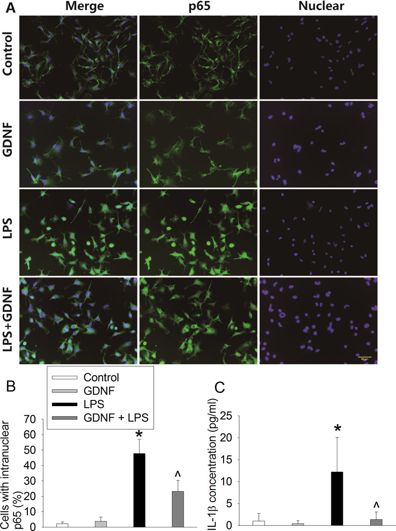
Methods: Four-month old male Fischer 344 rats were subjected to right carotid exposure under intravenous anesthesia. Some rats received intraperitoneal injection of 25 mg/kg/day amantadine for 3 days with the first dose at 15 min before the surgery or intracerebroventricular injection of GDNF or an anti-GDNF antibody at the end of surgery. One week later, rats were started to be tested by Barnes maze and fear conditioning. Hippocampus was harvested at 6 h, 24 h or 10 days after the surgery for biochemical analysis. C8-B4 cells, a microglial cell line, were pretreated with 1 ng/ml GDNF for 30 min before being exposed to 5 ng/ml lipopolysaccharide for 2 h.

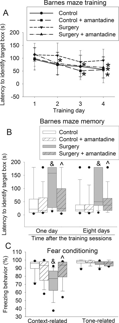
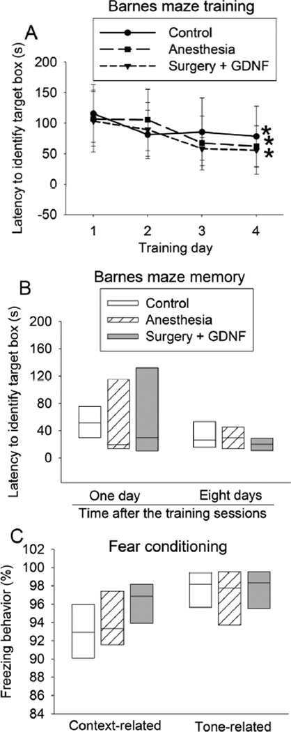
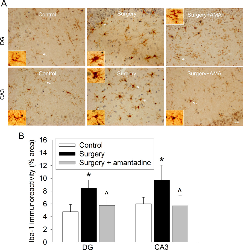
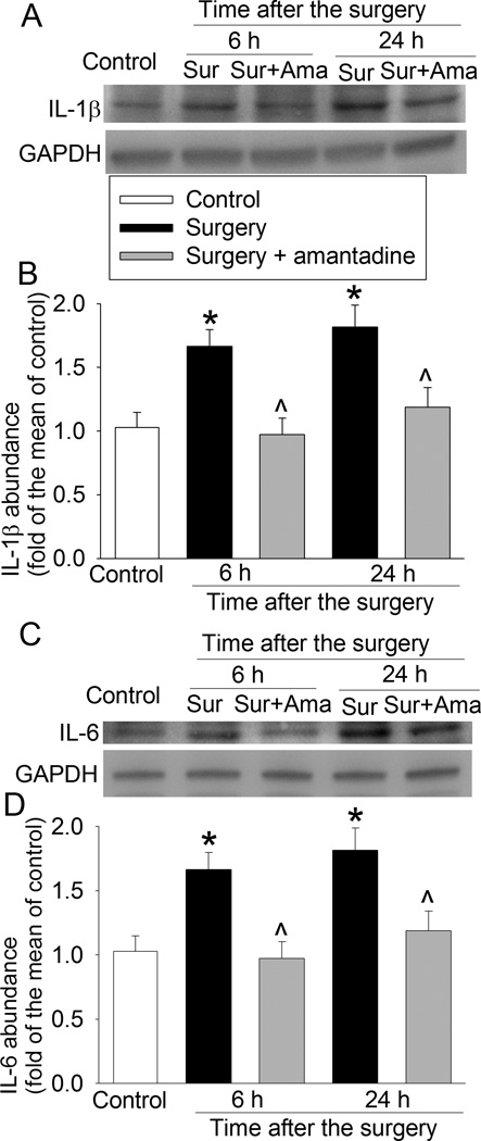
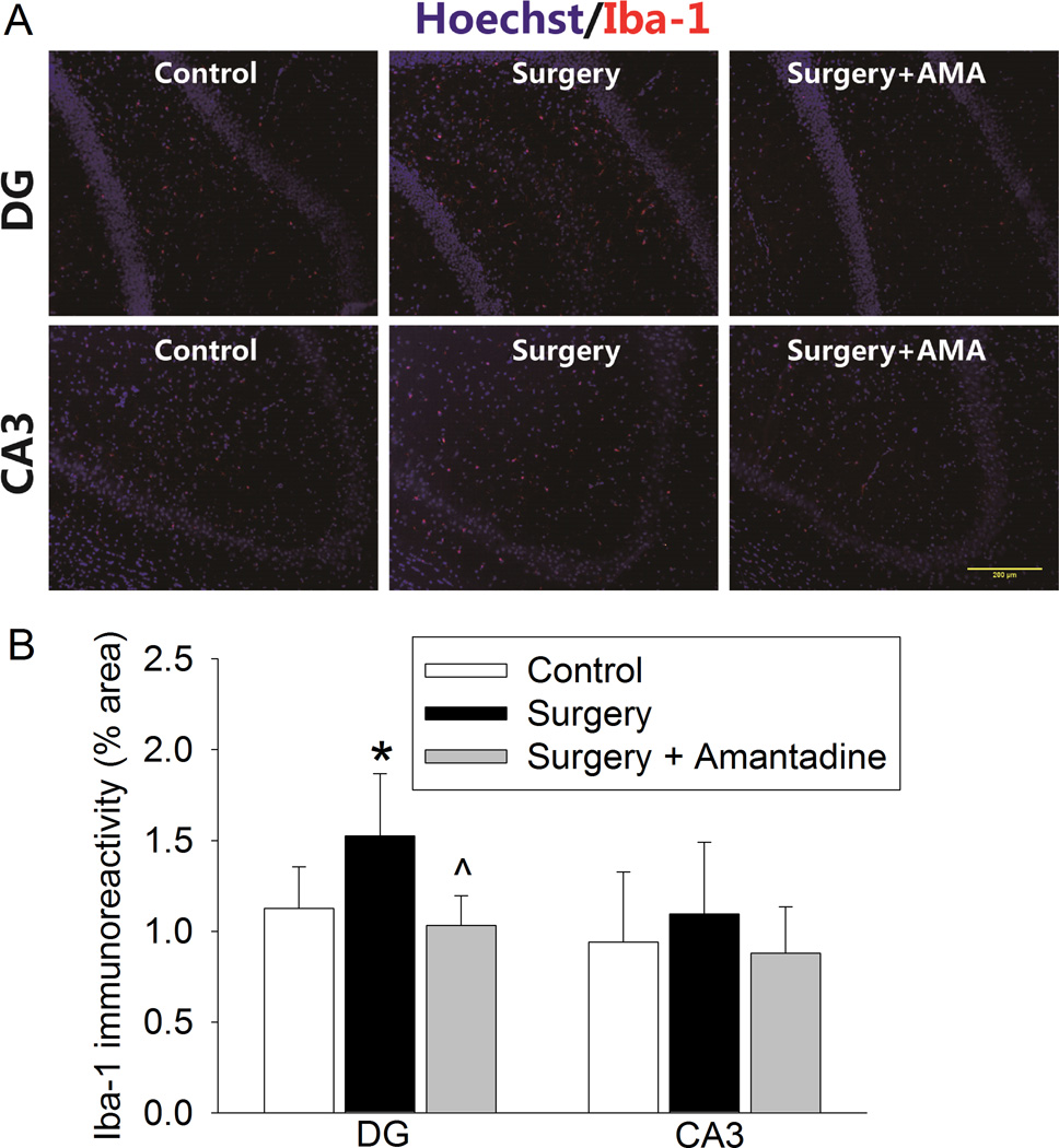
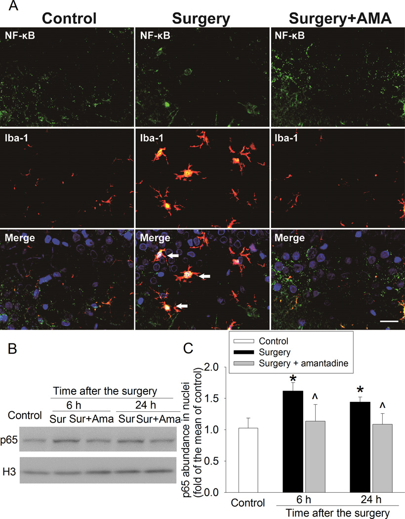
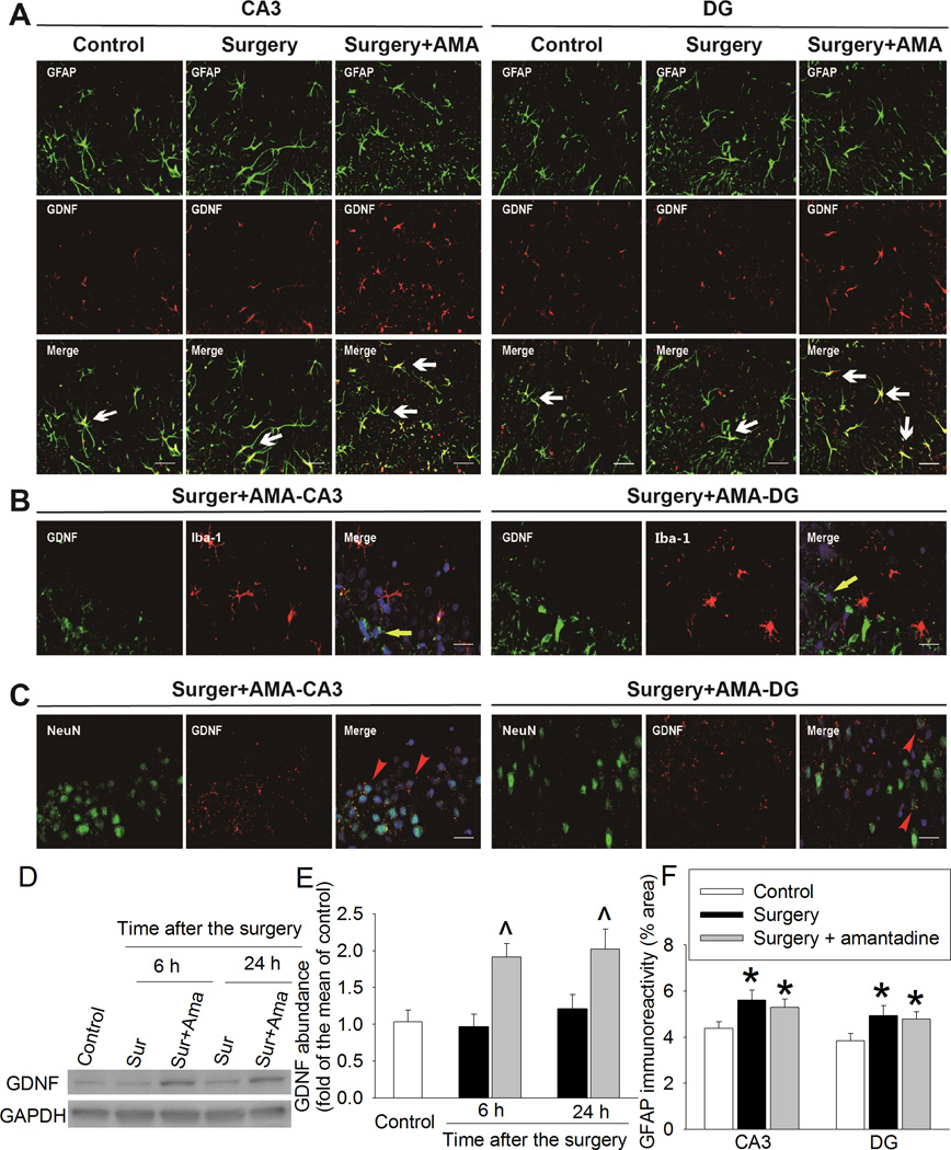
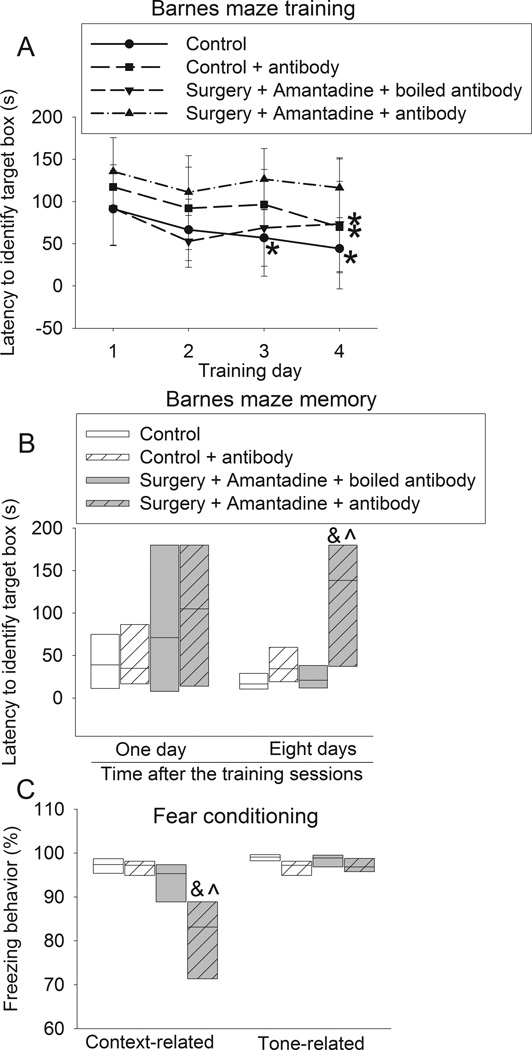
Results: Surgery increased the time to identify the target box in the Barnes maze when tested 1 day [22 (median) (11-66) (interquartile range) of control group vs. 158 (29-180) of surgery group, n = 15, P = 0.022) or 8 days after the training sessions and reduced context-related freezing behavior in the fear conditioning test. These effects were attenuated by amantadine (25 (14-90), n = 15, P = 0.029 compared with surgery group at 1 day after the training sessions in Barnes maze) and intracerebroventricular GDNF. Amantadine increased GDNF that was co-localized with glial fibrillary acidic protein, an astrocytic marker, in the hippocampus. Intracerebroventricular injection of an anti-GDNF antibody but not the denatured antibody blocked the effects of amantadine on cognition. Surgery induced neuroinflammation that was inhibited by amantadine. Lipopolysaccharide increased interleukin 1β production from C8-B4 cells. This effect was inhibited by GDNF.

Conclusions: Our results suggest that amantadine attenuated surgery-induced learning and memory impairment. This effect may be mediated by GDNF via inhibition of neuroinflammation.


Figures

Fig. 1
Fig. 1. Amantadine attenuated surgery-induced learning and memory impairment
Four-month old Fischer 344 rats were subjected to right carotid exploration under intravenous anesthesia with or without the treatment of amantadine. Barnes maze training sessions started 1 week after the surgery. A: Barnes maze training sessions. Results are mean ± S.D. (n = 15). * P < 0.05 compared with the corresponding data on day 1. B: Barnes maze memory phase. C: fear conditioning. Results in panels B and C are in box plot format (n = 15). ●: lowest or highest score (the score will not show up if it falls in the 95th percentile); between lines: 95th percentile of the data; inside boxes: 25th to 75th percentile including the median of the data. & P < 0.05 compared with the control group, ^ P < 0.05 compared with surgery alone group.
Fig. 2
Fig. 2. Glial cell line-derived neurotrophic factor (GDNF) attenuated surgery-induced learning and memory impairment
Four-month old Fischer 344 rats were subjected to right carotid exploration under intravenous anesthesia. Some rats received intra-cerebroventricular injection of GDNF. Barnes maze training sessions started 1 week after the surgery. A: Barnes maze training sessions. Results are mean ± S.D. (n = 8). * P < 0.05 compared with the corresponding data on day 1. B: Barnes maze memory phase. C: fear conditioning. Results in panels B and C are in box plot format (n = 8). Inside boxes: 25th to 75th percentile including the median of the data.
Fig. 3
Fig. 3. Amantadine inhibited surgery-induced ionized calcium binding adapter molecule 1 (Iba-1) expression at one day after the surgery
Four-month old Fischer 344 rats were subjected to right carotid exploration under intravenous anesthesia with or without the treatment of amantadine. Hippocampus was harvested at 24 h after the surgery. A: representative immunostaining images for Iba-1. Scale bars = 30 µm. B: graphic presentation of the percentage area that is Iba-1-postive staining in the CA3 and dentate gyrus (DG). Values are presented as mean ± S.D. (n = 6). * P < 0.05 compared with the control group, ^ P < 0.05 compared with surgery alone group.
Fig. 4
Fig. 4. Amantadine inhibited surgery-induced interleukin (IL)-6 and IL-1β expression
Four-month old Fischer 344 rats were subjected to right carotid exploration under intravenous anesthesia with or without the treatment of amantadine. Hippocampus was harvested at 6 h or 24 h after surgery. A: representative Western blot images of IL-1β. B: graphic presentation of IL-1β abundance. C: representative Western blot images of IL-6. D: graphic presentation of IL-6 abundance. Values are expressed as fold changes over the mean values of control rats and are presented as mean ± S.D. (n = 6). * P < 0.05 compared with the control group, ^ P < 0.05 compared with corresponding surgery only group. Ama: amantadine, GAPDH: glyceraldehyde 3-phosphate dehydrogenase, Sur: Surgery.
Fig. 5
Fig. 5. Amantadine inhibited surgery-induced ionized calcium binding adapter molecule 1 (Iba-1) expression at ten days after the surgery
Four-month old Fischer 344 rats were subjected to right carotid exploration under intravenous anesthesia with or without the treatment of amantadine. Hippocampus was harvested at 10 days after the surgery. A: representative immunostaining images. Scale bars = 200 µm. B: graphic presentation of the percentage area that is Iba-1-postive staining in the CA3 and dentate gyrus (DG). Values are presented as mean ± S.D. (n = 6). * P < 0.05 compared with the control group, ^ P < 0.05 compared with surgery alone group. AMA: amantadine.
Fig. 6
Fig. 6. Amantadine inhibited surgery-induced nuclear translocation of p65
Four-month old Fischer 344 rats were subjected to right carotid exploration under intravenous anesthesia with or without the treatment of amantadine. Hippocampus was harvested at 6 h or 24 h after surgery. A: representative immunostaining images of the hippocampal CA3 region at 24 h after the surgery. Scale bars = 30 µm. B: representative Western blot images. C: graphic presentation of p65 abundance in nuclei. Values are expressed as fold changes over the mean values of control rats and are presented as mean ± S.D. (n = 6). * P < 0.05 compared with control group, ^ P < 0.05 compared with corresponding surgery group. Ama: amantadine, Iba-1: ionized calcium binding adapter molecule 1, NF-κB: nuclear factor κ-light-chain-enhancer of activated B cells, Sur: Surgery.
Fig. 7
Fig. 7. Amantadine increased glial cell line-derived neurotrophic factor (GDNF) expression
Four-month old Fischer 344 rats were subjected to right carotid exploration under intravenous anesthesia with or without the treatment of amantadine. Hippocampus was harvested at 6 h or 24 h after surgery. A, B and C: representative immunostaining images of hippocampus harvested at 24 h after surgery. Scale bars = 30 µm. D: representative Western blot images. E: graphic presentation of GDNF abundance. Values are expressed as fold changes over the mean values of control rats and are presented as mean ± S.D. (n = 6). ^ P < 0.05 compared with corresponding surgery alone group. F: graphic presentation of the percentage area that is glial fibrillary acidic protein (GFAP)-positive staining in the CA3 and dentate gyrus (DG) at 24 h after the surgery. Values are presented as mean ± S.D. (n = 6). * P < 0.05 compared with the control group. Ama: amantadine, GAPDH: glyceraldehyde 3-phosphate dehydrogenase, Iba-1: ionized calcium binding adapter molecule 1, Sur: Surgery.
Fig. 8
Fig. 8. Glial cell line-derived neurotrophic factor (GDNF) inhibited lipopolysaccharide (LPS)-induced microglial activation
C8-B4 cells were pretreated with 1 ng/ml GDNF for 30 min before being exposed to 5 ng/ml lipopolysaccharide. A: representative immunostaining images of the cells after being exposed to lipopolysaccharide for 1 h. Nuclei were stained by Hoechst 33342. Scale bars = 50 µm. B: graphic presentation of the percentage cells that are positive staining of intra-nuclear p65 after being exposed to lipopolysaccharide for 1 h. C: interleukin (IL)-1β concentrations in culture medium that was harvested at 24 h after the cells were incubated with lipopolysaccharide for 2 h. Values are presented as mean ± S.D. (n = 6). * P < 0.05 compared with the control group, ^ P < 0.05 compared with the group treated with lipopolysaccharide only.
Fig. 9
Fig. 9. Anti-glial cell line-derived neurotrophic factor (GDNF) antibody blocked the effects of amantadine on learning and memory after surgery
Four-month old Fischer 344 rats were subjected to right carotid exploration under intravenous anesthesia with or without the treatment of amantadine. Some rats received intra-cerebroventricular injection of an anti-GDNF antibody. Barnes maze training sessions started 1 week after the surgery. A: Barnes maze training sessions. Results are mean ± S.D. (n = 8). * P < 0.05 compared with the corresponding data on day 1. B: Barnes maze memory phase. C: fear conditioning. Results in panels B and C are in box plot format (n = 8). Inside boxes: 25th to 75th percentile including the median of the data. & P < 0.05 compared with the control group, ^ P < 0.05 compared with surgery plus amantadine plus boiled antibody group.

References

    1. Terrando N, Brzezinski M, Degos V, Eriksson LI, Kramer JH, Leung JM, Miller BL, Seeley WW, Vacas S, Weiner MW, Yaffe K, Young WL, Xie Z, Maze M. Perioperative cognitive decline in the aging population. Mayo Clin. Proc. 2011;86:885–893. - PMC - PubMed
    1. Moller JT, Cluitmans P, Rasmussen LS, Houx P, Rasmussen H, Canet J, Rabbitt P, Jolles J, Larsen K, Hanning CD, Langeron O, Johnson T, Lauven PM, Kristensen PA, Biedler A, van Beem H, Fraidakis O, Silverstein JH, Beneken JE, Gravenstein JS. Long-term postoperative cognitive dysfunction in the elderly ISPOCD1 study. ISPOCD investigators. International Study of Post-Operative Cognitive Dysfunction. Lancet. 1998;351:857–861. - PubMed
    1. Monk TG, Weldon BC, Garvan CW, Dede DE, van der Aa MT, Heilman KM, Gravenstein JS. Predictors of cognitive dysfunction after major noncardiac surgery. Anesthesiology. 2008;108:18–30. - PubMed
    1. Newman MF, Kirchner JL, Phillips-Bute B, Gaver V, Grocott H, Jones RH, Mark DB, Reves JG, Blumenthal JA. Longitudinal assessment of neurocognitive function after coronary-artery bypass surgery. N. Engl. J. Med. 2001;344:395–402. - PubMed
    1. Steinmetz J, Christensen KB, Lund T, Lohse N, Rasmussen LS. Long-term consequences of postoperative cognitive dysfunction. Anesthesiology. 2009;110:548–555. - PubMed

Publication types

MeSH terms

Substances