. 2015 Mar;61(3):930-41.
doi: 10.1002/hep.27492. Epub 2015 Jan 28.

Axl activates autocrine transforming growth factor-β signaling in hepatocellular carcinoma

Affiliations

Axl activates autocrine transforming growth factor-β signaling in hepatocellular carcinoma

Patrick Reichl et al. Hepatology. 2015 Mar.

Abstract

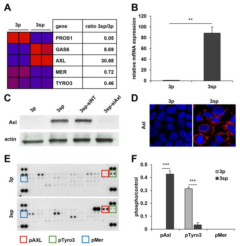
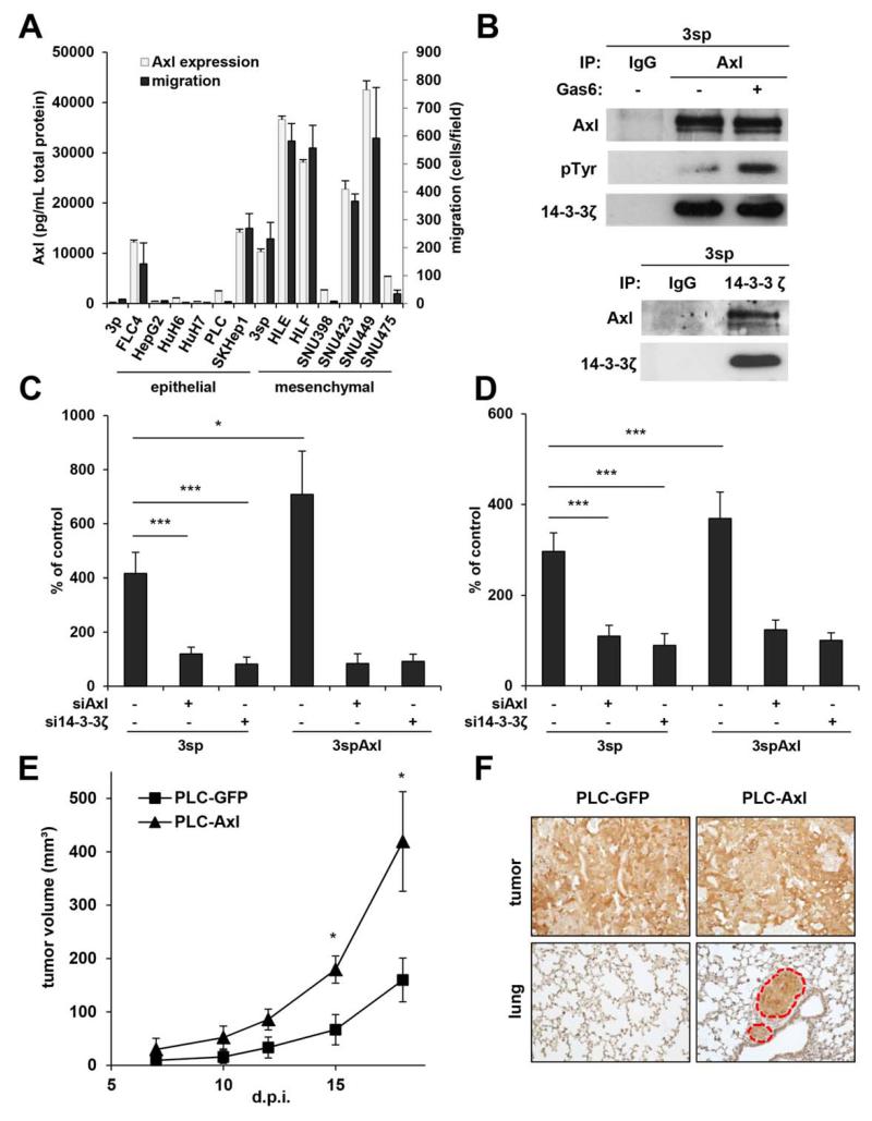
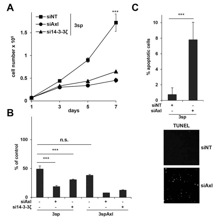
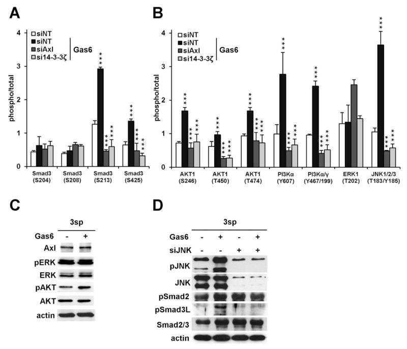
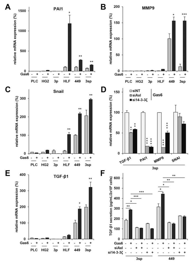
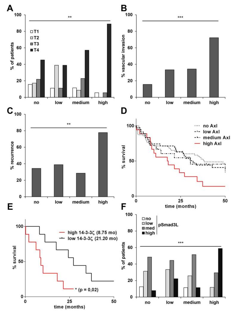
In hepatocellular carcinoma (HCC), intrahepatic metastasis frequently correlates with epithelial to mesenchymal transition (EMT) of malignant hepatocytes. Several mechanisms have been identified to be essentially involved in hepatocellular EMT, among them transforming growth factor (TGF)-β signaling. Here we show the up-regulation and activation of the receptor tyrosine kinase Axl in EMT-transformed hepatoma cells. Knockdown of Axl expression resulted in abrogation of invasive and transendothelial migratory abilities of mesenchymal HCC cells in vitro and Axl overexpression-induced metastatic colonization of epithelial hepatoma cells in vivo. Importantly, Axl knockdown severely impaired resistance to TGF-β-mediated growth inhibition. Analysis of the Axl interactome revealed binding of Axl to 14-3-3ζ, which is essentially required for Axl-mediated cell invasion, transendothelial migration, and resistance against TGF-β. Axl/14-3-3ζ signaling caused phosphorylation of Smad3 linker region (Smad3L) at Ser213, resulting in the up-regulation of tumor-progressive TGF-β target genes such as PAI1, MMP9, and Snail as well as augmented TGF-β1 secretion in mesenchymal HCC cells. Accordingly, high Axl expression in HCC patient samples correlated with elevated vessel invasion of HCC cells, higher risk of tumor recurrence after liver transplantation, strong phosphorylation of Smad3L, and lower survival. In addition, elevated expression of both Axl and 14-3-3ζ showed strongly reduced survival of HCC patients.

Conclusion: Our data suggest that Axl/14-3-3ζ signaling is central for TGF-β-mediated HCC progression and a promising target for HCC therapy.


Figures

Fig. 1
Fig. 1
Expression of TAM receptors and their ligands in epithelial and mesenchymal HCC cells. (A) Down-regulation (blue) and up-regulation (red) of Tyro3, Axl, Mer, and their ligands Gas6 and Protein S (PROS1) in epithelial 3p and mesenchymal 3sp cells as assessed by whole-genome expression analysis. (B) Axl mRNA levels in 3p and 3sp cells analyzed by qRT-PCR. (C) Western blot analysis of Axl expression in 3p and 3sp cells. Actin was used as loading control. (D) Localization of Axl as shown by confocal immunofluorescence analysis (red). (E) Phospho-RTK array analysis of 3p and 3sp cells. Rectangles indicate phosphorylation of TAM receptors. (F) Quantification of TAM phosphorylation in 3p and 3sp cells. Data are expressed as mean ± SD. **P < 0.01; ***P < 0.001.
Fig. 2
Fig. 2
Role of Axl and 14-3-3ζ in migration, transendothelial invasion, and metastasis of HCC cells. (A) Fourteen hepatoma cell lines were analyzed for Axl expression by ELISA and migration. (B) Immunoprecipitation against Axl with and without Gas6 stimulation (500 ng/mL; upper panel). Phosphorylation of Axl by Gas6 and interaction with 14-3-3ζ as shown by immunoblotting against phosphotyrosine and 14-3-3ζ. Lower panel: Immunoprecipitation of 14-3-3ζ and probing for Axl. (C) Transwell migration of 3sp and Axl-overexpressing 3spAxl cells after stimulation with Gas6 (500 ng/mL) and interference with Axl or 14-3-3ζ by siRNA. (D) Transendothelial invasion of 3sp and 3spAxl cells through a monolayer of HSECs after incubation with Gas6 (500 ng/mL) and interference with Axl or 14-3-3ζ by siRNA. Data are expressed as mean ± SD. (E) Tumor growth of Axl-overexpressing PLC/PRF/5 cells (PLC-Axl; n = 4), injected subcutaneously into NSG mice, as compared to control (PLC-GFP; n = 4). (F) Immunohistochemical analysis of GFP in primary subcutaneous tumors and lung metastasis 30 days postresection of subcutaneous tumors derived from PLC-GFP (n = 4) and PLC-Axl (n = 4). Red broken circles indicate metastatic nodules. Data are expressed as mean ± SEM. d.p.i., days postinjection; NT, nontarget. *P < 0.05; ***P < 0.001.
Fig. 3
Fig. 3
Role of Axl and 14-3-3ζ in TGF-β resistance. (A) Proliferation kinetics of 3sp cells incubated with TGF-β1 (2.5 ng/mL) after knockdown of Axl or 14-3-3ζ. (B) Cell survival of 3sp and 3spAxl cells after treatment with TGF-β1 (2.5 ng/mL) for 7 days and interference with Axl or 14-3-3ζ. (C) Induction of cell death by TGF-β1 in 3sp cells after knockdown of Axl as shown by TUNEL assay. Cells were treated with 10 ng/mL TGF-β1 for 24 hours. Lower panel shows images of TUNEL-positive cells by fluorescence microscopy. Data are expressed as mean ± SD. ***P < 0.001; n.s., nonsignificant.
Fig. 4
Fig. 4
Modulation of TGF-β signaling by the Gas6/Axl/14-3-3ζ axis. (A) Phosphorylation of Smad3 was assessed by phospho-specific antibody microarrays in untreated 3sp control cells (siNT) and 3sp cells with a knockdown of Axl or 14-3-3ζ (siAxl, si14-3-3ζ). Cells were treated with Gas6 (500 ng/mL) for 15 minutes or left untreated (siNT). (B) Phosphorylation of PI3K/AKT, ERK, and JNK by Gas6 in 3sp cells with and without interference with Axl or 14-3-3ζ as detected by array analysis. (C) Western blot analysis of ERK and AKT in 3sp cells with and without Gas6 stimulation (500 ng/mL). Actin was used as loading control. (D) Western blot analysis of JNK, phospho-Smad2 (pSmad2), phospho-Smad3L (pSmad3L), and total Smad2/3 after stimulation with Gas6 (500 ng/mL) and interference with JNK by siRNA (siJNK). Data are expressed as mean ± SD. ***P < 0.001.
Fig. 5
Fig. 5
Axl/14-3-3ζ activates tumor-promoting TGF-β target genes and autocrine TGF-β signaling. qRT-PCR analysis of epithelial PLC/PRF/5 (PLC), HepG2 (HG2), and 3p HCC cells as well as mesenchymal HLF, SNU449 (449), and 3sp cells after stimulation with Gas6 for 8 hours. (A-C) Analysis of PAI1, MMP9, and Snail. Untreated SNU449 were 100%. (D) Repression of TGF-β target gene expression in 3sp cells after stimulation with Gas6 (500 ng/mL) for 8 hours and knockdown of Axl or 14-3-3ζ. (E) Analysis of TGF-β1 mRNA expression. (F) Secretion of TGF-β1 protein was analyzed by ELISA in supernatants of 3sp and SNU449 cells with or without a knockdown of Axl or 14-3-3ζ. Cells were untreated or treated with 500 ng/mL Gas6 for 24 hours. Data are expressed as mean ± SD. *P < 0.05; **P < 0.01; ***P < 0.001.
Fig. 6
Fig. 6
Correlation of Axl and 14-3-3ζ levels with prognosis and survival of HCC patients. Immunohistochemical staining intensities of Axl were scored with no, low, medium, and high protein levels. (A) Correlation of staining intensities with tumor stages, (B) vascular invasion, (C) recurrence of disease. (D) Kaplan-Meier survival analysis of patients. (E) Kaplan-Meier survival analysis of patients expressing high Axl and either low or high 14-3-3ζ. (F) Correlation of Axl expression with phospho-Smad3L (pSmad3L) levels. Numbers in parentheses indicate median survival in months. **P < 0.01; ***P < 0.001.
Fig. 7
Fig. 7
Scheme depicting the molecular cooperation between Axl/14-3-3ζ and TGF-β signaling. Phosphorylation of Smad3L by Axl/14-3-3ζ and JNK induces the expression of tumor-progressive TGF-β target genes leading to autocrine TGF-β1 activation (circled arrow) and invasion of HCC by escape from TGF-β sensitivity.

Comment in

References

    1. El-Serag HB, Rudolph KL. Hepatocellular carcinoma: epidemiology and molecular carcinogenesis. Gastroenterology. 2007;132:2557–2576. - PubMed
    1. Forner A, Llovet JM, Bruix J. Hepatocellular carcinoma. Lancet. 2012;379:1245–1255. - PubMed
    1. Reichl P, Haider C, Grubinger M, Mikulits W. TGF-beta in epithelial to mesenchymal transition and metastasis of liver carcinoma. Curr Pharm Des. 2012;18:4135–4147. - PubMed
    1. Thiery JP, Acloque H, Huang RY, Nieto MA. Epithelial-mesenchymal transitions in development and disease. Cell. 2009;139:871–890. - PubMed
    1. Giannelli G, Villa E, Lahn M. Transforming growth factor-beta as a therapeutic target in hepatocellular carcinoma. Cancer Res. 2014;74:1890–1894. - PubMed

Publication types

MeSH terms