. 2014 Dec 15;464(3):401-11.
doi: 10.1042/BJ20141057.

A context-independent N-glycan signal targets the misfolded extracellular domain of Arabidopsis STRUBBELIG to endoplasmic-reticulum-associated degradation

Affiliations

A context-independent N-glycan signal targets the misfolded extracellular domain of Arabidopsis STRUBBELIG to endoplasmic-reticulum-associated degradation

Silvia Hüttner et al. Biochem J. .

Abstract

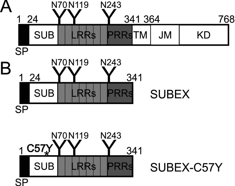
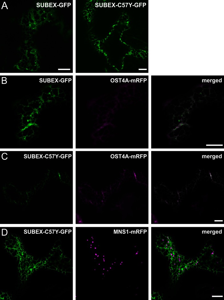
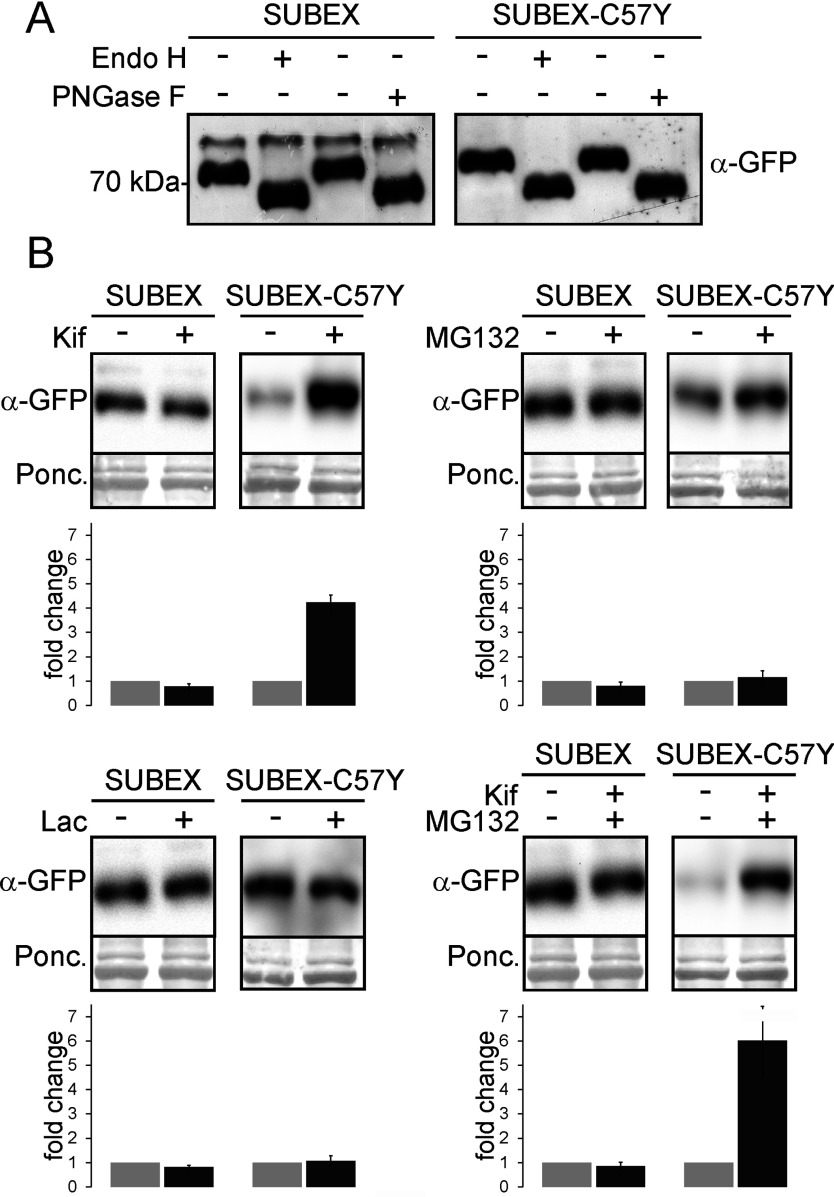
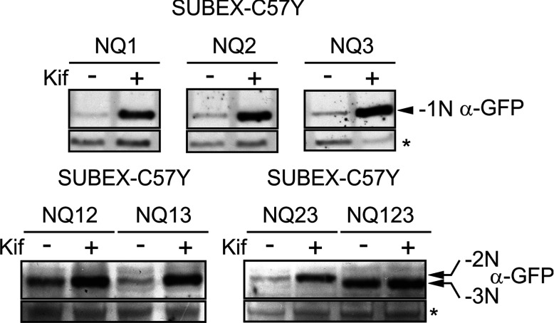
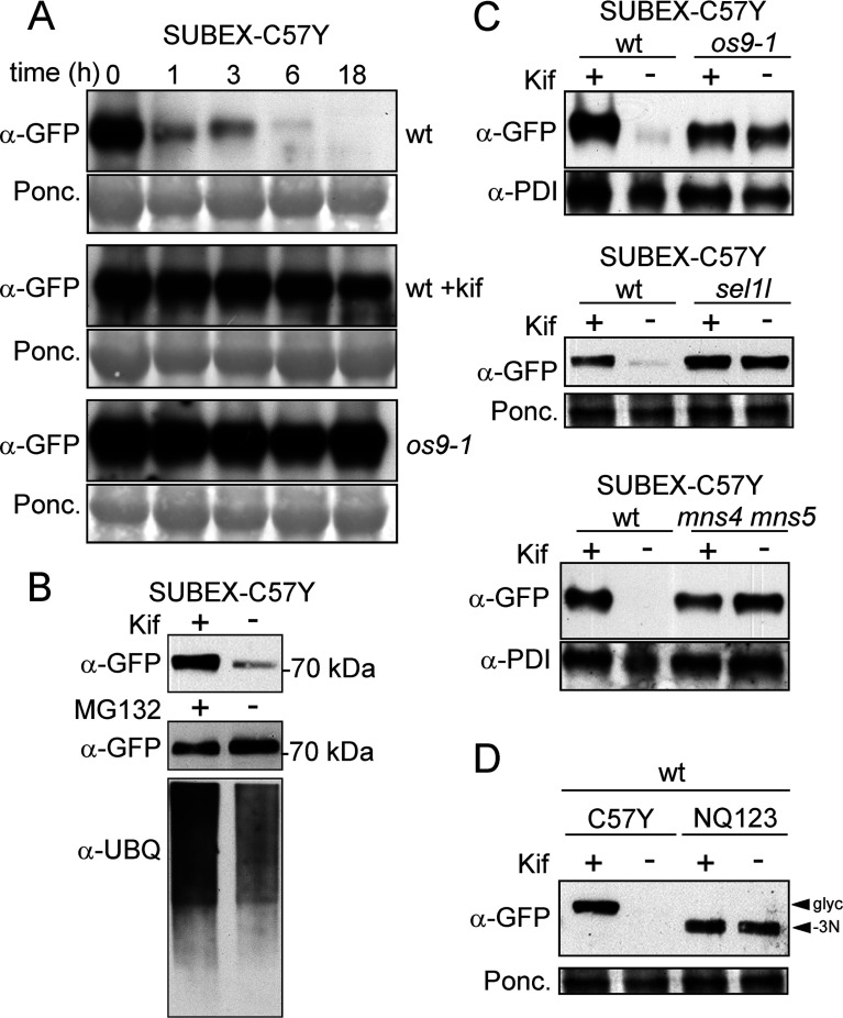
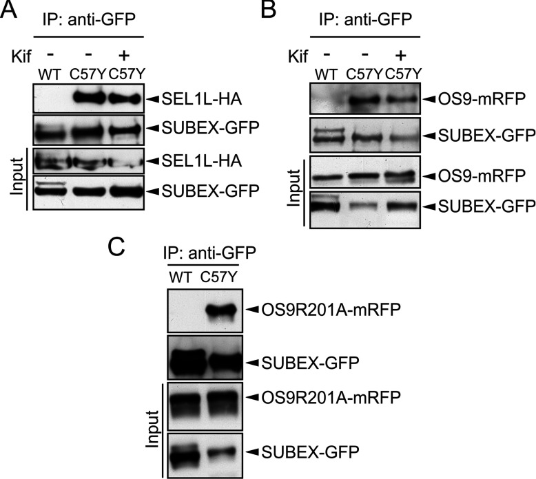
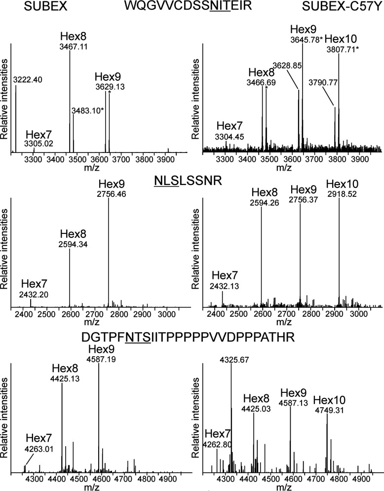
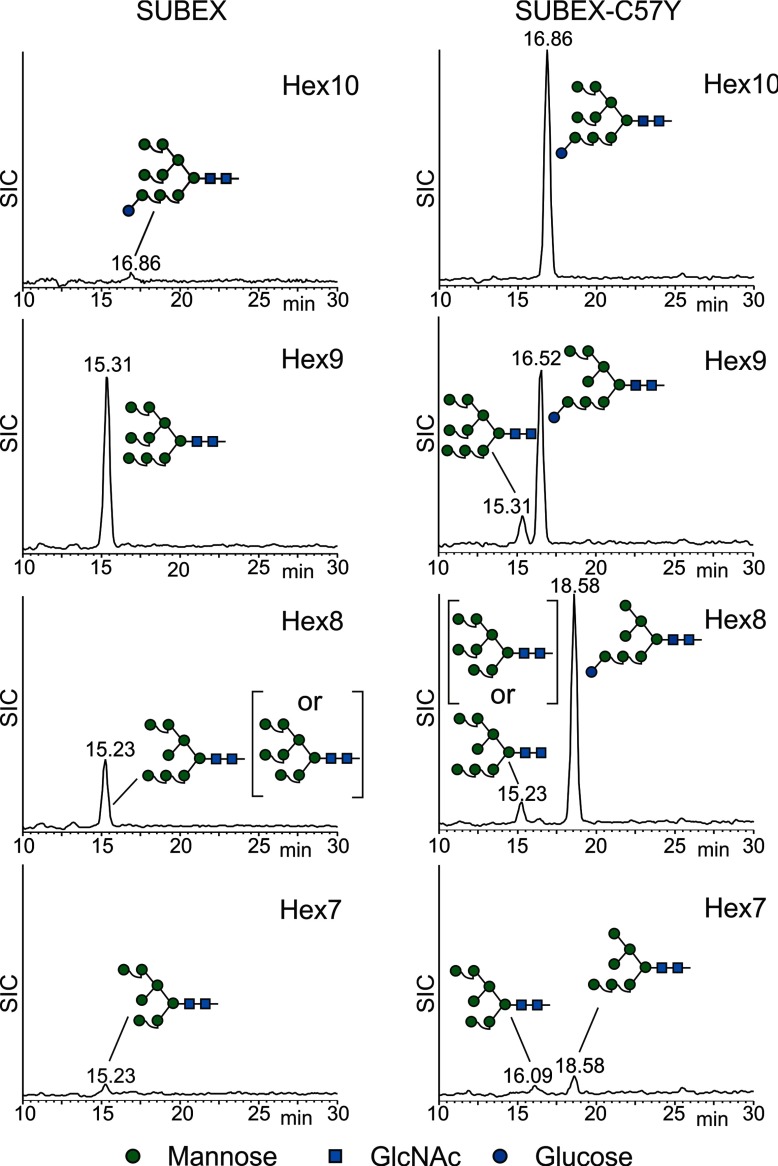
N-glycosylation of proteins plays an important role in the determination of the fate of newly synthesized glycoproteins in the endoplasmic reticulum (ER). Specific oligosaccharide structures recruit molecular chaperones that promote folding or mannose-binding lectins that assist in the clearance of improperly-folded glycoproteins by delivery to ER-associated degradation (ERAD). In plants, the mechanisms and factors that recognize non-native proteins and sort them to ERAD are poorly understood. In the present study, we provide evidence that a misfolded variant of the STRUBBELIG (SUB) extracellular domain (SUBEX-C57Y) is degraded in a glycan-dependent manner in plants. SUBEX-C57Y is an ER-retained glycoprotein with three N-glycans that is stabilized in the presence of kifunensine, a potent inhibitor of α-mannosidases. Stable expression in Arabidopsis thaliana knockout mutants revealed that SUBEX-C57Y degradation is dependent on the ER lectin OS9 and its associated ERAD factor SEL1L. SUBEX-C57Y was also stabilized in plants lacking the α-mannosidases MNS4 and MNS5 that generate a terminal α1,6-linked mannose on the C-branch of N-glycans. Notably, the glycan signal for degradation is not constrained to a specific position within SUBEX-C57Y. Structural analysis revealed that SUBEX-C57Y harbours considerable amounts of Glc1Man7GlcNAc2 N-glycans suggesting that the ER-quality control processes involving calnexin/calreticulin (CNX/CRT) and ERAD are tightly interconnected to promote protein folding or disposal by termination of futile folding attempts.


Figures

Figure 1
Figure 1. Domain structure of SUB and SUBEX variants
(A) Schematic representation of the full-length SUB protein. Different protein domains are indicated [22]. (B) Schematic representation of the truncated proteins SUBEX and SUBEX-C57Y, comprising only the extracellular domain of SUB. The C57Y mutation is indicated by an asterisk. N-glycosylation sites are represented by ‘Y’ and their amino acid positions are shown. JM, juxtamembrane domain; KD, kinase domain; LRR, leucine-rich repeat; PRR, proline-rich repeat; SP, signal peptide; SUB, SUB-domain; TM, transmembrane domain.
Figure 2
Figure 2. SUBEX and SUBEX-C57Y display ER-localization
(A) SUBEX-GFP and SUBEX-C57Y-GFP respectively were transiently expressed in N. benthamiana leaf epidermal cells and analysed by confocal microscopy. (B) Expression of SUBEX-GFP (green) together with the ER-marker OST4A-mRFP (magenta) showing co-location (merged image). (C) Expression of SUBEX-C57Y-GFP (green) together with the ER-marker OST4A-mRFP (magenta) showing co-location (merged image). (D) Co-expression of SUBEX-C57Y-GFP (green) and the Golgi-marker MNS1-mRFP (magenta) indicates specific ER-localization of SUBEX-C57Y-GFP. Scale bars=10 μm.
Figure 3
Figure 3. Stabilization of SUBEX-C57Y is glycan-dependent but proteasome-independent
(A) SUBEX-GFP and SUBEX-C57Y-GFP were transiently expressed in N. benthamiana plants. Protein extracts from leaves were digested with Endo H and PNGase F and subjected to SDS/PAGE followed by immunoblotting. The shift upon endoglycosidase digestion shows the presence of oligomannosidic N-glycans. (B) Leaves expressing SUBEX-GFP or SUBEX-C57Y-GFP were incubated for 5 h with the α-mannosidase inhibitor kifunensine (Kif), the proteasome inhibitors MG132 or lactacystin (Lac) or a combination thereof. Proteins were extracted and analysed by SDS/PAGE followed by immunoblotting. Ponceau S (Ponc.) staining served as a loading control. At the bottom of each blot, the quantification of the SUBEX and SUBEX-C57Y signals are shown (mean of three independent experiments; error bars=S.D.). The amount of SUBEX or SUBEX-C57Y without inhibitor was set to 1 (light grey bars) and the relative stabilization of inhibitor-treated samples is shown (dark grey bars).
Figure 4
Figure 4. Stabilizing effect of kifunensine on SUBEX-C57Y is dependent on the presence of N-glycans
Protein extracts from plants transiently expressing SUBEX-C57Y-GFP lacking one (NQ1, NQ2, NQ3), two (NQ12, NQ13, NQ23) or all three (NQ123) of its N-glycans were incubated with/without kifunensine (Kif) and subjected to SDS/PAGE followed by immunoblotting. The asterisk indicates an unspecific band used as loading control.
Figure 5
Figure 5. SUBEX-C57Y is stabilized in A. thaliana os9-1, sel1l and mns4 mns5 mutants
(A) Leaves from wild-type (wt) and os9-1 plants stably expressing SUBEX-C57Y-GFP were incubated with/without kifunensine (Kif). At the indicated time-points, proteins were extracted and analysed by immunoblotting. (B) Seedlings from wt plants stably expressing SUBEX-C57Y-GFP were incubated with/without Kif or MG132. Protein extracts were subjected to SDS/PAGE and immunoblotting. Detection of ubiquitin serves as a control for MG132 treatment. (C) Leaves from os9-1, sel1l and mns4 mns5 mutants stably expressing SUBEX-C57Y-GFP were incubated with or without Kif. (D) Leaves from wt plants stably expressing SUBEX-C57Y-GFP or the non-glycosylated SUBEX-C57Y-GFP variant NQ123 were incubated with/without Kif. Ponceau S (Ponc.) staining or anti-PDI detection served as loading controls.
Figure 6
Figure 6. SEL1L and OS9 interact with SUBEX-C57Y independent of glycan trimming or a functional OS9 MRH domain
(A) SUBEX-GFP and SUBEX-C57Y-GFP were transiently co-expressed with SEL1L-HA in N. benthamiana, in the presence or absence of 20 μM kifunensine (kif). (B) and (C) OS9-mRFP and OS9R201A-mRFP were co-expressed with SUBEX-GFP (WT) and SUBEX-C57Y-GFP (C57Y). SUBEX-GFP (WT) and SUBEX-C57Y-GFP (C57Y) were purified using GFP trap beads and co-purified proteins were analysed by immunoblotting with anti-HA or anti-mRFP antibodies. IP denotes the co-immunoprecipitated fraction.
Figure 7
Figure 7. SUBEX and SUBEX-C57Y differ in their N-glycan structures
SUBEX-GFP and SUBEX-C57Y-GFP were transiently expressed in N. benthamiana, purified, trypsin digested and the three glycopeptides analysed by LC-ESI-MS. The major peaks with masses corresponding to individual N-glycan structures are labelled. Hex7: Hex7HexNAc2; Hex8: Hex8HexNAc2; Hex9: Hex9HexNAc2; Hex10: Hex10HexNAc2. Ammonia adducts are indicated with an asterisk. The amino acid sequences of the peptides are indicated and the N-glycosylation sites are underlined.
Figure 8
Figure 8. SUBEX-C57Y carries increased amounts of N-glycans with a free α1,6-linked mannose residue at the C-branch
SUBEX-GFP and SUBEX-C57Y-GFP were transiently expressed in N. benthamiana, purified and the N-glycans were liberated and analysed by PGC-LC-ESI MS. Selected ion chromatograms (SICs) are shown. The assignment of structural isomers was based on reference glycans.

References

    1. Ellgaard L., Helenius A. Quality control in the endoplasmic reticulum. Nat. Rev. Mol. Cell Biol. 2003;4:181–191. doi: 10.1038/nrm1052. - DOI - PubMed
    1. Hegde R. S., Ploegh H. L. Quality and quantity control at the endoplasmic reticulum. Curr. Opin. Cell Biol. 2010;22:437–446. doi: 10.1016/j.ceb.2010.05.005. - DOI - PMC - PubMed
    1. Smith M. H., Ploegh H. L., Weissman J. S. Road to ruin: targeting proteins for degradation in the endoplasmic reticulum. Science. 2011;334:1086–1090. doi: 10.1126/science.1209235. - DOI - PMC - PubMed
    1. Howell S. H. Endoplasmic reticulum stress responses in plants. Annu. Rev. Plant Biol. 2013;64:477–499. doi: 10.1146/annurev-arplant-050312-120053. - DOI - PubMed
    1. Tintor N., Saijo Y. ER-mediated control for abundance, quality, and signaling of transmembrane immune receptors in plants. Front. Plant Sci. 2014;5:65. doi: 10.3389/fpls.2014.00065. - DOI - PMC - PubMed

Publication types

MeSH terms