. 2014 Nov;93(11):1124-32.
doi: 10.1177/0022034514552675. Epub 2014 Sep 24.

Transplantation of SHED prevents bone loss in the early phase of ovariectomy-induced osteoporosis

Affiliations

Transplantation of SHED prevents bone loss in the early phase of ovariectomy-induced osteoporosis

Y Liu et al. J Dent Res. 2014 Nov.

Abstract

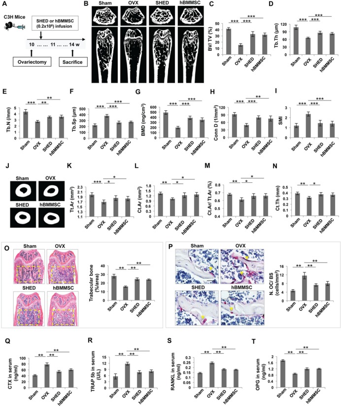
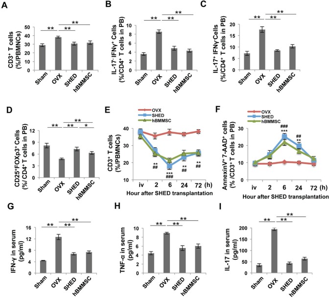
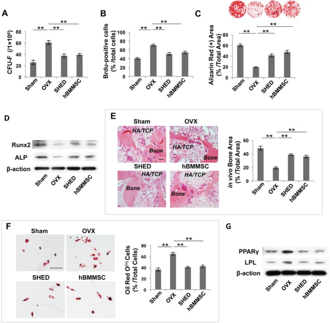
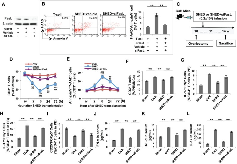
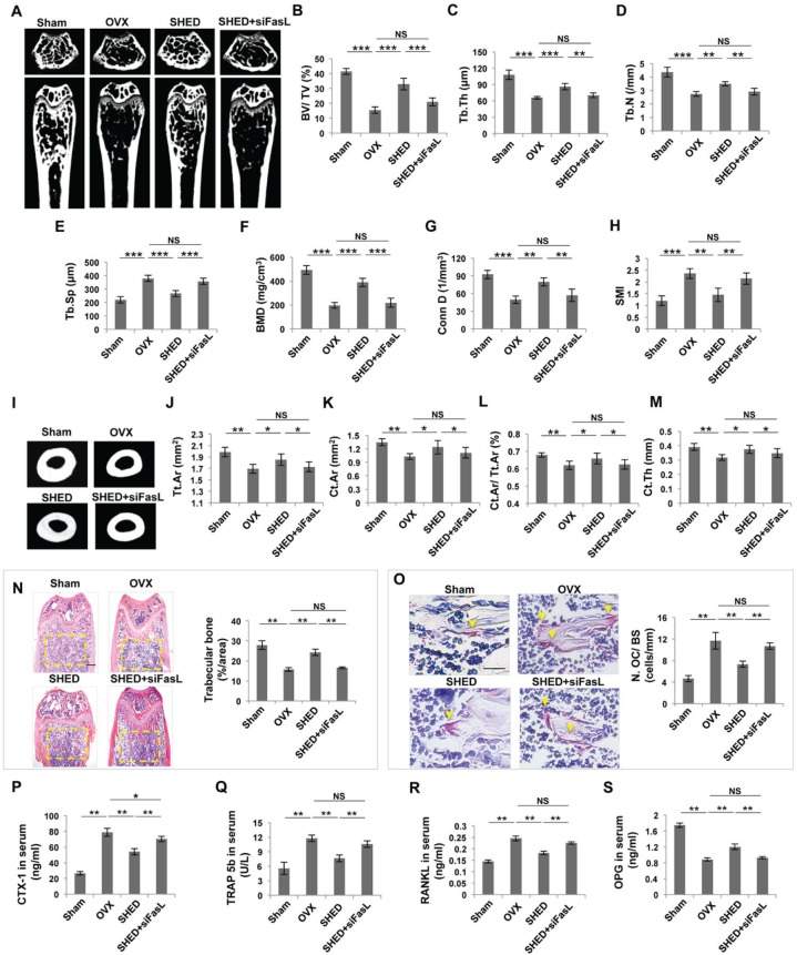
Stem cells from human exfoliated deciduous teeth (SHED) are a unique postnatal stem cell population, possessing multipotent differentiation capacity and immunomodulatory properties. However, the mechanism by which SHED treat immune diseases is not fully understood. Here we show that systemic transplantation of SHED via the tail vein ameliorates ovariectomy (OVX)-induced osteopenia by reducing T-helper 1 (Th1) and T-helper 17 (Th17) cell numbers in the recipient OVX mice. Mechanistically, SHED transplantation induces activated T-cell apoptosis in OVX mice via Fas ligand (FasL)-mediated Fas pathway activation, leading to up-regulation of regulatory T-cells (Tregs) and down-regulation of Th1 and Th17 cells. This SHED-mediated immunomodulation rescues OVX-induced impairment of bone marrow mesenchymal stem cells (BMMSCs) and activation of osteoclastogenesis, resulting in increased bone mass. In summary, SHED-mediated T-cell apoptosis via a FasL/Fas pathway results in immune tolerance and ameliorates the osteopenia phenotype in OVX mice.

Keywords: Fas ligand; apoptosis; deciduous teeth; immunotherapy; mesenchymal stem cells; regulatory T cells (Tregs).


Conflict of interest statement

The authors declare no potential conflicts of interest with respect to the authorship and/or publication of this article.

Figures

Figure 1.
Figure 1.
SHED transplantation blocked early osteoporotic phenotype development in OVX mice. (A) Schema indicating the experimental design for SHED or hBMMSC transplantation in OVX mice. (B) μCT images of trabecular bone structure in the femurs of sham, OVX, SHED-transplanted, and hBMMSC-transplanted mice. OVX mice showed a reduced trabecular bone volume compared with that in the sham group. SHED and hBMMSC transplantation partially rescued OVX-induced reduction of trabecular bone volume. (C-I) μCT analysis showed that OVX induced reduced bone volume (BV/TV), trabecular thickness (Tb.Th), trabecular number (Tb.N), bone mineral density (BMD), and connectivity density (Conn.D), along with increased trabecular space (Tb.Sp) and structure model index (SMI) in the femurs. (J) μCT analysis showed that the cortical bone volume in the femur was reduced in OVX mice. SHED and hBMMSC transplantation increased the cortical bone volume in the femurs of OVX mice. (K-N) μCT analysis showed that OVX resulted in reduced total cross-sectional area (Tt.Ar), cortical bone area (Ct.Ar), cortical bone fraction (Ct.Ar/Tt.Ar), and cortical thickness (Ct.Th) in the femurs of OVX mice. SHED and hBMMSC transplantation significantly increased Tt.Ar, Ct.Ar, Ct.Ar/Tt.Ar, and Ct.Th in OVX mice. (O) Hematoxylin and eosin (H&E) staining showed that OVX resulted in reduced trabecular bone area (yellow circled area) compared with the sham mice, as analyzed by ImageJ software. SHED and hBMMSC transplantation significantly increased the trabecular bone area. (P) TRAP staining showed that OVX resulted in increased TRAP+ cells (yellow arrows) in the femurs of OVX mice. SHED and hBMMSC transplantation reduced the number of TRAP+ cells in the femurs of OVX mice. (Q-R) The levels of CTX-1, TRAP 5b, and RANKL in serum were markedly decreased in both SHED and hBMMSC transplantation groups compared with the “OVX without transplantation” group, whereas OPG levels were markedly increased in both treatment groups. n = 5 in each group. Scale bar = 200 μm; *p < .05; **p < .01; Error bars: mean ± SD.
Figure 2.
Figure 2.
SHED transplantation impeded changes in the T-cell subset in OVX mice. (A-D) Flow cytometric analysis showed that the percentage of CD3+ T-cells and the percentage of Th1 and Th17 cells in CD4+ T-cells were increased in peripheral blood of OVX mice. However, the percentage of Tregs in CD4+ T-cells was reduced. SHED and hBMMSC transplantation reduced the levels of CD3+ T-cells, Th1, and Th17 cells in OVX mice at 4 wk post-transplantation. SHED transplantation exhibited a stronger capacity to up-regulate the levels of Tregs in OVX mice compared with the hBMMSC transplantation group. (E, F) The percentage of CD3+ T-cells in peripheral blood was examined in a time-course by flow cytometric analysis. SHED or hBMMSC transplantation reduced the number of CD3+ T-cells and increased AnnexinV+7AAD+ apoptotic CD3+ T-cells in OVX mice, reaching a peak at 6 h. (G-I) ELISA analysis showed that OVX mice had elevated levels of IFN-γ, TNF-α, and IL-17 in peripheral blood when compared with the sham-operated control group. After SHED or hBMMSC transplantation, the levels of IFN-γ, TNF-α, and IL-17 were markedly reduced. N = 5 in each group; *p < .05; **p < .01; ***p < .005. Error bars: mean ± SD.
Figure 3.
Figure 3.
SHED transplantation rescued BMMSC deficiency in OVX mice. (A) The number of CFU-F was markedly increased in OVX mice when compared with sham-operated control mice. SHED or hBMMSC transplantation markedly decreased the number of CFU-F. (B) The proliferation rate of mBMMSCs from the OVX group was significantly increased compared with that in the sham-operated control group, and SHED or hBMMSC transplantation reduced the number of Brdu-positive cells. (C) Alizarin Red staining showed that both SHED and hBMMSC transplantation increased mineralized nodule formation when compared with that in untreated OVX mice. (D) Western blot analysis showed that mBMMSCs derived from SHED-treated or hBMMSC-treated OVX mice expressed higher levels of Runx2 and ALP when cultured under osteogenic-inductive conditions. (E) When implanted into immunocompromised mice with HA/TCP as a carrier, mBMMSCs derived from OVX mice exhibited reduced capacities to generate new bone. After SHED or hBMMSC transplantation, mBMMSCs from the recipient mice showed increased capacity to form new bone. (F) SHED or hBMMSC transplantation decreased the adipogenic differentiation of mBMMSCs derived from recipient OVX mice when compared with mBMMSCs from untreated OVX mice, assessed by the decreased number of Oil Red O-positive cells. (G) Western blot analysis showed that the expression of adipogenic genes PPARγ and LPL was markedly decreased in mBMMSCs derived from SHED- or hBMMSC-treated OVX mice. n = 5 in each group. Scale bar = 200 μm; *p < .05; **p < .01; ***p < .005. Error bars: mean ± SD.
Figure 4.
Figure 4.
The FasL signaling pathway was required for SHED-mediated immunomodulation in OVX mice. (A) Western blot analysis showed that FasL siRNA knocked down FasL expression in SHED. (B) When co-cultured with T-cells, FasL knockdown SHED showed a reduced capacity to induce Annexin+ 7AAD+ double-positive apoptotic T-cells when compared with the control siRNA group. (C) Schema indicating the experimental design for SHED or FasL knockdown SHED transplantation and OVX procedure. (D, E) Flow cytometric analysis showed that SHED transplantation reduced the percentage of CD3+ T-cells in peripheral blood. FasL knockdown SHED failed to reduce the number of CD3+ T-cells and increased AnnexinV+7AAD+ apoptotic CD3+ T-cells at in OVX mice. (F-I) Flow cytometric analysis showed that SHED transplantation reduced the percentage of CD3+ T-cells and the percentage of Th1 and Th17 in CD4+ T-cells in peripheral blood at 4 wk post-transplantation. However, SHED transplantation increased the percentage of Tregs in CD4+ T-cells in peripheral blood. FasL knockdown SHED failed to reduce the levels of CD3+ T-cells, Th1, and Th17 cells and also failed to increase the levels of Tregs in OVX mice when compared with the SHED-treated OVX group. (J-L) ELISA analysis showed that FasL knockdown SHED failed to decrease the levels of IFN-γ, TNF-α, and IL-17 in the serum of peripheral blood when compared with the SHED-treated OVX group. n = 5 in each group; *p < .05; **p < .01; ***p < .005. Error bars: mean ± SD.
Figure 5.
Figure 5.
Transplantation of FasL knockdown SHED failed to ameliorate the osteoporotic phenotype in OVX mice. (A) μCT image showed that OVX mice had reduced trabecular bone volume in the femurs compared with the sham mice. SHED, but not FasL knockdown SHED, transplantation increased trabecular bone volume in the femurs of OVX mice. (B-H) μCT analysis showed that SHED, but not FasL knockdown SHED, transplantation increased bone volume (BV/TV), trabecular thickness (Tb.Th), trabecular number (Tb.N), bone mineral density (BMD), and connectivity density (Conn.D), along with reduced trabecular space (Tb.Sp) and structure model index (SMI) in the femurs of OVX mice. (I) μCT images showed that SHED, but not FasL knockdown SHED, transplantation increased cortical bone volume in the femurs of OVX mice. (J-M) μCT analysis showed that SHED, but not FasL knockdown SHED, transplantation improved total cross-sectional area (Tt.Ar), cortical bone area (Ct.Ar), cortical bone fraction (Ct.Ar/Tt.Ar), and cortical thickness (Ct.Th) in the femurs of OVX mice. (N) Hematoxylin and eosin (H&E) staining showed that SHED, but not FasL knockdown SHED, transplantation increased trabecular bone area (yellow circled area) in distal femurs of OVX mice, as analyzed by ImageJ software. (O) TRAP staining showed that SHED, but not FasL knockdown SHED, transplantation reduced the number of TRAP+ cells (yellow arrows) in the femurs of OVX mice. (P-S) ELISA analysis showed that FasL knockdown SHED transplantation failed to regulate the serum levels of CTX-1, TRAP 5b, RANKL, and OPG in OVX mice in comparison with the SHED transplantation group. n = 5 in each group. Scale bar = 200 μm; *p < .05; **p < .01; ***p < .005. Error bars: mean ± SD.

References

    1. Aggarwal S, Pittenger MF. (2005). Human mesenchymal stem cells modulate allogeneic immune cell responses. Blood 105:1815-1822. - PubMed
    1. Akiyama K, Chen C, Wang D, Xu X, Qu C, Yamaza T, et al. (2012). Mesenchymal-stem-cell-induced immunoregulation involves FAS-ligand-/FAS-mediated T cell apoptosis. Cell Stem Cell 10:544-555. - PMC - PubMed
    1. Cenci S, Weitzmann MN, Roggia C, Namba N, Novack D, Woodring J, et al. (2000). Estrogen deficiency induces bone loss by enhancing T-cell production of TNF-alpha. J Clin Invest 106:1229-1237. - PMC - PubMed
    1. Cenci S, Toraldo G, Weitzmann MN, Roggia C, Gao Y, Qian WP, et al. (2003). Estrogen deficiency induces bone loss by increasing T cell proliferation and lifespan through IFN-gamma-induced class II transactivator. Proc Natl Acad Sci USA 100:10405-10410. - PMC - PubMed
    1. Chen C, Akiyama K, Yamaza T, You YO, Xu X, Li B, et al. (2014). Telomerase governs immunomodulatory properties of mesenchymal stem cells by regulating FAS ligand expression. EMBO Mol Med 6:322-334. - PMC - PubMed

Publication types

MeSH terms