. 2014 Sep 25:4:6466.
doi: 10.1038/srep06466.

Real-time imaging of pulvinus bending in Mimosa pudica

Affiliations

Real-time imaging of pulvinus bending in Mimosa pudica

Kahye Song et al. Sci Rep. .

Abstract

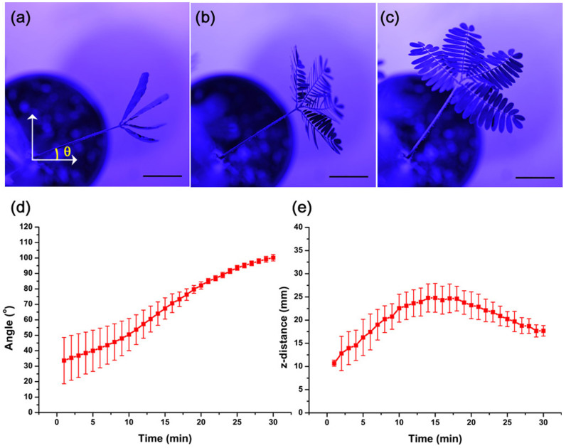
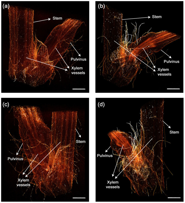
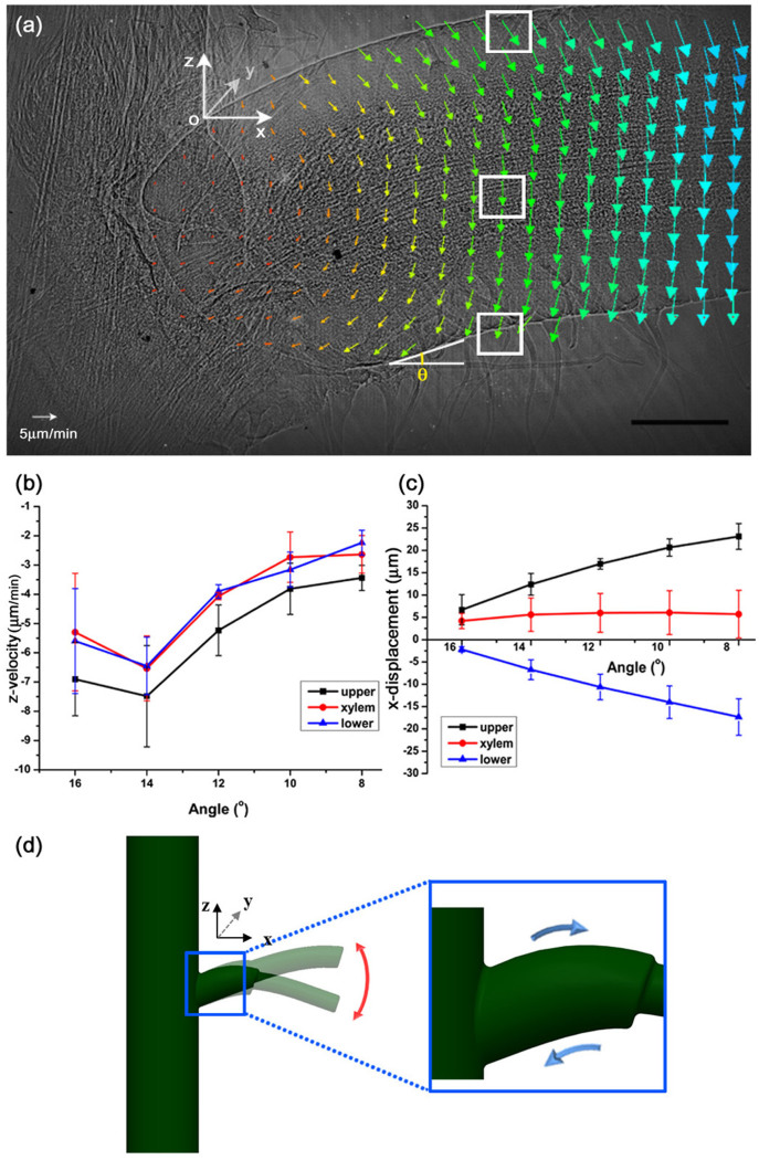
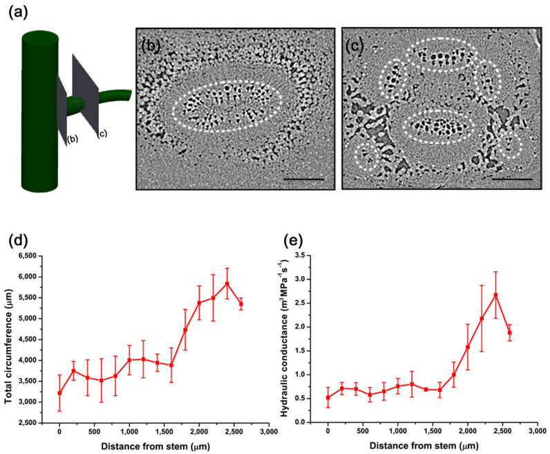
Mimosa pudica is a plant that rapidly shrinks its body in response to external stimuli. M. pudica does not perform merely simple movements, but exhibits a variety of movements that quickly change depending on the type of stimuli. Previous studies have investigated the motile mechanism of the plants from a biochemical perspective. However, an interdisciplinary study on the structural characteristics of M. pudica should be accompanied by biophysical research to explain the principles underlying such movements. In this study, the structural characteristics and seismonastic reactions of M. pudica were experimentally investigated using advanced bio-imaging techniques. The results show that the key factors for the flexible movements by the pulvinus are the following: bendable xylem bundle, expandable/shrinkable epidermis, tiny wrinkles for surface modification, and a xylem vessel network for efficient water transport. This study provides new insight for better understanding the M. pudica motile mechanism through structural modification.


Conflict of interest statement

The authors declare no competing financial interests.

Figures

Figure 1
Figure 1. M. pudica motion in response to an external stimulus.
M. pudica rotates its pulvinus and opens its leaves in response to 430 nm LED light. Top view images of M. pudica at the angles (θ) 23.2° (a), 33.9° (b) and 55.2° (c). The angles (d) and z-distance (e) of the pulvinus change with time lapsed after LED exposure. The error bars indicate the standard error (n = 3). Scale bar, 20 mm.
Figure 2
Figure 2. 3D flexible structures of the straight and bending pulvinus.
(a–d) X-ray tomograms show the internal morphological structure of the two different pulvini. 3D reconstructed images of the straight (a, c) and the bending (b, d) pulvini. The pulvinus reconstructed in a flank view (a, b) and a 130° rotated view around the vertical axis (c, d). The xylem vessels inside the pulvinus are straight or bent depending on the morphological condition. Scale bar, 2 mm.
Figure 3
Figure 3. Expandable and contractible epidermis of the pulvinus.
(a) Displacement of the epidermis and xylem of the pulvinus were obtained using PIV technique with X-ray microscopic images. A reference point O was set at the upper part of the pulvinus near the stem. The bending angle (θ) at the lower pulvinus depicted in (a) indicates the state of the bending motion. The upper epidermis moves in a positive direction, whereas the lower epidermis moves in a negative direction along the x-axis. (b) The z-directional velocities of the upper and lower epidermis and the xylems in the ROIs (white boxes) show a similar trend. (c) Their x-directional displacements exhibit different trend as the angle θ decreases. (d) A schematic diagram of the upper and lower epidermis of the pulvinus, which expands and contracts during bending. Error bars indicate standard error (n = 3). Scale bar, 500 μm.
Figure 4
Figure 4. Spread of wrinkles on the pulvinus surface.
(a) A typical X-ray image showing a large number of tiny wrinkles on the surface of the pulvinus. (b–d) As the angle (θ), which is depicted in the pulvinus bending motion, decreases, the tiny wrinkles spread out. Inverse images in the ROI at the angles (b) 14.0°, (c) 10.7° and (d) 8.8° were magnified to clearly show the wrinkles in 2D X-ray images. (e–g) The image intensity profiles were measured around the wrinkles (dashed circles in b–d) and (h) their FWHMs were calculated. The FWHM values exponentially increased with an increasing pulvinus bending angle. The scale bars in (a) and (b–d) indicate 1 mm and 100 μm, respectively.
Figure 5
Figure 5. Morphological characteristics of xylem vessels in the pulvinus.
Cross-sectional X-ray images of the pulvinus branched from the main stem to the petiole side. (a) A schematic diagram shows the cross-sectional imaging positions 200 μm (b) and 2600 μm (c) from the main stem. In the pulvinus, one xylem bundle (b) and six reorganized, independent bundles were observed (c). A xylem bundle in the cross-sectional images is depicted as a white dashed circle. (d) The total circumference and (e) hydraulic conductance of the xylem vessels vary with the distance from the main stem. The circumference and hydraulic conductance rapidly increase beyond a specific point due to larger surface area and greater number of xylem vessels. Error bars indicate standard error (n = 3). Scale bar, 200 μm.

References

    1. Elbaum R., Zaltzman L., Burgert I. & Fratzl P. The role of wheat awns in the seed dispersal unit. Science 316, 884–886 (2007). - PubMed
    1. Dawson C., Vincent J. F. V. & Rocca A. M. How pine cones open. Nature 390, 668–668 (1997).
    1. Skotheim J. M. & Mahadevan L. Physical limits and design principles for plant and fungal movements. Science 308, 1308–1310 (2005). - PubMed
    1. Edwards J., Whitaker D., Klionsky S. & Laskowski M. J. Botany: a record-breaking pollen catapult. Nature 435, 164 (2005). - PubMed
    1. Dumais J. & Forterre Y. e. “Vegetable Dynamicks”: The Role of Water in Plant Movements. Annu. Rev. of Fluid Mech. 44, 453–478 (2012).

Publication types

LinkOut - more resources