Comparative Study
. 2014 Dec;88(23):13845-57.
doi: 10.1128/JVI.02086-14. Epub 2014 Sep 24.

Variation of the specificity of the human antibody responses after tick-borne encephalitis virus infection and vaccination

Affiliations
Comparative Study

Variation of the specificity of the human antibody responses after tick-borne encephalitis virus infection and vaccination

Johanna Jarmer et al. J Virol. 2014 Dec.

Abstract

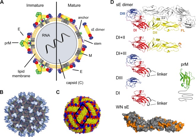
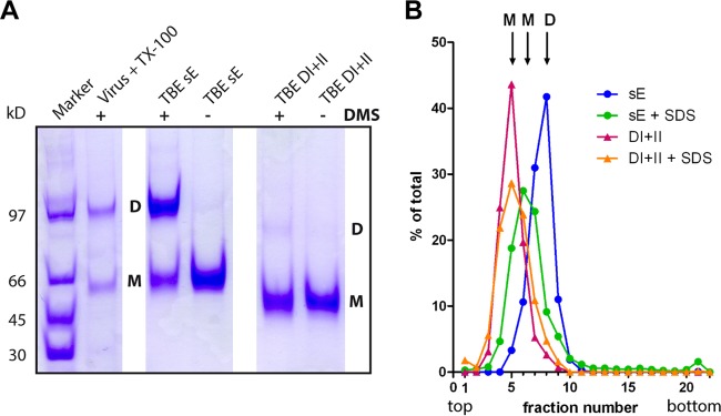
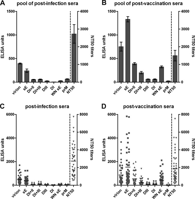
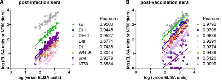
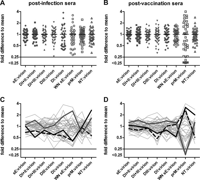
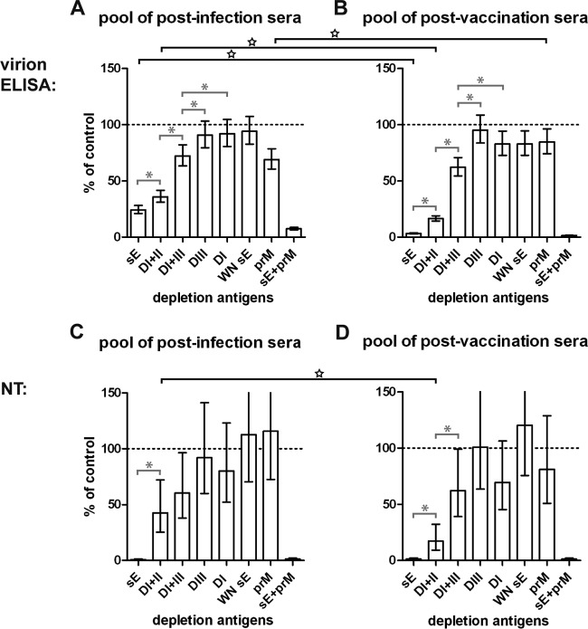
Tick-borne encephalitis (TBE) virus is an important human-pathogenic flavivirus endemic in large parts of Europe and Central and Eastern Asia. Neutralizing antibodies specific for the viral envelope protein E are believed to mediate long-lasting protection after natural infection and vaccination. To study the specificity and individual variation of human antibody responses, we developed immunoassays with recombinant antigens representing viral surface protein domains and domain combinations. These allowed us to dissect and quantify antibody populations of different fine specificities in sera of TBE patients and vaccinees. Postinfection and postvaccination sera both displayed strong individual variation of antibody titers as well as the relative proportions of antibodies to different domains of E, indicating that the immunodominance patterns observed were strongly influenced by individual-specific factors. The contributions of these antibody populations to virus neutralization were quantified by serum depletion analyses and revealed a significantly biased pattern. Antibodies to domain III, in contrast to what was found in mouse immunization studies with TBE and other flaviviruses, did not play any role in the human neutralizing antibody response, which was dominated by antibodies to domains I and II. Importantly, most of the neutralizing activity could be depleted from sera by a dimeric soluble form of the E protein, which is the building block of the icosahedral herringbone-like shell of flaviviruses, suggesting that antibodies to more complex quaternary epitopes involving residues from adjacent dimers play only a minor role in the total response to natural infection and vaccination in humans.

Importance: Tick-borne encephalitis (TBE) virus is a close relative of yellow fever, dengue, Japanese encephalitis, and West Nile viruses and distributed in large parts of Europe and Central and Eastern Asia. Antibodies to the viral envelope protein E prevent viral attachment and entry into cells and thus mediate virus neutralization and protection from disease. However, the fine specificity and individual variation of neutralizing antibody responses are currently not known. We have therefore developed new in vitro assays for dissecting the antibody populations present in blood serum and determining their contribution to virus neutralization. In our analysis of human postinfection and postvaccination sera, we found an extensive variation of the antibody populations present in sera, indicating substantial influences of individual-specific factors that control the specificity of the antibody response. Our study provides new insights into the immune response to an important human pathogen that is of relevance for the design of novel vaccines.


Figures

FIG 1
FIG 1
Schematic models and structures of immature and mature flavivirus particles and recombinant antigens employed. (A) Schematic representation of a virion with the immature form on the left and the mature form after prM cleavage on the right side. The nucleocapsid, surrounded by a lipid bilayer, is composed of C proteins and contains the viral RNA. In immature virions, prM and E form heterodimers. During virus secretion from host cells, prM is cleaved and E proteins are rearranged into homodimers. sE dimer, soluble form of the E dimer, lacking the stem and anchor regions. Reproduced from reference under the CC BY license. (B) Surface representation of an immature dengue 2 virus as determined by cryoelectron microscopy. A total of 60 trimers of E-prM heterodimers protrude from the surface of immature virions. prM proteins at the tip of the spikes are colored in light gray. E proteins are shown in darker gray. Reprinted from reference with permission of the publisher. (C) E protein arrangement on the surface of mature dengue 2 virus particles. Three E dimers form a raft (encircled in black) of the herringbone-like lattice covering the virion surface. Reproduced from reference under the CC BY license. (D) Ribbon diagrams of the TBE sE dimer and other recombinant antigens used in the study (DI+II, DI+III, DIII, DI, and prM). In the surface representation of WN sE, amino acids conserved between TBE and WN viruses are highlighted in orange. Color code: DI, red; DII, yellow; DIII, blue; the fusion loop (FL) is highlighted in orange. prM (displayed is the structure of the dengue 2 prM [80], assuming that, like E, the overall structure of the TBE prM protein will be similar) is shown in green and black. Gly linkers introduced in constructs of DI and DI+III are shown as black loops.
FIG 2
FIG 2
Blocking ELISA of recombinant antigens with MAbs. The MAbs recognize the following antigenic sites: IN3 and 4G2, flavivirus fusion loop (16); IC3, TBE DI; A3 and A5, TBE DII; B4, TBE DIII (34); 13A10, TBE prM (81); E16 and E24, WN DIII (82). The antigens analyzed are indicated in different colors, as shown at the bottom of the panel.
FIG 3
FIG 3
Determination of the oligomeric state of TBE DI+II. (A) SDS-PAGE of recombinant TBE sE and DI+II as well as full-length E obtained from virus particles solubilized with Triton X-100 without (−) and with (+) cross-linking by DMS. Positions of monomers (M) and dimers (D) are indicated. Staining with Coomassie brilliant blue. (B) Sedimentation analysis of TBE antigens. The antigens are color coded as indicated in the panel. Monomeric controls were generated by SDS treatment (+SDS) of the antigens. Sedimentation is from left to right. The antigen content in each fraction was determined by ELISA. Positions of monomers (M) and dimers (D) are indicated.
FIG 4
FIG 4
ELISAs and NTs of TBE postinfection and postvaccination sera. Analysis of serum pools (A, B) and individual sera (C, D) in TBE virion and subunit ELISAs as well as in neutralization tests. Error bars in panels A and B represent standard errors of the means calculated from the results from at least 3 independent experiments. Black horizontal lines in panels C and D represent the means of individual sera.
FIG 5
FIG 5
Correlations of individual serum reactivities in different assays. Linear regression between virion ELISA and subunit ELISA units as well as with NT titers. Pearson correlation coefficients (r) are indicated. (A) Postinfection sera; (B) postvaccination sera.
FIG 6
FIG 6
Variation of reactivities of individual sera. (A, B) Ratios of subunit ELISA units and NT titers in relation to virion ELISA units, expressed as fold difference from the mean of all sera. (C, D) Ratios obtained for each individual serum are connected by gray lines. Patterns of three selected sera are highlighted. (A, C) Postinfection sera; (B, D) postvaccination sera.
FIG 7
FIG 7
Depletion of antibody subsets from serum pools with recombinant antigens. Postdepletion analysis of the pools of postinfection sera (A, C) and postvaccination sera (B, D) in virion ELISA (A, B) and NT (C, D). The reactivity of depleted serum pools is given as a percentage of the mean of non- and mock-depleted controls. Depletion is significant when the error bar does not reach the 100% mark (dashed line). Error bars represent confidence intervals of 2 to 4 independent experiments. Gray capped lines with asterisks within each of the panels indicate significant differences between depletions of the same serum pool with different antigens. Black capped lines with stars spanning the panels indicate significant differences between depletions of the postinfection and postvaccination pools with the same antigen. Antigens used for depletion are indicated below the panels.
FIG 8
FIG 8
Depletion of antibody subsets from individual sera with recombinant antigens. Postdepletion analysis of three postinfection sera (I-16, I-9, I-29) and three postvaccination sera (V-208, V-207, V-229) in virion ELISA (A) and NT (B). The reactivity of depleted sera is given as the percentage of the mean of non- and mock-depleted controls. Depletion is significant when the error bar does not reach the 100% mark (dashed line). Error bars represent confidence intervals of 2 to 4 independent experiments. Gray capped lines with asterisks within each of the panels indicate significant differences between depletions of the same serum with different antigens. Black stars indicate significant differences between depletions of an individual serum and the respective serum pool with the same antigen. Antigens used for depletion are indicated below the panels.

References

    1. Simmonds P, Becher P, Collett MS, Gould EA, Heinz FX, Meyers G, Monath T, Pletnev A, Rice CM, Stiasny K, Thiel HJ, Weiner A, Bukh J. 2011. Family Flaviviridae. In King AMQ, Adams MJ, Lefkowitz EJ, Carstens EB. (ed), Virus taxonomy, vol IX Report of the International Committee on Taxonomy of Viruses Elsevier Academic Press, San Diego, CA.
    1. Monath TP, Gershman M, Staples JE, Barrett ADT. 2013. Yellow fever vaccine, p 870–968 In Plotkin SA, Orenstein WA, Offit PA. (ed), Vaccines, 6th ed. Elsevier Saunders, Philadelphia, PA.
    1. Halstead SB, Jacobson J, Dubischar-Kastner K. 2013. Japanese encephalitis vaccine, p 312–351 In Plotkin SA, Orenstein WA, Offit PA. (ed), Vaccines, 6th ed. Elsevier Saunders, Philadelphia, PA.
    1. Barrett PN, Portsmouth D, Ehrlich HJ. 2013. Tick-borne encephalitis virus vaccines, p 773–788 In Plotkin SA, Orenstein WA, Offit PA. (ed), Vaccines, 6th ed. Elsevier Saunders, Philadelphia, PA.
    1. Halstead SB, Thomas SJ. 2013. Dengue vaccines, p 1042–1051 In Plotkin SA, Orenstein WA, Offit PA. (ed), Vaccines, 6th ed. Elsevier Saunders, Philadelphia, PA.

Publication types