. 2014 Dec;88(24):14017-29.
doi: 10.1128/JVI.02126-14. Epub 2014 Sep 24.

Vaccinia virus mutations in the L4R gene encoding a virion structural protein produce abnormal mature particles lacking a nucleocapsid

Affiliations

Vaccinia virus mutations in the L4R gene encoding a virion structural protein produce abnormal mature particles lacking a nucleocapsid

Desyree Murta Jesus et al. J Virol. 2014 Dec.

Abstract

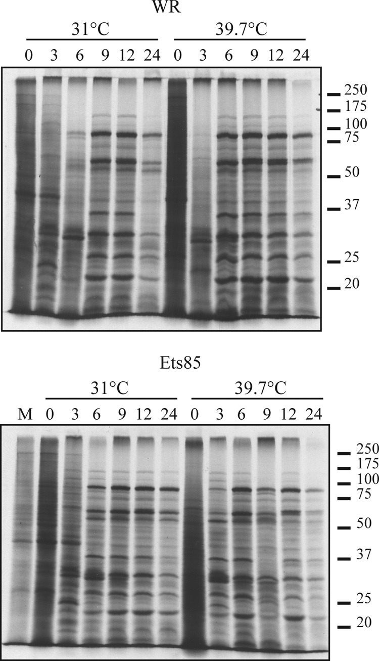
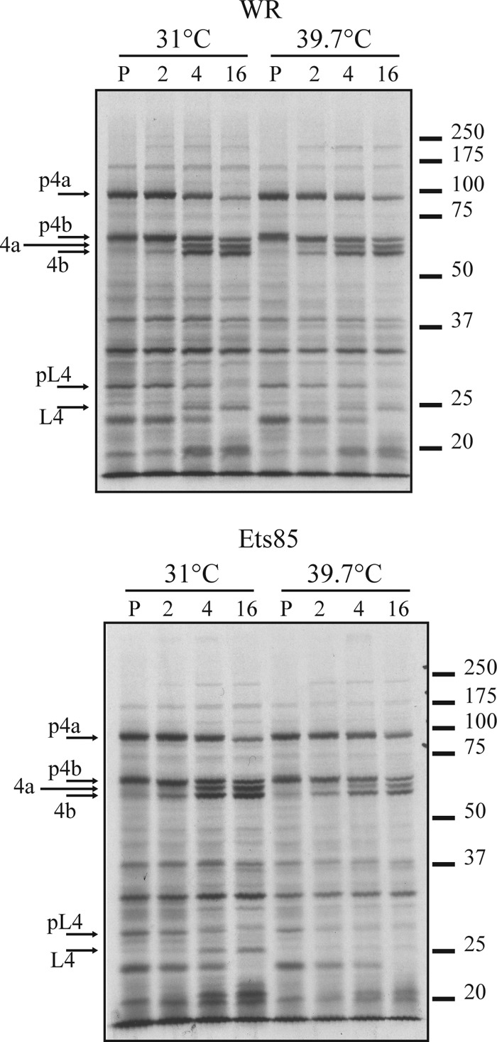
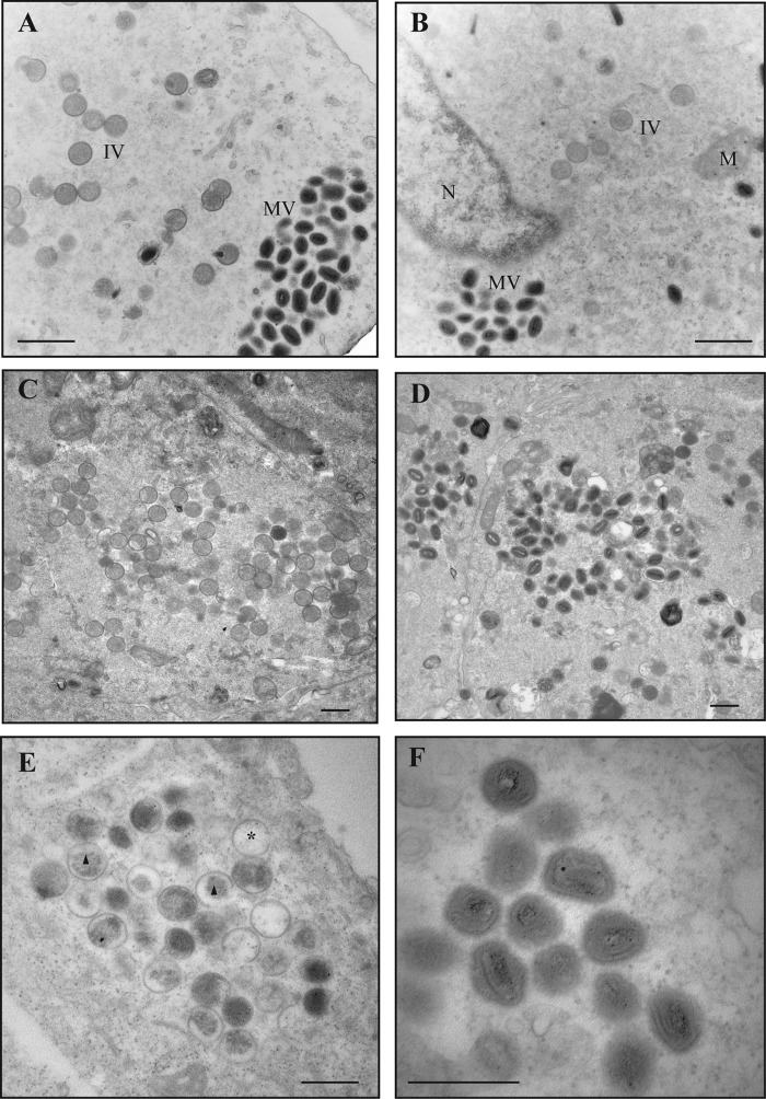
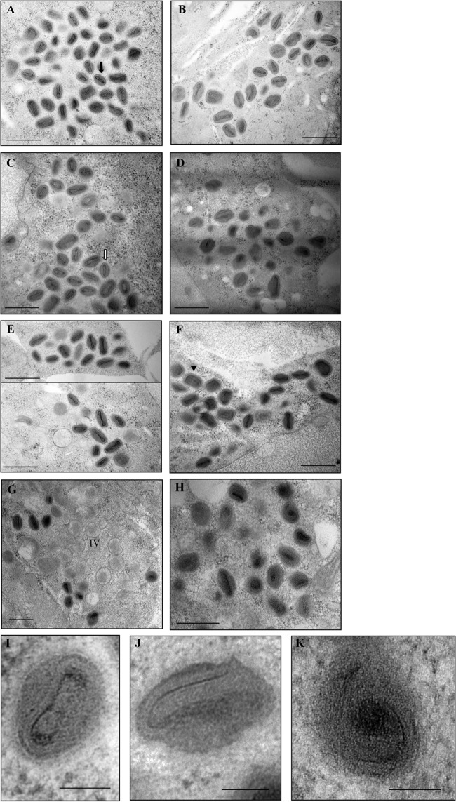
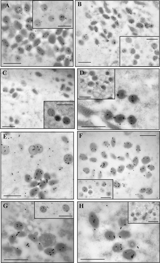
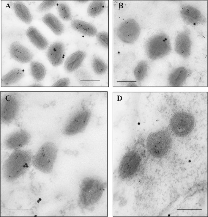
Electron micrographs from the 1960s revealed the presence of an S-shaped tubular structure in the center of the vaccinia virion core. Recently, we showed that packaging of virus transcription enzymes is necessary for the formation of the tubular structure, suggesting that the structure is equivalent to a nucleocapsid. Based on this study and on what is known about nucleocapsids of other viruses, we hypothesized that in addition to transcription enzymes, the tubular structure also contains the viral DNA and a structural protein as a scaffold. The vaccinia virion structural protein L4 stands out as the best candidate for the role of a nucleocapsid structural protein because it is abundant, it is localized in the center of the virion core, and it binds DNA. In order to gain more insight into the structure and relevance of the nucleocapsid, we analyzed thermosensitive and inducible mutants in the L4R gene. Using a cryo-fixation method for electron microscopy (high-pressure freezing followed by freeze-substitution) to preserve labile structures like the nucleocapsid, we were able to demonstrate that in the absence of functional L4, mature particles with defective internal structures are produced under nonpermissive conditions. These particles do not contain a nucleocapsid. In addition, the core wall of these virions is abnormal. This suggests that the nucleocapsid interacts with the core wall and that the nucleocapsid structure might be more complex than originally assumed.

Importance: The vaccinia virus nucleocapsid has been neglected since the 1960s due to a lack of electron microscopy techniques to preserve this labile structure. With the advent of cryo-fixation techniques, like high-pressure freezing/freeze-substitution, we are now able to consistently preserve and visualize the nucleocapsid. Because vaccinia virus early transcription is coupled to the viral core structure, detailing the structure of the nucleocapsid is indispensable for determining the mechanisms of vaccinia virus core-directed transcription. The present study represents our second attempt to understand the structure and biological significance of the nucleocapsid. We demonstrate the importance of the protein L4 for the formation of the nucleocapsid and reveal in addition that the nucleocapsid and the core wall may be associated, suggesting a higher level of complexity of the nucleocapsid than predicted. In addition, we prove the utility of high-pressure freezing in preserving the vaccinia virus nucleocapsid.


Figures

FIG 1
FIG 1
Localization of vaccinia virus core proteins in the virions. Cells were infected at an MOI of 10 with vE6i in the presence of IPTG and incubated at 37°C. At 24 h postinfection, cells were processed for immunoelectron microscopy as described in Materials and Methods. Ultrathin sections were probed with antibodies against L4, A3, A4, and F17 (top) or only against L4 (bottom) and visualized in an electron microscope. Scale bar, 100 nm.
FIG 2
FIG 2
Analysis of L4 sequence. The protein sequences were obtained from www.poxvirus.org and aligned using the software programs BioEdit and ClustalW Multiple. The gray box highlights residue 138 (proline) that is mutated in the mutant Ets85. Vaccinia virus (VACV) WR and variola, monkeypox, ectromelia, and cowpox viruses are representatives of the genus Orthopoxvirus; deerpox virus is a member of the genus Cervidpoxvirus; molluscum contagiosum virus is a member of the genus Molluscipoxvirus. Amino acid identity relative to the WR vaccinia virus is 98% for members of genus Orthopoxvirus, 63% for deerpox virus, and 43% for molluscum contagiosum. Asterisk (*), identical amino acid; colon (:), conserved substitutions; period (.), semiconserved substitutions.
FIG 3
FIG 3
Characterization of the Ets85 mutant. (A) BSC-40 cells were infected with WR or Ets85 and incubated at 31°C or 39.7°C. After 7 days of infection, dishes were stained with crystal violet. (B) Cells were infected at an MOI of 10 with WR or Ets85 and incubated at the permissive or nonpermissive temperature. Samples were harvested at various times postinfection. Titers were determined by plaque assay at 31°C. The experiment was done three times. A representative experiment is shown.
FIG 4
FIG 4
Accumulation of L4 during infection. Cells were infected with WR or Ets85 at an MOI of 10 and incubated at 31°C or 39.7°C. Samples were harvested at various times postinfection and analyzed by SDS-PAGE, followed by Western blotting using antibody against L4.
FIG 5
FIG 5
Viral protein synthesis during WR and Ets85 infection. Cells were infected at an MOI of 10 with WR or Ets85 and incubated at 31°C or 39.7°C. At various times postinfection, cells were pulse-labeled for 30 minutes with [35S]methionine. Samples were then harvested and analyzed by SDS-PAGE and autoradiography. Molecular masses in kilodaltons are indicated on the right. The numbers at the top indicate the time (in hours) postinfection.
FIG 6
FIG 6
Analysis of protein processing during infection. Cells were infected at an MOI of 10 with WR or Ets85 and incubated at 31°C or 39.7°C. At 8 h postinfection, cells were pulse-labeled for 30 minutes with [35S]methionine. Some cells were harvested after the 30-min pulse (P), and some were incubated at 31°C or 39.7°C for 2, 4, or 16 h in medium lacking [35S]methionine. Samples were then harvested and analyzed by SDS-PAGE and autoradiography. Molecular masses in kilodaltons are indicated on the right.
FIG 7
FIG 7
Viral morphogenesis: conventional fixation. Cells were infected with WR or Ets85 at an MOI of 10 and incubated at 31°C or 39.7°C. At 24 h postinfection, cells were fixed with 2% glutaraldehyde and 1% osmium tetroxide and processed for transmission electron microscopy as described in Materials and Methods. (A) WR at 31°C. (B) Ets85 at 31°C. (C and D) WR at 39.7°C. (E and F) Ets85 at 39.7°C. MV, mature virus particles; IV, immature virus particles; M, mitochondria; N, nucleus; asterisk, empty IV; triangle, IV with nonhomogeneous viroplasm. Scale bar, 500 nm.
FIG 8
FIG 8
Viral morphogenesis: high-pressure freezing/freeze-substitution. Cells were infected with WR, Ets85, or vL4i at an MOI of 10. For Ets85, cells were incubated at 31°C or 39.7°C. For vL4i, cells were incubated at 37°C in the presence or absence of 200 μM IPTG. At 24 h postinfection, cells were processed by high-pressure freezing and freeze-substitution as described in Materials and Methods. Ultrathin sections were visualized in an electron microscope. (A) WR at 31°C. (B) WR at 39.7°C. (C) Ets85 at 31°C. (D) Ets85 at 39.7°C. (E) WR at 37°C. (F) vL4i in the presence of IPTG. (G and H) vL4i in the absence of IPTG. (I) Higher magnification of a wild-type mature particle. (J and K) Higher magnification of mature particles produced in mutant infections under nonpermissive conditions. Arrows and arrowheads indicate mutually perpendicular sections of representative particles in which the nucleocapsid can be visualized. Filled arrow, transverse section (A); open arrow, sagittal section (C); arrowhead, coronal section (F). Scale bars, 500 nm (A to H) and 100 nm (I to K).
FIG 9
FIG 9
Immunogold labeling of core proteins. Cells were infected with WR or Ets85 at an MOI of 10 and incubated at 31°C or 39.7°C. At 24 h postinfection, cells were processed for immunoelectron microscopy. Ultrathin sections were probed with antibodies against L4 (A to D) or A3 (E to H) and visualized in an electron microscope (Hitachi H-7000). (A and E) WR at 31°C. (B and F) WR at 40°C. (C and G) Ets85 at 31°C. (D and H) Ets85 at 39.7°C. The insets show the labeling of immature virions. Scale bar, 500 nm.
FIG 10
FIG 10
Immunogold labeling of DNA. Cells were infected with WR or Ets85 at an MOI of 10 and incubated at 31°C or 39.7°C. At 24 h postinfection, cells were processed for immunoelectron microscopy. Sections of 70- to 90-nm thickness were probed with antibodies against double-strand DNA (5-nm gold particles) or against the viral protein D13 (20-nm gold) and visualized in an electron microscope (Hitachi H-7000). (A) WR at 31°C. (B) WR at 40°C. (C) Ets85 at 31°C. (D) Ets85 at 39.7°C. Scale bar, 200 nm.

References

    1. Moss B. 2007. Poxviridae: the viruses and their replication, 2906–2945 In Knipe DM, Howley PM, Griffin DE, Lamb RA, Martin MA, Roizman B, Straus SE. (ed), Fields virology, 5th ed. Lippincott Williams & Wilkins, Philadelphia, PA.
    1. Dales S, Siminovitch L. 1961. The development of vaccinia virus in Earle's L strain cells as examined by electron microscopy. J. Biophys. Biochem. Cytol. 10:475–503. 10.1083/jcb.10.4.475. - DOI - PMC - PubMed
    1. Cairns J. 1960. The initiation of vaccinia infection. Virology 11:603–623. 10.1016/0042-6822(60)90103-3. - DOI - PubMed
    1. Vanslyke JK, Lee P, Wilson EM, Hruby DE. 1993. Isolation and analysis of vaccinia virus previrions. Virus Genes 7:311–324. 10.1007/BF01703388. - DOI - PubMed
    1. Ansarah-Sobrinho C, Moss B. 2004. Role of the I7 protein in proteolytic processing of vaccinia virus membrane and core components. J. Virol. 78:6335–6343. 10.1128/JVI.78.12.6335-6343.2004. - DOI - PMC - PubMed

Publication types

MeSH terms

LinkOut - more resources