Comparative Study
. 2014 Dec;88(24):14057-69.
doi: 10.1128/JVI.02320-14. Epub 2014 Sep 24.

Drosophila C virus systemic infection leads to intestinal obstruction

Affiliations
Comparative Study

Drosophila C virus systemic infection leads to intestinal obstruction

Stanislava Chtarbanova et al. J Virol. 2014 Dec.

Abstract

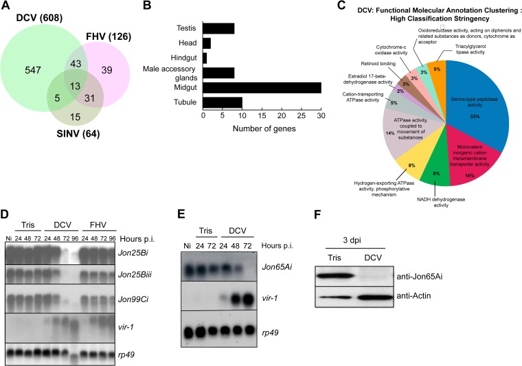
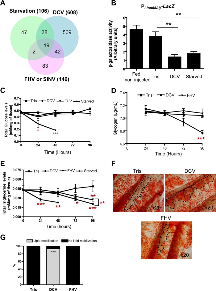
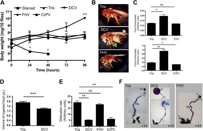
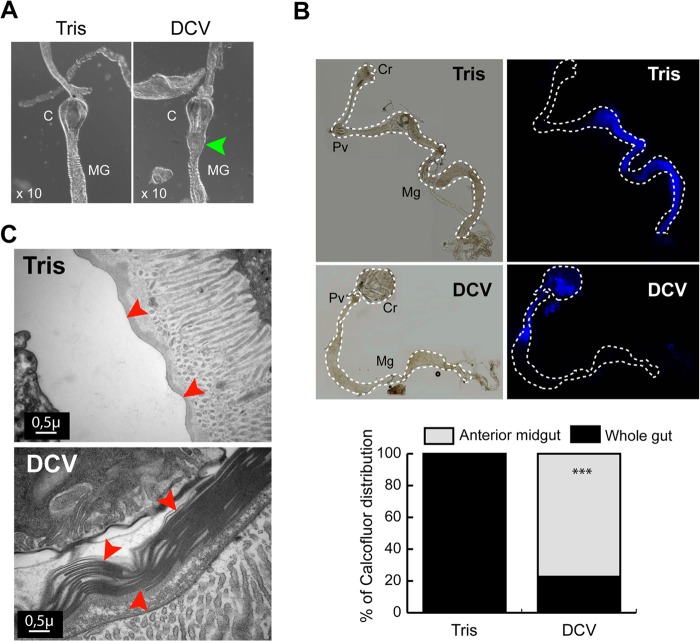
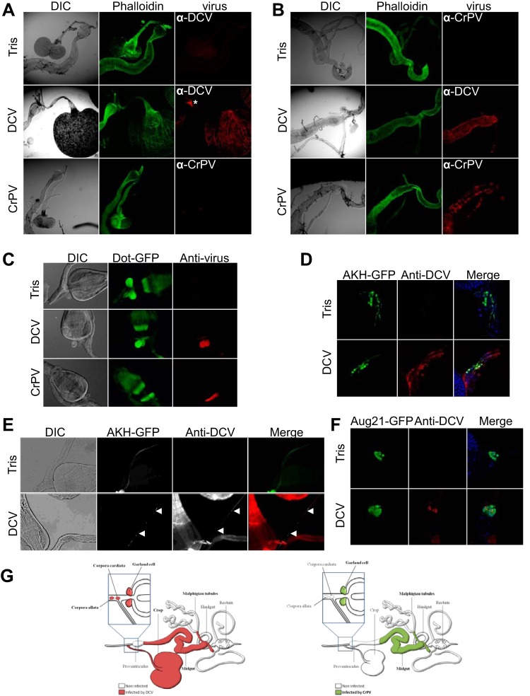
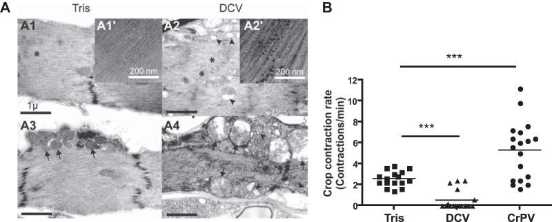
Drosophila C virus (DCV) is a positive-sense RNA virus belonging to the Dicistroviridae family. This natural pathogen of the model organism Drosophila melanogaster is commonly used to investigate antiviral host defense in flies, which involves both RNA interference and inducible responses. Although lethality is used routinely as a readout for the efficiency of the antiviral immune response in these studies, virus-induced pathologies in flies still are poorly understood. Here, we characterize the pathogenesis associated with systemic DCV infection. Comparison of the transcriptome of flies infected with DCV or two other positive-sense RNA viruses, Flock House virus and Sindbis virus, reveals that DCV infection, unlike those of the other two viruses, represses the expression of a large number of genes. Several of these genes are expressed specifically in the midgut and also are repressed by starvation. We show that systemic DCV infection triggers a nutritional stress in Drosophila which results from intestinal obstruction with the accumulation of peritrophic matrix at the entry of the midgut and the accumulation of the food ingested in the crop, a blind muscular food storage organ. The related virus cricket paralysis virus (CrPV), which efficiently grows in Drosophila, does not trigger this pathology. We show that DCV, but not CrPV, infects the smooth muscles surrounding the crop, causing extensive cytopathology and strongly reducing the rate of contractions. We conclude that the pathogenesis associated with systemic DCV infection results from the tropism of the virus for an important organ within the foregut of dipteran insects, the crop.

Importance: DCV is one of the few identified natural viral pathogens affecting the model organism Drosophila melanogaster. As such, it is an important virus for the deciphering of host-virus interactions in insects. We characterize here the pathogenesis associated with DCV infection in flies and show that it results from the tropism of the virus for an essential but poorly characterized organ in the digestive tract, the crop. Our results may have relevance for other members of the Dicistroviridae, some of which are pathogenic to beneficial or pest insect species.


Figures

FIG 1
FIG 1
Gene repression in DCV-infected flies. (A) Venn diagram showing the number of repressed genes in DCV-, FHV-, and SINV-infected flies. (B) DCV represses midgut-specific genes. The diagram shows the number of tissue-specific genes that are downregulated by a factor of at least two in DCV-infected flies. (C) Functional molecular annotation clustering of the DCV-repressed genes. Genes repressed by a factor of at least 2-fold were classified in different categories using the DAVID clustering tool. (D and E) Quantitative validation of the microarray analysis for four members of the Jonah family of serine proteases. vir-1 is a marker gene induced after viral infection, and rp49 is used as a loading control. Ni, noninfected. (F) Jon65Ai is repressed at the protein level. The Western blot is of proteins extracted from 10 dissected guts showing that the Jon65Ai protein is repressed 3 days after infection.
FIG 2
FIG 2
Transcriptional repression of Jon65Ai and LysE during DCV infection. (A) X-Gal staining of dissected Drosophila tissues from Jon65Ai-LacZ flies. The β-galactosidase activity is detectable in the posterior midgut of Jon65Ai-LacZ flies. OV, ovaries; MT, Malpighian tubules; aMG, anterior midgut; pMG, posterior midgut; Cr, crop. (B) β-Galactosidase activity in extracts from Jon65Ai-LacZ flies significantly decreases 3 dpi in DCV- but not FHV- or CrPV-infected flies. The graph represents means ± standard deviations (SD) from three independent experiments. (C) X-Gal staining of dissected Drosophila tissues from LysE-LacZ transgenic flies. The β-galactosidase activity is detectable in the anterior midgut of LysE-LacZ flies. (D) β-Galactosidase activity significantly decreases 3 dpi in DCV- but not FHV-infected flies. The graph represents means ± SD from three independent experiments. ns, nonsignificant. (E to G) Analysis of Jon65Ai promoter region. Transgenic flies containing the LacZ gene under the control of truncated fragments of the Jon65Ai promoter (from bp −773 [E], −483 [F], or −236 [G]) were challenged with DCV, and β-galactosidase activity in single flies was measured 72 h later. Results correspond to the means ± SD from three independent experiments. Representative results for at least two independent transgenic lines are shown.
FIG 3
FIG 3
Infection by DCV triggers nutritional stress in Drosophila. (A) Venn diagram showing the overlap of downregulated genes between DCV-, FHV-, and SINV-infected and starved flies. (B) DCV infection and starvation downregulate the β-galactosidase activity in Jon65Ai-lacZ flies. The graph represents means ± SD from three independent experiments. (C) Quantification of glucose levels in virus-infected flies. The graph represents means ± SD from three independent experiments for each time point. (D) Glycogen levels decrease after DCV, but not FHV, infection. The graph represents means ± SD from three independent experiments for each time point. (E) Viral infection is accompanied by a decrease in triglyceride levels. The graph represents means ± SD from three independent experiments for each time point. (F) Lipid mobilization from fat body to oenocytes in Drosophila adults 3 days after infection with DCV but not FHV. Oenocytes are circled with dashed lines. (G) Quantification of lipid mobilization in infected flies. Tris mock-infected flies, n = 16; DCV, n = 14; FHV, n = 14.
FIG 4
FIG 4
DCV triggers an intestinal obstruction. (A) Increase in body weight is observed in DCV- but not FHV- or CrPV-infected flies. Groups of 10 flies (5 males and 5 females) were weighed at intervals of 24 h after starvation or injection with Tris (control), DCV, FHV, or CrPV. The graph represents means ± SD from three independent experiments. (B) The characteristic abdominal swelling observed in DCV-infected flies is indicated by the arrowhead. (C) Higher levels of incorporated food in DCV-infected flies. Groups of 10 flies (5 males and 5 females) were injected with Tris, DCV, FHV, or CrPV and kept on tracking medium for 4 days, and the incorporated food was measured by a colorimetric assay. Graphs represent means ± SD from three independent experiments, each performed in triplicate. (D) Measurement of the ingested volume of food in Tris-injected controls and DCV-infected flies using the CAFE assay. The graph represents means ± SD from two independent experiments. At least 15 individual flies were tested in each experiment. (E) Decreased defecation rates after DCV infection. Groups of 10 flies were injected with Tris, DCV, FHV, or CrPV and kept on tracking medium. Three days later, flies were transferred to empty vials, and blue spots corresponding to excretion were counted 5 h later. The graph represents means ± SD from three independent experiments, each performed in triplicate. (F) Dissected digestive tracts of Tris-, DCV-, and FHV-injected flies kept on tracking medium. The blue dye is equally distributed through the whole gut of Tris-injected controls and FHV-infected flies. The blue food accumulates preferentially in the crop and anterior part of the midgut in DCV-infected flies. C, cardia; Cr, crop; Mg, midgut.
FIG 5
FIG 5
Alterations of the cardia and peritrophic matrix in DCV-infected flies. (A) Morphological changes of the Drosophila cardia during DCV infection. Digestive tracts were dissected 3 dpi from Tris- and DCV-infected flies. The posterior part of the cardia is swollen in DCV-infected flies (arrowhead). (B) Dissected digestive tracts from Tris- and DCV-injected flies, fed for 6 h on Calcofluor. The dye is equally distributed throughout the gut in Tris-injected controls. The position of midguts is indicated by dashed lines. Cr, crop; PV, proventriculus; Mg, midgut. The quantification of the intestinal obstruction of Tris-injected and DCV-infected flies is shown on the right. Tris, n = 19; DCV, n = 22. (C) Electron micrographs showing transversal sections of the proventriculus in Tris-injected and DCV-infected flies 3 days postinfection. Layers of peritrophic matrix (arrowheads) accumulate in the proventriculus of DCV-infected flies.
FIG 6
FIG 6
DCV-induced pathology does not result from activation of immune defense pathways. (A) Jon65Ai is repressed in the gut of Dcr-2 mutant flies. Western blot analysis of proteins extracted from the guts of Tris- and DCV-injected flies 72 h postinfection. (B) Increased body weight in Dcr-2 mutant flies after infection with DCV. The graph represents means ± SD from three independent experiments. All Dcr-2 mutants were dead at 96 h postinfection. (C) Increase of the incorporated food in Dcr-2 mutant flies after infection with DCV. The graph represents means ± SD from two independent experiments. (D) Lipids are mobilized from FB to oenocytes in Dcr-2 mutant flies, as shown by oil red O staining of dissected carcasses from Dcr-2 mutant flies. The dashed lines show the location of the oenocytes. (E) Table showing the development of DCV-induced symptoms in mutants for other antiviral immune pathways, Jak/STAT (Hopscotch), Toll (Dif), and IMD (imd).
FIG 7
FIG 7
DCV and CrPV have different tropisms for the Drosophila digestive tract. Immunostaining of the anterior (A) and posterior (B) parts of the midgut with anti-DCV and anti-CrPV antisera 3 dpi. Only DCV infects the muscles of the crop (A), whereas both DCV and CrPV are detectable in the visceral muscles surrounding the posterior midgut (B). The asterisk indicates the Drosophila ring gland. DIC, differential interference contrast. (C) Garland cells are infected by both DCV and CrPV. UAS-GFP expression was specifically triggered in garland cells with the Dot-Gal4 driver. (D) DCV infects corpora cardiacum (CC) cells. UAS-GFP expression was triggered in CC cells with the Akh-Gal4 driver. (E) Presence of DCV particles in a CC neuron innervating the crop muscle (arrowheads). (F) Corpora allatum (CA) cells are infected by DCV. UAS-GFP expression was triggered in CA cells with the Aug21-Gal4 driver. (G) Schematic summary representation of the tissue tropism of DCV and CrPV in the Drosophila digestive tract. Ten to 15 dissected flies were analyzed, and representative images are shown.
FIG 8
FIG 8
DCV infection of the crop triggers cytopathology and affects its function. (A) Electron micrographs showing the crop muscle of Tris-injected (A1 and A3) or DCV-infected (A2 and A4) Oregon-R flies 4 days postinfection. The presence of DCV particles is noticeable in the cytoplasm of the crop muscle cells (compare the magnified inset of A2′ to that of A1′). Disorganized myofibrils (asterisks), swollen mitochondria (arrows), and vacuolization of the sarcoplasmic reticulum (arrowheads) are present in the DCV-infected muscle cells surrounding the crop (compare A2 and A4 with A1 and A3). (B) Measurement of crop contraction rates in Tris-, DCV-, and CrPV-injected flies. The graph represents means ± SD from two independent experiments. At least 6 individual flies were tested per condition in each experiment.

References

    1. Ferrandon D, Imler JL, Hetru C, Hoffmann JA. 2007. The Drosophila systemic immune response: sensing and signalling during bacterial and fungal infections. Nat. Rev. Immunol. 7:862–874. 10.1038/nri2194. - DOI - PubMed
    1. Lindsay SA, Wasserman SA. 2014. Conventional and non-conventional Drosophila Toll signaling. Dev. Comp. Immunol. 42:16–24. 10.1016/j.dci.2013.04.011. - DOI - PMC - PubMed
    1. Kleino A, Silverman N. 2014. The Drosophila IMD pathway in the activation of the humoral immune response. Dev. Comp. Immunol. 42:25–35. 10.1016/j.dci.2013.05.014. - DOI - PMC - PubMed
    1. Honti V, Csordas G, Kurucz E, Markus R, Ando I. 2014. The cell-mediated immunity of Drosophila melanogaster: hemocyte lineages, immune compartments, microanatomy and regulation. Dev. Comp. Immunol. 42:47–56. 10.1016/j.dci.2013.06.005. - DOI - PubMed
    1. Pean CB, Dionne MS. 2014. Intracellular infections in Drosophila melanogaster: host defense and mechanisms of pathogenesis. Dev. Comp. Immunol. 42:57–66. 10.1016/j.dci.2013.04.013. - DOI - PubMed

Publication types

MeSH terms

LinkOut - more resources