. 2014 Nov 7;281(1794):20141550.
doi: 10.1098/rspb.2014.1550.

Early evolution of limb regeneration in tetrapods: evidence from a 300-million-year-old amphibian

Affiliations

Early evolution of limb regeneration in tetrapods: evidence from a 300-million-year-old amphibian

Nadia B Fröbisch et al. Proc Biol Sci. .

Abstract

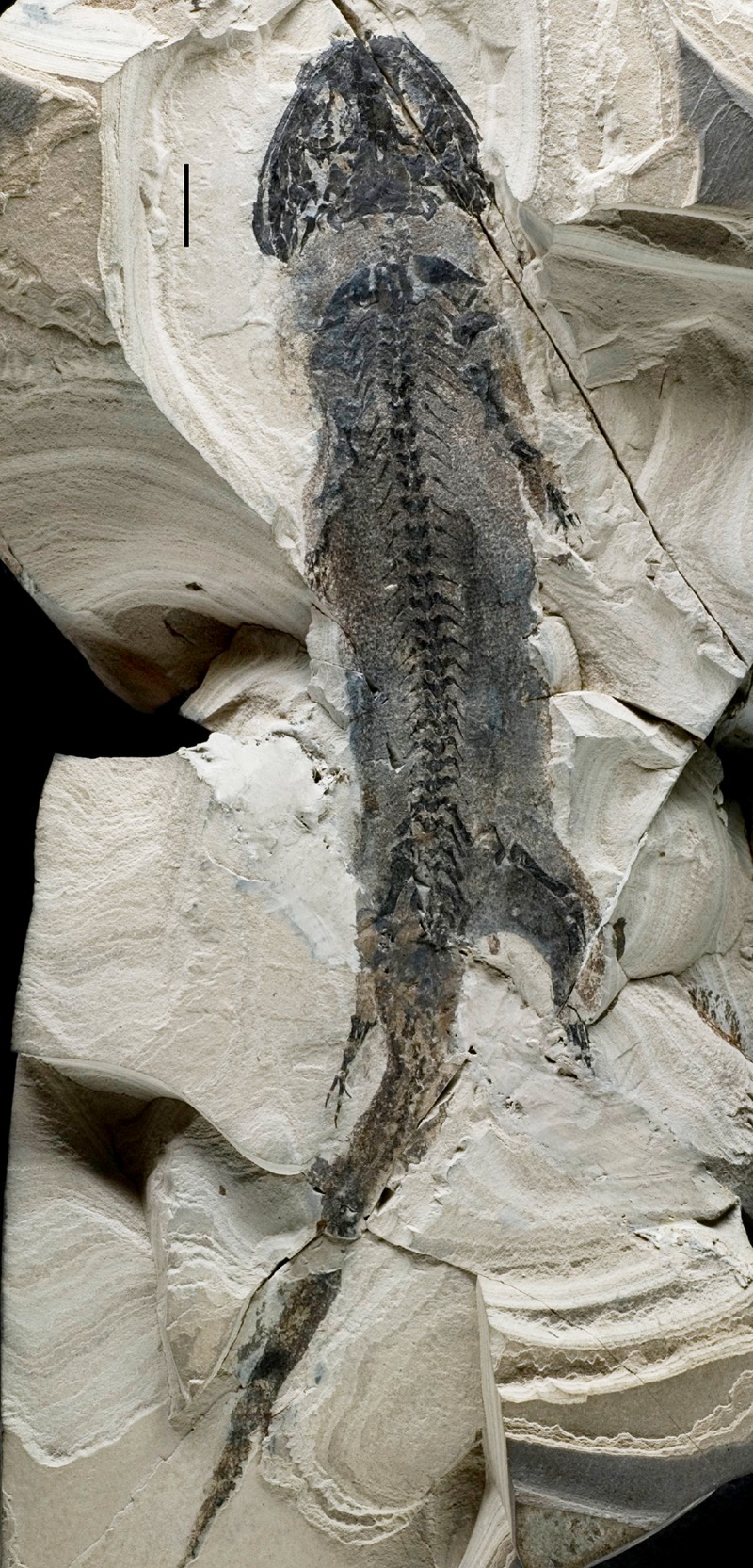
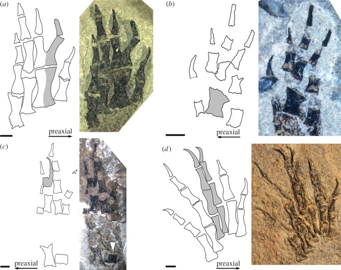
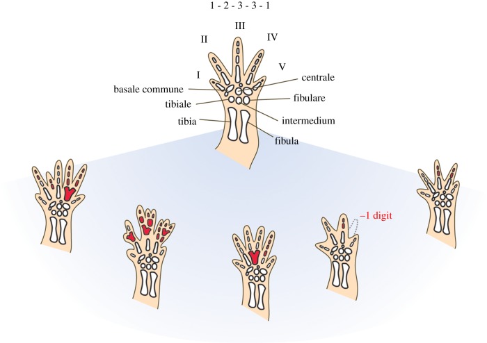
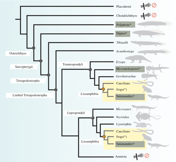
Salamanders are the only tetrapods capable of fully regenerating their limbs throughout their entire lives. Much data on the underlying molecular mechanisms of limb regeneration have been gathered in recent years allowing for new comparative studies between salamanders and other tetrapods that lack this unique regenerative potential. By contrast, the evolution of animal regeneration just recently shifted back into focus, despite being highly relevant for research designs aiming to unravel the factors allowing for limb regeneration. We show that the 300-million-year-old temnospondyl amphibian Micromelerpeton, a distant relative of modern amphibians, was already capable of regenerating its limbs. A number of exceptionally well-preserved specimens from fossil deposits show a unique pattern and combination of abnormalities in their limbs that is distinctive of irregular regenerative activity in modern salamanders and does not occur as variants of normal limb development. This demonstrates that the capacity to regenerate limbs is not a derived feature of modern salamanders, but may be an ancient feature of non-amniote tetrapods and possibly even shared by all bony fish. The finding provides a new framework for understanding the evolution of regenerative capacity of paired appendages in vertebrates in the search for conserved versus derived molecular mechanisms of limb regeneration.

Keywords: Dissorophoidea; Palaeozoic; Temnospondyli; amphibian; fossil; limb regeneration.


Figures

Figure 1.
Figure 1.
Whole specimen of Micromelerpeton credneri. Specimen MB.Am.1210 showing the exceptional quality of preservation of fossil amphibians from the fossil lake deposits of Lake Odernheim. Note the preservation of ‘skin shadow’, external gills, retinal pigments and scalation patterns. Scale bar equals 1 cm.
Figure 2.
Figure 2.
Examples for abnormalities caused by regeneration in Micromelerpeton credneri. Drawing (left) and photo (right) of some of the exemplar autopods displaying regeneration in Micromelerpeton. The normal condition is four digits in the hand with the phalangeal formula 2-2-3-3 and five digits in the foot with the phalangeal formula 2-2-3-4-3. (a) Right hand of specimen SSN 1102 showing enlarged metacarpal and proximal fusion of the first phalanges. (b) Left hand of specimen MB.Am. 1183 showing a fused metacarpal. (c) Left foot of specimen MB.Am. 1183 showing spur-like branching of the phalangeal element and an underdeveloped fibula (white arrow). (d) Left foot of specimen SSN GwK-34 showing a centrally positioned adventitious digit, note that both central digits are thinner than normal digits. Scale bar equals 1 mm. See also the electronic supplementary material figure S1.
Figure 3.
Figure 3.
Range of patterns and combinations of abnormalities caused by regeneration. Regeneration causes a distinct pattern and combination of abnormalities in the limbs of salamanders including extra digits formed by branching or insertion of adventitious digits, missing digits caused by fusion or failure to regenerate, and an increase or reduction of phalangeal elements within digits. Different abnormalities within one limb or between different limbs of the same individual can occur. Hindlimbs of the salamander Nothophthalmus viridescens are depicted in this figure with the normal morphology of five digits and a phalangeal formula of 1-2-3-3-1 on top. Abnormal regions are highlighted in red. Data are based on the study of Stock & Bryant [38].
Figure 4.
Figure 4.
Regenerative capacity in vertebrates depicted in a phylogenetic framework. Taxa capable of limb regeneration are highlighted in grey with asterisk. Frogs only show regenerative capacity of the limbs until metamorphic climax (denoted by asterisks (*)). Taxa for which the lack of regenerative capacity in the limbs has been demonstrated are indicated with prohibition signs. Lissamphibians are highlighted in yellow in two alternative positions within the phylogeny marked by a star to represent alternative hypotheses for lissamphibian origins (see text). The phylogenetic distribution of regenerative capacity in paired appendages suggests the potential presence of plesiomorphic features of appendage regeneration in Osteichthyes.

References

    1. Alvarado AS. 2000. Regeneration in metazoans: why does it happen? BioEssays 22, 578–590. (10.1002/(SICI)1521-1878(200006)22:6<578::AID-BIES11>3.0.CO;2-#) - DOI - PubMed
    1. Bely AE, Nyberg KG. 2010. Evolution of animal regeneration: re-emergence of a field. Trends Ecol. Evol. 25, 161–170. (10.1016/j.tree.2009.08.005) - DOI - PubMed
    1. Brockes JP, Kumar A, Velloso CP. 2001. Regeneration as an evolutionary variable. J. Anat. 199, 3–11. (10.1046/j.1469-7580.2001.19910003.x) - DOI - PMC - PubMed
    1. Gardiner DM, Bryant SV. 2007. Tetrapod limb regeneration. In Fins into limbs (ed. Hall BK.), pp. 163–182. Chicago, IL: University of Chicago Press.
    1. Simon A, Tanaka EM. 2013. Limb regeneration. Wiley Interdisc. Rev. Dev. Biol. 2 291–300. (10.1002/wdev.73) - DOI - PubMed

Publication types