. 2014 Sep 24;6(255):255ra130.
doi: 10.1126/scitranslmed.3009027.

Characterization of the molecular mechanisms underlying increased ischemic damage in the aldehyde dehydrogenase 2 genetic polymorphism using a human induced pluripotent stem cell model system

Affiliations

Characterization of the molecular mechanisms underlying increased ischemic damage in the aldehyde dehydrogenase 2 genetic polymorphism using a human induced pluripotent stem cell model system

Antje D Ebert et al. Sci Transl Med. .

Abstract

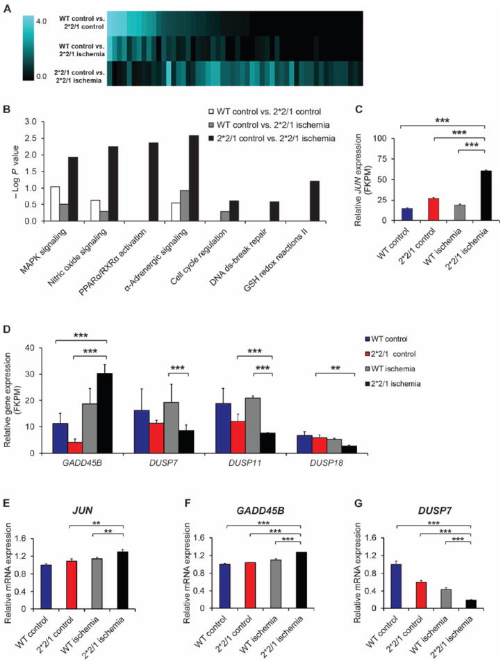
Nearly 8% of the human population carries an inactivating point mutation in the gene that encodes the cardioprotective enzyme aldehyde dehydrogenase 2 (ALDH2). This genetic polymorphism (ALDH2*2) is linked to more severe outcomes from ischemic heart damage and an increased risk of coronary artery disease (CAD), but the underlying molecular bases are unknown. We investigated the ALDH2*2 mechanisms in a human model system of induced pluripotent stem cell-derived cardiomyocytes (iPSC-CMs) generated from individuals carrying the most common heterozygous form of the ALDH2*2 genotype. We showed that the ALDH2*2 mutation gave rise to elevated amounts of reactive oxygen species and toxic aldehydes, thereby inducing cell cycle arrest and activation of apoptotic signaling pathways, especially during ischemic injury. We established that ALDH2 controls cell survival decisions by modulating oxidative stress levels and that this regulatory circuitry was dysfunctional in the loss-of-function ALDH2*2 genotype, causing up-regulation of apoptosis in cardiomyocytes after ischemic insult. These results reveal a new function for the metabolic enzyme ALDH2 in modulation of cell survival decisions. Insight into the molecular mechanisms that mediate ALDH2*2-related increased ischemic damage is important for the development of specific diagnostic methods and improved risk management of CAD and may lead to patient-specific cardiac therapies.


Conflict of interest statement

Competing interests: J.C.W. is a cofounder of Stem Cell Theranostics. D.M.-R. is the founder of ALDEA Pharmaceuticals. However, she has no role in the company, and the research in her laboratory is supported only by the NIH and is not disclosed to the company.

Figures

Fig. 1
Fig. 1. Analysis of a human model system for the ALDH2*2/1 polymorphism reveals that reduced ALDH2 enzymatic activity correlates with increased levels of ROS and 4HNE in ALDH2*2/1 cells
(A) Genotyping of skin biopsy–derived fibroblasts confirms the ALDH2 wild type (wt) or heterozygous 2*2/1 mutation. In ALDH2*2/1, the G peak corresponding to the single remaining wt allele is strongly reduced and overlaid by the A peak of the mutated allele. (B) Enzymatic activity of ALDH2 in lysates from ALDH2*2/1 human fibroblasts (n = 5) is strongly reduced compared to that in wt control (n = 4). ALDH2-specific small molecules were used as a control for specificity. The ALDH2 activator Alda-1 enhances enzymatic activity, whereas the ALDH2 inhibitor 4HNE has the opposite effect. (C) Total baseline ROS are quantified via cellular hydrogen peroxide (H2O2) levels in an Amplex Red–dependent fluorescent readout. ALDH2*2/1 fibroblasts show elevated ROS levels. Data represent n = 5 per group. (D) ALDH2 expression levels in wt and ALDH2*2/1 are comparable, as assessed by Western blot (n = 5 per group). (E) Quantification of expression levels shown in (D) for n = 5 per group. Differences between groups are not significant. (F) Significantly increased levels of 4HNE in ALDH2*2/1 human fibroblasts are confirmed by Western blot, normalizing via β-actin as loading control. (G) Quantification of Western blot analysis for n = 5 per group. Experiments were performed in the presence or absence of 20 μM Alda-1, 80 μM 4HNE, or control vehicle [dimethyl sulfoxide (DMSO)] as indicated. Data are expressed as means ± SEM. ns, not significant. *P < 0.05, calculated by Student’s t test.
Fig. 2
Fig. 2. ALDH2*2/1 fibroblasts show reduced cell proliferation and viability due to cell cycle arrest
Cell proliferation and viability were determined in a 96-well plate, high-content readout format. (A) Quantification of cell proliferation 48 hours after plating using a fluorescent, membrane permeable nuclear dye, Syto-60. Experiments were performed in triplicates and n = 5 per group. (B) Viability in ALDH2*2/1 fibroblasts was quantified via spectrophotometric measurement of XTT [2,3-bis-(2-methoxy-4-nitro-5-sulfophenyl)-2H-tetrazolium-5-carboxanilide] absorbance. Experiments were performed in triplicates and n = 5 per group. (C) Representative confocal images show cell cycle arrest in ALDH2*2/1 fibroblasts analyzed via BrdU incorporation and costaining with both DAPI as a nuclear marker and the mitochondrial structural protein TOM20. Scale bar, 100 μM. (D) Quantification and statistical analysis (n = 5 cell lines per group) analyzing a total of 42 images for wt and 44 images for ALDH2*2/1. Experiments were performed in the presence or absence of 20 μM Alda-1, 80 μM 4HNE, or control vehicle (DMSO) as indicated. Data are expressed as means ± SEM. *P < 0.05 and **P < 0.01, calculated by Student’s t test.
Fig. 3
Fig. 3. Increased ischemic damage in ALDH2*2/1 iPSC-CMs is caused by enhanced apoptosis
(A and B) Total ROS in ALDH2*2/1 iPSC-CMs are quantified via cellular H2O2 in an Amplex Red–dependent fluorescent readout. ALDH2*2/1 iPSC-CMs show elevated ROS levels, especially post-ischemia, which can be ameliorated by Alda-1–mediated activation of ALDH2 before ischemic challenge. Data represent n = 5 per group. (C and D) Viability of ALDH2*2/1 iPSC-CMs is significantly reduced compared to wt iPSC-CMs after ischemia. Alda-1–mediated activation of ALDH2 can rescue this phenotype. Absorbance of XTT was measured ina 96-well plate under a high-content readout format (n = 3 per group performed in duplicate). (E to G) Ischemic damage in ALDH2*2/1 iPSC-CMs is due to apoptosis, as shown by TUNEL staining. After ischemia, ALDH2*2/1 iPSC-CMs express significantly increased numbers of TUNEL-positive nuclei, compared to wt control. (E) Representative confocal images showing TUNEL and DAPI staining, as well as costaining for cardiac troponin T (TNNT2). Scale bar, 50 μM. (F) Quantification of TUNEL/DAPI ratio for wt control (n = 7 images), wt ischemia (n = 8), ALDH2*2/1 control (n = 9), and ALDH2*2/1 ischemia (n = 11). (G) Post-ischemic ALDH2*2/1 iPSC-CMs show fragmented DNA. All data are expressed as means ± SEM. ns, not significant. *P < 0.05 and **P < 0.01, calculated by Student’s t test.
Fig. 4
Fig. 4. Transcriptome analysis identifies signaling mechanisms underlying the proapoptotic phenotype of ALDH2*2/1 iPSC-CMs after ischemia
(A) Changes in the transcriptome of ALDH2*2/1 and wt iPSC-CMs after ischemia, as identified by whole-genome RNA sequencing and subsequent IPA. (B) IPA mapping for significantly altered pathways [−log (P value) >1.5] indicates marked profile changes comparing the baseline control (wt control versus ALDH2*2/1 control) iPSC-CMs to post-ischemia. MAPK, mitogen-activated protein kinase; PPARα/RXRα, peroxisome proliferator-activated receptor α and retinoid X receptor α; ds, double-stranded; GSH, glutathione. (C) Whole-genome RNA sequencing reveals that highly enhanced JUN expression was associated with the ALDH2*2/1 genotype, especially after ischemia. FKPM, fragments per kilobase of exon per million. (D) Changes in the expression levels of GADD45B, DUSP7, DUSP11, and DUSP18 as determined by whole-genome RNA sequencing. FKPM, fragments per kilobase of exon per million. (E) Validation by quantitative real-time polymerase chain reaction (qRT-PCR) confirms significantly increased JUN expression in ALDH2*2/1 iPSC-CMs after ischemia. Experiments performed in triplicate and n = 5 per group. (F and G) qRT-PCR–based validation also confirmed up-regulation of GADD45B as well as down-regulation of DUSP7 in post-ischemic ALDH2*2/1 iPSC-CMs. Experiments were performed in triplicate and n = 2 per group. Relative mRNA expression levels of each gene were determined by 2−ΔΔC, and expression was normalized to human 18S expression. Data are expressed as means ± SEM. **P < 0.01 and ***P < 0.001, as calculated by Student’s t test.
Fig. 5
Fig. 5. JNK inhibition recovers ROS scavenging in ALDH2*2/1 iPSC-CMs and restores cell cycle progression in ALDH2*2/1 fibroblasts
(A) Selective inhibition of JNK, the upstream regulator of c-Jun, by JIP decreases ROS levels in ALDH2*2/1 iPSC-CMs after ischemia (n = 3 per group). (B) ALDH2*2/1 and wt iPSC-CMs were challenged with H2O2 before ischemia in the presence or absence of control vehicle (DMSO) or JIP as indicated. In contrast to the wt control, ALDH2*2/1 iPSC-CMs cannot scavenge increased endogenous ROS during ischemia. This effect is enhanced if an external ROS challenge, H2O2, is applied to ALDH2*2/1 iPSC-CMs before ischemia. JIP-mediated JNK inhibition during H2O2 challenge can restore ROS scavenging in post-ischemic ALDH2* 2/1 iPSC-CMs. (C) Western blot analysis confirms reduced levels of phosphorylated JNK (p-JNK) upon JIP treatment in post-ischemic ALDH2*2/1 iPSC-CMs, compared to wt. Activation of ALDH2 and hence lower ROS levels also reduce p-JNK levels. Data are normalized for β-actin as loading control. (D) Quantification of Western blot analysis for seven experiments performed in ALDH2*2/1 or wt iPSC-CMs (n = 2 per group). See also table S6. (E) Viability ofALDH2*2/1 fibroblasts is rescued by inhibition of JNK via the chemical inhibitors SP600125 and JIP. Experiments were performed as n = 5 per group in triplicate. (F) siRNA-mediated knockdown of JUN confirms the rescue of cell viability in ALDH2*2/1 fibroblasts. Experiments were performed as n = 5 per group in triplicate. (G and H) Cell proliferation and cell cycle arrest can be overcome by JNK inhibition. (G) Representative confocal images show BrdU staining and costaining for total number of nuclei with DAPI, as well as the mitochondrial marker TOM20. Scale bar, 100 μM. (H) Quantification and statistical analysis. Experiments were performed in three independent cell lines per group, quantifying 10 images each. All data are expressed as means ± SEM. *P < 0.05, **P < 0.01, and ***P < 0.001, as calculated by Student’s t test.
Fig. 6
Fig. 6. JNK inhibition rescues the proapoptotic phenotype in ALDH2*2/1 iPSC-CMs
(A and B) Inhibition of JNK via two selective inhibitors, SP600125 and JIP, increases viability in ALDH2*2/1 iPSC-CMs and wt iPSC-CMs after ischemia and reverses the proapoptotic phenotype of post-ischemic ALDH2*2/1 iPSC-CMs (n = 3 per group). (C and D) Inhibition of JNK significantly reduces apoptotic damage in ALDH2*2/1 iPSC-CMs after ischemia. (C) Representative confocal images showing TUNEL and DAPI staining, as well as costaining for TNNT2. Scale bar, 50 μM. (D) Quantification of TUNEL/DAPI ratio for n = 10 images per group. (E) Mechanistic schema of the cell survival cascade regulated by ALDH2. Loss of ALDH2 activity, such as that occurring in the ALDH2*2 genotype, leads to elevated 4HNE and increased ROS, which activates JNK and leads to downstream proapoptotic signaling events. These events are enhanced during ischemic challenge, which triggers additional ROS. In ischemic ALDH2*2/1 iPSC-CMs, accumulating 4HNE and unscavenged ROS build up over time, causing higher levels of JNK activity and downstream c-Jun–dependent activation of apoptotic genes. ALDH2 activation via Alda-1 as well as JNK inhibition via JIP can rescue the proapoptotic phenotype in post-ischemic ALDH2*2/1 iPSC-CMs, whereas 4HNE inhibits ALDH2. All data are expressed as means ± SEM. *P < 0.05, **P < 0.01, and ***P < 0.001, calculated by Student’s t test.

References

    1. Larson HN, Weiner H, Hurley TD. Disruption of the coenzyme binding site and dimer interface revealed in the crystal structure of mitochondrial aldehyde dehydrogenase “Asian” variant. J Biol Chem. 2005;280:30550–30556. - PMC - PubMed
    1. Chen CH, Budas GR, Churchill EN, Disatnik MH, Hurley TD, Mochly-Rosen D. Activation of aldehyde dehydrogenase-2 reduces ischemic damage to the heart. Science. 2008;321:1493–1495. - PMC - PubMed
    1. Guo YJ, Chen L, Bai YP, Li L, Sun J, Zhang GG, Yang TL, Xia J, Li YJ, Chen XP. The ALDH2 Glu504Lys polymorphism is associated with coronary artery disease in Han Chinese: Relation with endothelial ADMA levels. Atherosclerosis. 2010;211:545–550. - PubMed
    1. Takagi S, Iwai N, Yamauchi R, Kojima S, Yasuno S, Baba T, Terashima M, Tsutsumi Y, Suzuki S, Morii I, Hanai S, Ono K, Baba S, Tomoike H, Kawamura A, Miyazaki S, Nonogi H, Goto Y. Aldehyde dehydrogenase 2 gene is a risk factor for myocardial infarction in Japanese men. Hypertens Res. 2002;25:677–681. - PubMed
    1. Takeuchi F, Yokota M, Yamamoto K, Nakashima E, Katsuya T, Asano H, Isono M, Nabika T, Sugiyama T, Fujioka A, Awata N, Ohnaka K, Nakatochi M, Kitajima H, Rakugi H, Nakamura J, Ohkubo T, Imai Y, Shimamoto K, Yamori Y, Yamaguchi S, Kobayashi S, Takayanagi R, Ogihara T, Kato N. Genome-wide association study of coronary artery disease in the Japanese. Eur J Hum Genet. 2012;20:333–340. - PMC - PubMed

Publication types

MeSH terms

Associated data