. 2014 Nov 14;289(46):32144-32152.
doi: 10.1074/jbc.M114.589739. Epub 2014 Sep 24.

Origins of fluorescence in evolved bacteriophytochromes

Affiliations

Origins of fluorescence in evolved bacteriophytochromes

Shyamosree Bhattacharya et al. J Biol Chem. .

Abstract

Use of fluorescent proteins to study in vivo processes in mammals requires near-infrared (NIR) biomarkers that exploit the ability of light in this range to penetrate tissue. Bacteriophytochromes (BphPs) are photoreceptors that couple absorbance of NIR light to photoisomerization, protein conformational changes, and signal transduction. BphPs have been engineered to form NIR fluorophores, including IFP1.4, Wi-Phy, and the iRFP series, initially by replacement of Asp-207 by His. This position was suggestive because its main chain carbonyl is within hydrogen-bonding distance to pyrrole ring nitrogens of the biliverdin chromophore, thus potentially functioning as a crucial transient proton sink during photoconversion. To explain the origin of fluorescence in these phytofluors, we solved the crystal structures of IFP1.4 and a comparison non-fluorescent monomeric phytochrome DrCBDmon. Met-186 and Val-288 in IFP1.4 are responsible for the formation of a tightly packed hydrophobic hub around the biliverdin D ring. Met-186 is also largely responsible for the blue-shifted IFP1.4 excitation maximum relative to the parent BphP. The structure of IFP1.4 revealed decreased structural heterogeneity and a contraction of two surface regions as direct consequences of side chain substitutions. Unexpectedly, IFP1.4 with Asp-207 reinstalled (IFPrev) has a higher fluorescence quantum yield (∼9%) than most NIR phytofluors published to date. In agreement, fluorescence lifetime measurements confirm the exceptionally long excited state lifetimes, up to 815 ps, in IFP1.4 and IFPrev. Our research helps delineate the origin of fluorescence in engineered BphPs and will facilitate the wide-spread adoption of phytofluors as biomarkers.

Keywords: Fluorescence; Photoreceptor; Phytochrome; Protein Engineering; Spectroscopy; Structural Biology.


Figures

FIGURE 1.
FIGURE 1.
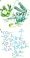
Structure of IFP1.4. A, architecture of IFP1.4 is almost identical to previously published DrCBD structures. A protein knot keeps the PAS domain and N-terminal residues (lime) packed against the GAF domain (mint). BV (cyan), covalently attached at Cys-24, is nestled in the GAF domain. B, polar contacts to BV (dotted lines) are unchanged, whereas new and formerly observed hydrophobic contacts with the chromophore (starbursts; green for IFP1.4, blue for DrCBDmon) stabilize the D ring. In particular, Met-186 and Val-288 are absent in the wild-type counterpart DrCBDmon.
FIGURE 2.
FIGURE 2.
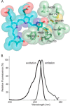
The evolved hydrophobic hub leads to excitation wavelength shift. A, hydrophobic interactions of conserved and novel (Met-186, Val-188) side chains restrict the D ring rotation. Notably, Val-288 adopts two rotamers, one of which interacts preferentially with Met-174, whereas the other stabilizes the C-ring methyl group (13). B, a single amino acid change of V186M is sufficient to blue-shift the excitation wavelength maximum to 692 from 701. Scans shown are from 5-nm step-size excitation (monitored at 713 nm) and emission (excited at 645 nm) scans.
FIGURE 3.
FIGURE 3.
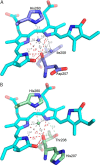
Network of polar contacts involving BV ring nitrogens. A, in DrCBDmon, the carbonyl backbone of His-207 forms direct polar contacts (red) with the A, B, and C ring nitrogens of BV, all of which are protonated. The pyrrole-water is a central part of this network, interacting with each of the ring nitrogens and the carbonyl. A second non-conserved water participates in the polar interaction network. B, in IFP1.4 the novel hydrogen-bond donor Thr-208 may reduce the likelihood of proton transfer from the chromophore to the 207 carboxylate.
FIGURE 4.
FIGURE 4.
Contraction of surface residues in IFP1.4. A, packing of the Met-54 pushes PAS domain α2 outward in some DrCBD variants (Wi-Phy 3S7Q shown in blue) but can adopt variable positions in the family of DrCBD structures (dimeric DrCBD (PDB code 2O9C) and high resolution DrCBD D207H (PDB code 3S7O) shown in sand). In fluorescent IFP1.4 (lime), the Val-54 side chain cannot accomplish this displacement so α2 adopts the more compact position closer to β2. B, substitution V195M in IFP1.4 (mint) pulls residues 195–197 of the GAF domain inward, closing a surface cleft seen in DrCBDmon (blue) and displacing glycerol (GOL) as well as disrupting the interaction between 196 main chain and Arg-172 side chain.
FIGURE 5.
FIGURE 5.
Fluorescence decay traces of dark-adapted phytochromes. Emission decays of purified IFP1.4, IFPrev, DrCBDmon, and DrCBDmon variants (in 30 mm Tris, pH 8.0) excited at 660 nm and monitored at 714 nm. Solid lines show the exponential fits of the data.

References

    1. Ying B. W., Fourmy D., Yoshizawa S. (2007) Substitution of the use of radioactivity by fluorescence for biochemical studies of RNA. RNA 13, 2042–2050 - PMC - PubMed
    1. Shimomura O., Johnson F. H., Saiga Y. (1962) Extraction, purification and properties of aequorin, a bioluminescent protein from the luminous hydromedusan, Aequorea. J. Cell Comp. Physiol. 59, 223–239 - PubMed
    1. Heim R., Prasher D. C., Tsien R. Y. (1994) Wavelength mutations and posttranslational autoxidation of green fluorescent protein. Proc. Natl. Acad. Sci. U.S.A. 91, 12501–12504 - PMC - PubMed
    1. Stepanenko O. V., Verkhusha V. V., Kuznetsova I. M., Uversky V. N., Turoverov K. K. (2008) Fluorescent proteins as biomarkers and biosensors: throwing color lights on molecular and cellular processes. Curr. Protein. Pept. Sci. 9, 338–369 - PMC - PubMed
    1. Jöbsis F. F. (1974) Intracellular metabolism of oxygen. Am. Rev. Respir. Dis. 110, 58–63 - PubMed

Publication types

MeSH terms