. 2014 Nov 7;289(45):31111-20.
doi: 10.1074/jbc.M114.600320. Epub 2014 Sep 24.

Increased spinal cord Na⁺-K⁺-2Cl⁻ cotransporter-1 (NKCC1) activity contributes to impairment of synaptic inhibition in paclitaxel-induced neuropathic pain

Affiliations

Increased spinal cord Na⁺-K⁺-2Cl⁻ cotransporter-1 (NKCC1) activity contributes to impairment of synaptic inhibition in paclitaxel-induced neuropathic pain

Shao-Rui Chen et al. J Biol Chem. .

Abstract

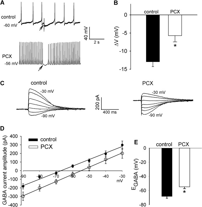
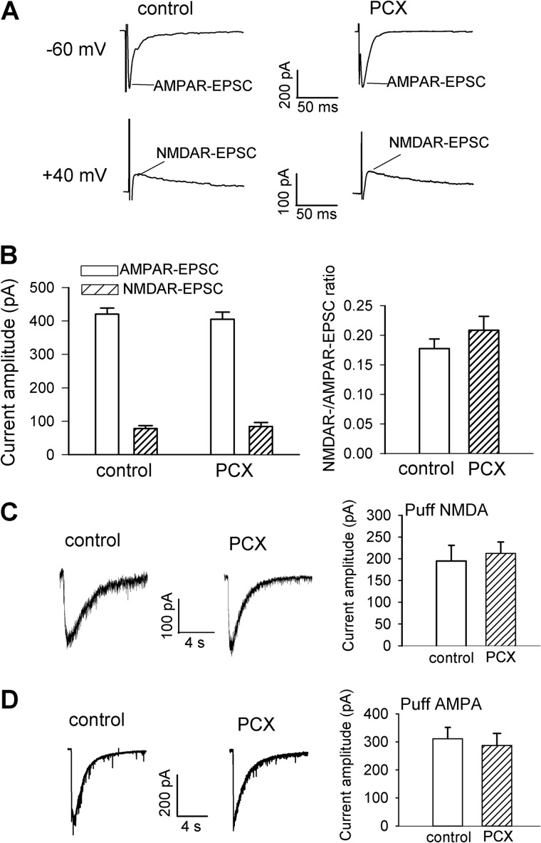
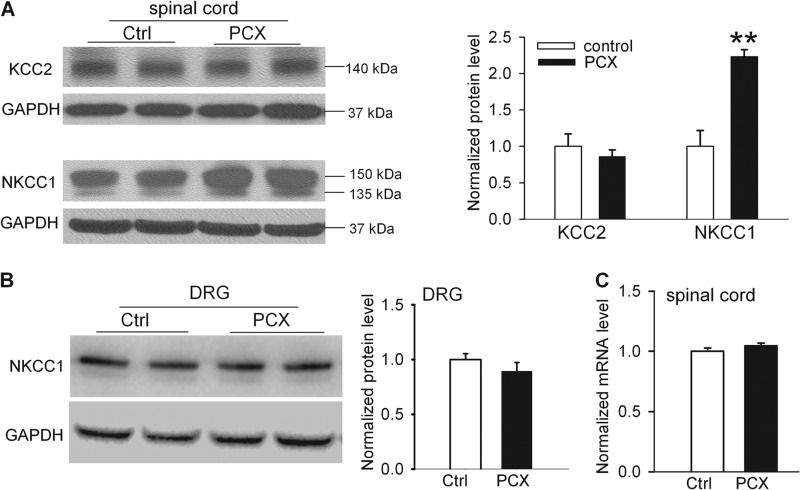
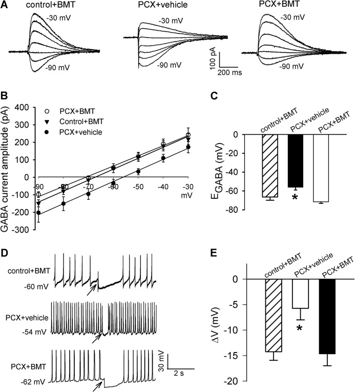
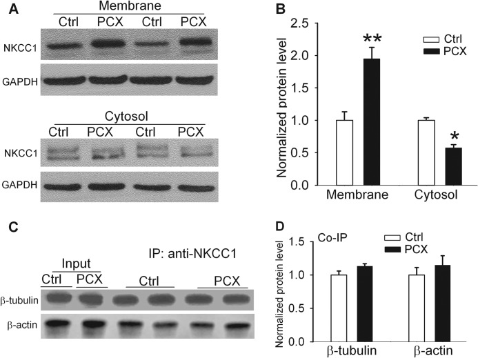
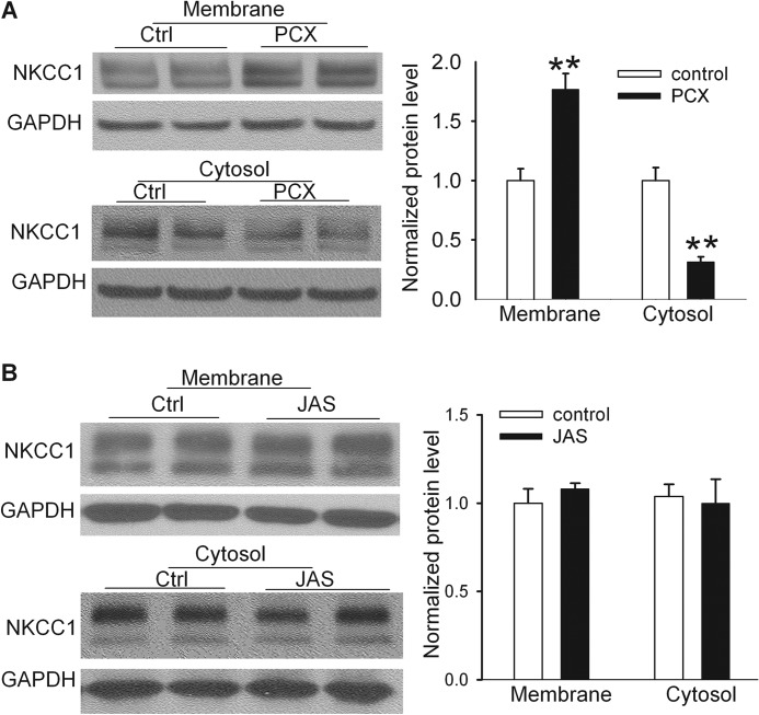
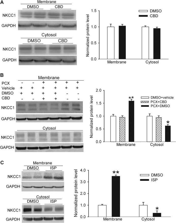
Microtubule-stabilizing agents, such as paclitaxel (Taxol), are effective chemotherapy drugs for treating many cancers, and painful neuropathy is a major dose-limiting adverse effect. Cation-chloride cotransporters, such as Na(+)-K(+)-2Cl(-) cotransporter-1 (NKCC1) and K(+)-Cl(-) cotransporter-2 (KCC2), critically influence spinal synaptic inhibition by regulating intracellular chloride concentrations. Here we show that paclitaxel treatment in rats significantly reduced GABA-induced membrane hyperpolarization and caused a depolarizing shift in GABA reversal potential of dorsal horn neurons. However, paclitaxel had no significant effect on AMPA or NMDA receptor-mediated glutamatergic input from primary afferents to dorsal horn neurons. Paclitaxel treatment significantly increased protein levels, but not mRNA levels, of NKCC1 in spinal cords. Inhibition of NKCC1 with bumetanide reversed the paclitaxel effect on GABA-mediated hyperpolarization and GABA reversal potentials. Also, intrathecal bumetanide significantly attenuated hyperalgesia and allodynia induced by paclitaxel. Co-immunoprecipitation revealed that NKCC1 interacted with β-tubulin and β-actin in spinal cords. Remarkably, paclitaxel increased NKCC1 protein levels at the plasma membrane and reduced NKCC1 levels in the cytosol of spinal cords. In contrast, treatment with an actin-stabilizing agent had no significant effect on NKCC1 protein levels in the plasma membrane or cytosolic fractions of spinal cords. In addition, inhibition of the motor protein dynein blocked paclitaxel-induced subcellular redistribution of NKCC1, whereas inhibition of kinesin-5 mimicked the paclitaxel effect. Our findings suggest that increased NKCC1 activity contributes to diminished spinal synaptic inhibition and neuropathic pain caused by paclitaxel. Paclitaxel disrupts intracellular NKCC1 trafficking by interfering with microtubule dynamics and associated motor proteins.

Keywords: Chloride Transport; GABA Receptor; Neuroscience; Neurotransmitter; Neurotransmitter Receptor; Pain; Synaptic Plasticity; Trafficking.


Figures

FIGURE 1.
FIGURE 1.
Paclitaxel treatment diminishes GABA-mediated synaptic inhibition and causes a depolarizing shift in EGABA in spinal dorsal horn neurons. A and B, representative perforated recordings and mean data show changes in membrane potentials in response to puff application of 100 μm GABA to lamina II neurons in vehicle-treated control and paclitaxel (PCX)-treated rats (n = 16 and 18 neurons, respectively). Arrows indicate the time when GABA was puffed to the neuron. C and D, original current traces and mean current-voltage plot data show the GABA-elicited currents recorded at different holding potentials in lamina II neurons from vehicle-treated control and paclitaxel-treated rats (n = 18 and 19 neurons, respectively). E, mean data show the difference in the GABA reversal potentials of lamina II neurons recorded in D. *, p < 0.05 compared with the control group. Error bars represent the S.E.
FIGURE 2.
FIGURE 2.
Paclitaxel treatment does not significantly alter AMPAR and NMDAR activities of spinal dorsal horn neurons. A and B, original current traces and mean data show the amplitude and ratio of evoked NMDAR-EPSCs to AMPAR-EPSCs of lamina II neurons recorded from vehicle-treated control and paclitaxel (PCX)-treated rats (n = 19 neurons in each group). The holding potential for recording AMPAR-EPSCs and NMDAR-EPSCs is indicated on the left. C, representative traces and mean data of the amplitude of NMDAR currents elicited by puff application of 100 μm NMDA to lamina II neurons recorded from vehicle-treated control and paclitaxel-treated rats (n = 11 and 12 neurons, respectively). D, representative traces and mean data of the amplitude of AMPAR currents elicited by puff application of 20 μm AMPA to lamina II neurons recorded from vehicle-treated control and paclitaxel-treated rats (n = 12 neurons in each group). Error bars represent the S.E.
FIGURE 3.
FIGURE 3.
Paclitaxel treatment increases NKCC1, but not KCC2, protein levels in the spinal cords. A, original gel images (left) and quantification (right) of KCC2 (∼140 kDa) and NKCC1(∼150 kDa and 135 kDa) in the dorsal spinal cord from vehicle-treated control and paclitaxel (PCX)-treated rats (n = 8 rats in each group). B, original gel blots (left) and quantification (right) of protein levels of NKCC1 in the DRG from vehicle-treated control and paclitaxel-treated rats (n = 6 rats in each group). GAPDH in the same sample was used as a loading control. C, mean data show the NKCC1 mRNA level in the dorsal spinal cord from vehicle-treated control and paclitaxel-treated rats (n = 8 rats in each group). **, p < 0.01 compared with the control group. Error bars represent the S.E.
FIGURE 4.
FIGURE 4.
NKCC1 inhibition restores the level of GABA-mediated synaptic inhibition in spinal cords of paclitaxel-treated rats. A and B, representative traces of GABA-induced currents recorded at different holding potentials and mean current-voltage plot data show the GABA reversal potentials of lamina II neurons from spinal cords of control rats pretreated with 20 μm bumetanide (BMT) and spinal cords of paclitaxel (PCX)-treated rats pretreated with vehicle (0.02% DMSO) or 20 μm bumetanide (n = 14, 12, and 18 neurons, respectively). C, mean data show the difference in the GABA reversal potentials of the same neurons recorded in B. D and E, original perforated recordings and mean data show changes in membrane potentials in response to puff application of 100 μm GABA to lamina II neurons in the spinal cords of control rats pretreated with bumetanide and spinal cords of paclitaxel-treated rats pretreated with vehicle (0.02% DMSO) or bumetanide (n = 14, 14, and 15 neurons in each group). Arrows indicate the time when GABA was puffed to the neuron. *, p < 0.05 compared with the control group. Error bars represent the S.E.
FIGURE 5.
FIGURE 5.
Inhibition of NKCC1 at the spinal level attenuates pain hypersensitivity of paclitaxel-treated rats. A and B, time course of the mean effects of intrathecal injection of bumetanide or vehicle on tactile allodynia (A) and mechanical hyperalgesia (B) of paclitaxel-treated rats. Bumetanide (BMT; 2.5, 10, and 20 ng; n = 10 rats in each group) or vehicle (0.1% DMSO, n = 7 rats) was administered in rats 12–15 days after paclitaxel treatment. *, p < 0.05 compared with respective baseline control (time 0). Error bars represent the S.E.
FIGURE 6.
FIGURE 6.
Effects of systemic paclitaxel treatment on NKCC1 subcellular redistribution and association of NKCC1 with microtubules in the spinal cord. A and B, original gel blots and quantification of NKCC1 protein levels in the plasma membrane and cytosolic fractions of spinal cords obtained from vehicle-treated control and paclitaxel (PCX)-treated rats (n = 8 rats in each group). C and D, original gel images and quantification of protein levels of β-tubulin and β-actin in the NKCC1 immunoprecipitates (IP) of spinal cords obtained from vehicle-treated control and paclitaxel-treated rats (n = 6 rats in each group). *, p < 0.05; **, p < 0.01 compared with the control group. Error bars represent the S.E.
FIGURE 7.
FIGURE 7.
Paclitaxel causes NKCC1 subcellular redistribution in the spinal cord through stabilizing microtubules but not actin. A, original blot images and quantification of NKCC1 protein levels in the plasma membrane and cytosolic fractions of spinal cord slices treated with vehicle (Ctrl, Cremophor EL/ethanol 1:1; n = 8 rats) or paclitaxel (PCX, 10 μm, n = 8 rats) for 3 h. B, original gel images and quantification of NKCC1 protein levels in the plasma membrane and cytosolic fractions of spinal cord slices treated with vehicle control (0.1% DMSO, n = 6 rats) or jasplakinolide (JAS, 125 nm, n = 6 rats) for 3 h. **, p < 0.01 compared with the control group. Error bars represent the S.E.
FIGURE 8.
FIGURE 8.
Paclitaxel causes NKCC1 subcellular redistribution in the spinal cord through increased dynein activity. A, original blot images and quantification of NKCC1 protein levels in the plasma membrane and cytosolic fractions of spinal cord slices treated with vehicle (Ctrl, 0.1% DMSO) or ciliobrevin D (CBD, 50 μm) for 3 h. n = 6 rats in each group. B, original gel images and quantification of NKCC1 protein levels in the plasma membrane and cytosolic fractions of spinal cord slices treated with DMSO plus vehicle (Cremophor EL/ethanol), paclitaxel (PCX, 10 μm) plus ciliobrevin D, or PCX plus DMSO for 3 h. n = 6 rats in each group. C, original blot images and quantification of NKCC1 protein levels in the plasma membrane and cytosolic fractions of spinal cord slices with vehicle (Ctrl, 0.1% DMSO) or ispinesib (ISP, 1 μm). * p < 0.05; ** p < 0.01, compared with the vehicle control group. Error bars represent the S.E.

References

    1. Grisold W., Cavaletti G., Windebank A. J. (2006) Peripheral neuropathies from chemotherapeutics and targeted agents: diagnosis, treatment, and prevention. Neurol. Oncol. 14, 45–54 - PMC - PubMed
    1. Lee J. J., Swain S. M. (2006) Peripheral neuropathy induced by microtubule-stabilizing agents. J. Clin. Oncol. 24, 1633–1642 - PubMed
    1. Scripture C. D., Figg W. D., Sparreboom A. (2006) Peripheral neuropathy induced by paclitaxel: recent insights and future perspectives. Curr. Neuropharmacol. 4, 165–172 - PMC - PubMed
    1. Rowinsky E. K., Chaudhry V., Cornblath D. R., Donehower R. C. (1993) Neurotoxicity of Taxol. J. Natl. Cancer Inst. Monogr. 15, 107–115 - PubMed
    1. Polomano R. C., Bennett G. J. (2001) Chemotherapy-evoked painful peripheral neuropathy. Pain Med. 2, 8–14 - PubMed

Publication types

LinkOut - more resources