. 2014 Nov 27;124(23):3409-19.
doi: 10.1182/blood-2014-07-587642. Epub 2014 Sep 24.

Disruption of hSWI/SNF complexes in T cells by WAS mutations distinguishes X-linked thrombocytopenia from Wiskott-Aldrich syndrome

Affiliations

Disruption of hSWI/SNF complexes in T cells by WAS mutations distinguishes X-linked thrombocytopenia from Wiskott-Aldrich syndrome

Koustav Sarkar et al. Blood. .

Abstract

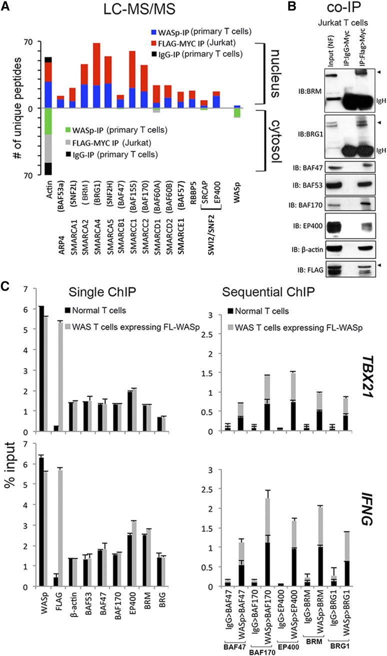
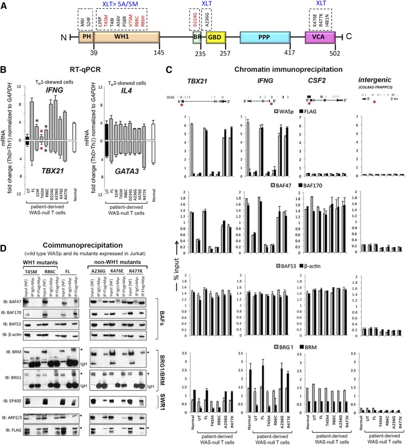
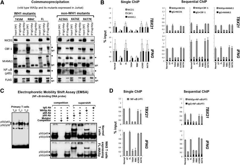
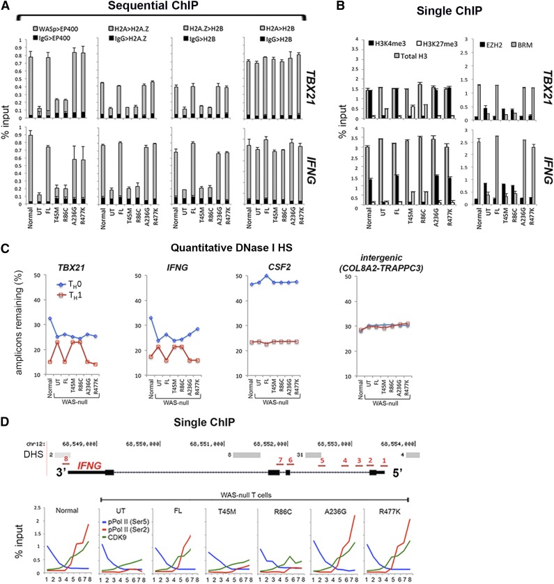
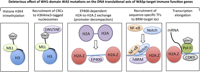
Wiskott-Aldrich syndrome (WAS), an immunodeficiency disorder, and X-linked thrombocytopenia (XLT), a bleeding disorder, both arise from nonsynonymous mutations in WAS, which encodes a hematopoietic-specific WASp. Intriguingly, XLT evolves into WAS in some patients but not in others; yet the biological basis for this cross-phenotype (CP) effect remains unclear. Using human T-helper (TH) cells expressing different disease-causing WAS mutations, we demonstrated that hSWI/SNF-like complexes require nuclear-WASp to execute their chromatin-remodeling activity at promoters of WASp-target, immune function genes during TH1 differentiation. Hot-spot WAS mutations Thr45Met and Arg86Cys, which result in XLT-to-WAS disease progression, impair recruitment of hBRM- but not BRG1-enriched BAF complexes to IFNG and TBX21 promoters. Moreover, promoter enrichment of histone H2A.Z and its catalyzing enzyme EP400 are both impaired. Consequently, activation of Notch signaling, a hBRM-regulated event, and its downstream effector NF-κB are both compromised, along with decreased accessibility of nucleosomal DNA and inefficient transcription-elongation of WASp-target TH1 genes. In contrast, patient mutations Ala236Gly and Arg477Lys that manifest in XLT without progressing to WAS do not disrupt chromatin remodeling or transcriptional reprogramming of TH1 genes. Our study defines an indispensable relationship between nuclear-WASp- and hSWI/SNF-complexes in gene activation and reveals molecular distinctions in TH cells that might contribute to disease severity in the XLT/WAS clinical spectrum.


Figures

Figure 1
Figure 1
Characterizing physiologic WASp:SWI/SNF associations in vivo. (A) Mass spectrometry. Actual number of polypeptides of WASp-associated, chromatin-remodeling complexes (CRC) captured from cytosolic and nuclear fractions of TH1-skewed, primary TH (endogenous WASp) or Jurkat TH (Flag/Myc-tagged transfected WASp) analyzed by LC-MS/MS after immunoaffinity purification with anti-WASp, anti-FLAG/MYC (sequential 2-step purification), or -IgG antibodies, as described. Searches from 3 to 4 biological replicates were combined to generate a MultiConsensus report of peptides and proteins identified from the WASp proteome after applying the filtering criteria previously described (supplemental Figure 1). (B) Selective validation of MS-generated WASp-associated CRC proteome by co-IP. Protein complexes isolated by 2-step IP (1st:Flag, 2nd:Myc) from the nuclear fraction of TCR-activated, TH1-skewed, Jurkat TH stably expressing Flag/Myc-tagged WASp were resolved by sequential western blotting with the same gel with indicated antibodies. This image is part of the full gel image shown in Figure 2D. (C) MNase ChIP-qPCR. Chromatin enrichment profiles of the indicated proteins, at 5′UTR (promoter region) of the indicated genes in TH1-skewed, normal, or WASnull TH cells stably transfected with Flag/Myc-tagged, full-length (FL) WASp. For sequential ChIP, 2 rounds of conventional ChIPs were performed in the indicated sequence (eg, WASp>BAF47 denotes 1stChIP:WASp, 2ndChIP:BAF47; IgG>BAF47 denotes1stChIP:IgG, 2ndChIP:BAF47). The displayed ChIP values (mean ± SEM) are percentages of total nuclear input chromatin and were derived after subtracting the background values obtained with isotype IgG antibody, the latter not shown for single ChIPs. Data were generated from 3 biological replicates. The genomic location of PCR primer/probes is shown in Figure 2C.
Figure 2
Figure 2
Characterizing the effects of pathogenic WAS missense mutations on WASp:SWI/SNF associations in vivo. (A) Multidomain structure of WASp is shown along with the indicated pathogenic mutations within its different domains. Recurring “hot spot” mutations are indicated in red along with their reported clinical severity grades (stable XLT vs XLT>WAS 5A/5M progressive disease; 5A, grade 5 with autoimmunity; 5M, grade 5 with malignancy). See supplemental Figure 3A for a detailed description of the mutations and their corresponding disease severity grades. (B) RT-qPCR quantitation of candidate TH1 or TH2 genes in WASnull T-cell line (HTLV-1–immortalized) reconstituted with transfected FL-WASp or the indicated mutants after CD3/28 activation under TH1 or TH2 skewing or TH0 nonskewing conditions. Normal CD4 TH cell line (HTLV-1 immortalized) is the control. UT, untranfected WASnull T cells. The mRNA copy numbers derived from the control TH0 cells are not shown but were subtracted from the displayed final mRNA values of the TH1- or TH2-skewed cells. Absolute copy numbers adjusted to GAPDH are displayed as fold change (up or down) in TH1 or TH2 cells compared with their TH0 controls. Data represent the average from at least 3 biological replicates, with bars indicating SEM. Wilcoxon nonparametric test using GraphPad InStat software determined the P values comparing the data between FL and mutants (red asterisk, P < .01; black asterisk, P > .01 but ≤ .05). In data where the differences did not reach statistical significance (ie, P > .05), an asterisk is not shown. (C). MNase ChIP-qPCR assays were performed for the indicated proteins as described in the legend to Figure 1C. The genomic location of PCR primer/probes is indicated by a red asterisk in the gene diagram shown at the top. For TBX21, the 5′ UTR primers were designed within the genomic region that also contains a GAS (γ-activated sequence) site (5′-TTCAGGCAA-3′ at about −770 bp from first coding ATG). For IFNG, the primers are located between −200 to −250 bp from first coding ATG, a region known to contain functional promoter elements. The intergenic region between COL8A2 and TRAPPC3 genes on Chr.1, which does not contain known protein-coding genes, served as a negative control. See supplemental Table 1 for primer/probe details. (D). Nuclear fraction of Jurkat (TH1-skewed, TCR-activated) cells stably expressing Flag/Myc dual-tagged FL-WASp or its indicated WASp mutants were sequentially IPed in the indicated combination (eg, Flag>Myc denotes first IP: Flag, second IP: Myc) and analyzed by sequential western blotting of the same gel with the indicated antibodies.
Figure 3
Figure 3
Characterizing the effects of pathogenic WAS missense mutations on WASp:Notch:NF-κB signaling module in vivo. (A) CoIP/western assay. The same gel shown in Figure 2D was sequentially reprobed with the indicated Notch signaling components. (B,D) Single and sequential ChIP-qPCR assays were performed for the indicated proteins as described in the legend to Figure 1C. (C) Left panel: Electrophoretic mobility shift assay (EMSA) with NF-κB oligo performed on the nuclei isolated from primary human CD4+ TH cells, nonskewed TH0, or TH1- or TH2-skewed and CD3/28-activated cells. Right panel: EMSA/Supershift assay with NF-κB oligo performed on TH1-skewed normal TH cells (endogenous WASp) or WAS-null TH cells stably expressing Flag-tagged, transfected FL-WASp. The data are representative of 2 biological replicates. White arrowheads indicate location of the shifted bands.
Figure 4
Figure 4
Characterizing the effects of pathogenic WAS missense mutations on histone H2A-to-H2A.Z exchange and transcription elongation in vivo. (A-B) Single and sequential MNase-ChIP assays performed on nuclear extracts from TH1-skewed, WASnull TH cells stably expressing the indicated WASp mutants. Other details are as described in the legend to Figures 2C and 3B. Data represent the averages of triplicates, with bars indicating the standard error from at least 3 biological replicates. (C) PCR-based quantitation of the absolute DNaseI hypersensitivity at the indicated gene promoter loci in nonskewed TH0 or TH1-skewed, normal, and WASnull TH cells stably expressing the indicated WASp mutants, or controls (UT, untransfected control; FL, full-length WASp transfected) is shown. TH nuclei were treated with DNaseI, and the number of amplicons lost was quantified by RT-qPCR and is displayed as percent of DNaseI-untreated control. Data represent the average of triplicate values from 1 experiment. (D) MNase-ChIP assays performed on nuclear extracts from TH1-skewed, WASnull TH cells stably expressing the indicated WASp mutants. The location of 8 different genomic positions (position 1 is near TSS at 5′UTR; position 8 is at 3′UTR immediately after the last coding exon) within the IFNG locus, where the ChIP-qPCR primer/probes were designed is indicated and numbered (in red). DNaseI HS (DHS) profile for the primary human peripheral TH1 cells (in gray) publicly available from the ENCODE–University of Washington was aligned alongside our custom track to give context to the location of our ChIP-qPCR primer/probes. “Normal” denotes an HTLV-1–immortalized CD4+T cell line generated from a healthy donor. UT, untransfected WAS-null patient-derived T cells line.
Figure 5
Figure 5
A working model for nuclear-WASp actions in reprogramming transcription. Step 1: H3K4me3 trimethylation. Because nuclear-WASp physically and functionally associates with the hMLL/COMPASS complex and histone H3K4 trimethylation,, the model proposes that WH1 mutants disrupt recruitment of MLL-enriched complex and the subsequent inscription of H3K4me3 mark at WASp-target gene promoters. Step 2: Recruitment of CRCs to H3K4me3-tagged nucleosome. Because the human SWR1-like protein EP400 favors binding promoters that are enriched with H3K4me3-marked nucleosomes, the model proposes that WASp regulates EP400 binding to chromatin in TH1 cells through its previously described effect on H3K4me3 modification. WH1 mutations disrupt this function of WASp at its target loci. Step 3: EP400-dependent H2A-to-H2A.Z exchange. Recruitment of EP400 to H3K4me3-marked chromatin catalyzes local H2A-to-H2A.Z exchange, which promotes promoter decompaction. Because the augmented recruitment of SWI/SNF occurs at genomic sites containing H2A.Z-tagged nucleosomes, the model proposes that WH1 mutants disrupt the initial H2A.Z-driven chromatin-remodeling and consequently also the deposition of SWI/SNF-like BAF subunits, which in turn affects higher-order chromatin reorganization and RNA polymerase II recruitment., Step 4: Recruitment of sequence-specific transcription factors (TFs). WH1 mutants disrupt recruitment of Notch-signal transduction components and NF-κB(p65) at the hBRM target loci in TH1 cells. Step 5: Transcription elongation. EP400-dependent H2A.Z deposition functions also relieve the RNA Pol II “pause” at +1 nucleosome., We propose that WH1 mutants, through their effects on EP400 and H2A-to-H2A.Z exchange, disrupt recruitment of elongating Pol II (CTD-Ser2) and CDK9 (PTEF-b subunit), protein complexes that actuate productive 5′>3′ transcription elongation. The disease model of WAS/XLT proposes that certain WAS mutations could impair one or multiple steps in this processive event, leading to a spectrum of defects that could manifest in either total loss of gene transcription or some gradations of it, which we postulate lends the immunologic basis for cross-phenotype effects and symptom heterogeneity in XLT/WAS.

References

    1. Symons M, Derry JM, Karlak B, et al. Wiskott-Aldrich syndrome protein, a novel effector for the GTPase CDC42Hs, is implicated in actin polymerization. Cell. 1996;84(5):723–734. - PubMed
    1. Taylor MD, Sadhukhan S, Kottangada P, et al. Nuclear role of WASp in the pathogenesis of dysregulated TH1 immunity in human Wiskott-Aldrich syndrome. Sci Transl Med. 2010;2(37):37ra44. - PMC - PubMed
    1. Sadhukhan S, Sarkar K, Taylor M, Candotti F, Vyas YM. Nuclear role of WASp in gene transcription is uncoupled from its ARP2/3-dependent cytoplasmic role in actin polymerization. J Immunol. 2014;193(1):150–160. - PMC - PubMed
    1. Villa A, Notarangelo L, Macchi P, et al. X-linked thrombocytopenia and Wiskott-Aldrich syndrome are allelic diseases with mutations in the WASP gene. Nat Genet. 1995;9(4):414–417. - PubMed
    1. Solovieff N, Cotsapas C, Lee PH, Purcell SM, Smoller JW. Pleiotropy in complex traits: challenges and strategies. Nat Rev Genet. 2013;14(7):483–495. - PMC - PubMed

Publication types

MeSH terms

Substances

Supplementary concepts