. 2014 Oct 7;111(40):14542-7.
doi: 10.1073/pnas.1402814111. Epub 2014 Sep 24.

Novel coding, translation, and gene expression of a replicating covalently closed circular RNA of 220 nt

Affiliations

Novel coding, translation, and gene expression of a replicating covalently closed circular RNA of 220 nt

Mounir Georges AbouHaidar et al. Proc Natl Acad Sci U S A. .

Erratum in

Abstract

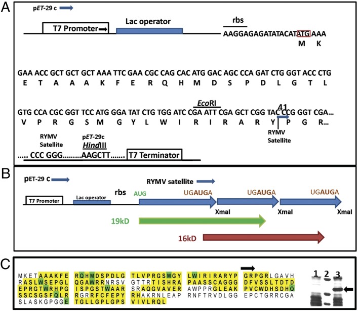
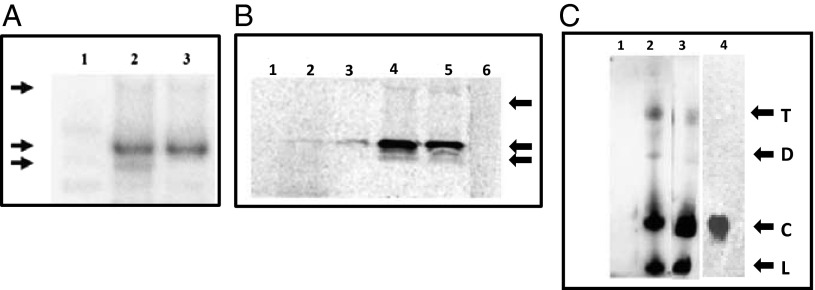
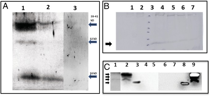
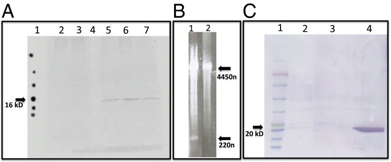
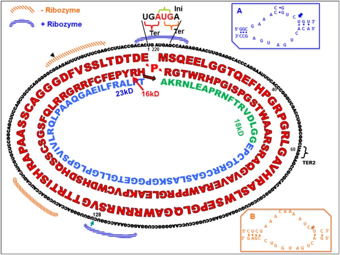
The highly structured (64% GC) covalently closed circular (CCC) RNA (220 nt) of the virusoid associated with rice yellow mottle virus codes for a 16-kDa highly basic protein using novel modalities for coding, translation, and gene expression. This CCC RNA is the smallest among all known viroids and virusoids and the only one that codes proteins. Its sequence possesses an internal ribosome entry site and is directly translated through two (or three) completely overlapping ORFs (shifting to a new reading frame at the end of each round). The initiation and termination codons overlap UGAUGA (underline highlights the initiation codon AUG within the combined initiation-termination sequence). Termination codons can be ignored to obtain larger read-through proteins. This circular RNA with no noncoding sequences is a unique natural supercompact "nanogenome."

Keywords: circular RNA translation; hammerhead ribozyme; leaky termination codons; sobemovirus.


Conflict of interest statement

The authors declare no conflict of interest.

Figures

Fig. 1.
Fig. 1.
pET52 is constructed by the insertion of a head-to-tail trimer of the scRYMV satellite sequence into the empty pET29(C) plasmid at the XmaI site. (A) Detailed diagram depicting part of the nucleotide and amino acid sequences for 19-kDa scRYMV/thrombin fusion protein. Ribosome binding site (rbs) with Shine/Dalgarno sequence, and T7 promoter and terminator are also depicted. (B) Schematic diagram showing the translation of 19 kDa (starting at AUG) (green) and 16 kDa [starting at UGAUGA (brown) protein]. Thick blue arrowheads indicate the start of each monomeric form of the scRYMV head-to-tail trimer sequence with (XmaI site) at nucleotide 41 in scRYMV sequence (Fig. 6 includes numbering). (C) Identification of the scRYMV/thrombin fusion 19-kDa protein. The prominent band of 19-kDa protein purified from SDS/PAGE of total E. coli proteins containing pET52, was subjected to MS analysis. LC-MS/MS recovered peptides are shown in yellow. Modified amino acids are shown in green (Materials and Methods and Table S1). The 163/227 aa (72% coverage) are shown. Arrow indicates the start of scRYMV sequence in the 19-kDa fusion protein. (Right) Identification of the 19-kDa scRYMV/thrombin fusion protein (arrow). Coomassie brilliant blue-R250 staining of total E. coli expressed proteins from pET29 (empty plasmid control, lane 1) and pET52 (19-kDa protein, lane 3). Lane 2: molecular size markers from top to bottom: 23, 18, and 14 kDa, respectively.
Fig. 2.
Fig. 2.
In vitro transcription/translation and immunoprecipitation of translated products using antiserum raised against the scRYMV ORF-encoded protein. (A) Lane 2 shows 19-kDa and 16-kDa proteins from reaction using pET52 DNA, whereas lane 3 shows only the 19 kDa from reaction using the truncated pETdimer DNA. Lane 1: Negative control reaction using pET29 (empty vector). Arrows on left indicate molecular size of proteins, from top to bottom: 36 kDa, 19 kDa, and 16 kDa. (B) In vitro translation reaction depicting the 16-kDa protein from the scRYMV circular RNA (purified from denaturing polyacrylamide gels) is shown in lane 3, whereas that of the reaction from total RNA of RYMV-infected rice is shown in lane 2. Lane 5 demonstrates the 16-kDa product from reaction using total viral RNA extracted from RYMV virus particles, and lane 4 shows the enhanced 16-kDa signal from reaction using RYMV total viral RNA but supplemented with the same amount of gel-purified circular RNA as that used in the reaction of lane 3. Lanes 1 and 6 represent negative control reactions using healthy rice and the endogenous empty lysate, respectively. Arrows on right depict the position of molecular weight markers 25, 16, and 14 kDa from top to bottom, respectively. (C) Northern analysis to detect the nature of scRYMV RNA species. Denaturing 4–20% PAGE in presence of 8 M urea was carried out. Lanes 2 and 3 demonstrate presence of linear (marked as “L”), circular (“C”), dimer (“D”), and trimer (“T”) forms of the scRYMV RNA in total RNA preparations from RYMV-infected rice and RYMV particles, respectively. Lane 1 shows the negative control of total RNA from healthy rice. Lane 4: 7 M urea-PAGE stained with ethidium bromide and showing the purified circular RNA extracted from the band corresponding to circular RNA (shown in lane 3). This purified circular RNA is used for in vitro translation. Arrows indicate the positions of RNA size markers: 220 (marked as “L” or “C”), 440 (“D”), and 660 (“T”) nt.
Fig. 3.
Fig. 3.
SDS/PAGE, Western detection, and time course of expression of the scRYMV-encoded proteins. (A) Western analysis using antibodies specific to the scRYMV ORF-encoded protein to detect the 16-kDa proteins among total plant proteins from RYMV infected (lane 1), healthy uninfected plant (lane 3), and purified RYMV virus (lane 2). Arrows on right indicate the positions of the 39-, 32-, and 16-kDa proteins (from top to bottom). (B) Western analysis time course of the appearance of the scRYMV 16 kDa during infection in rice plants. Lane 1 contains total proteins extracted from noninfected rice plants (negative control). Lane 2 contains total proteins from RYMV-infected plants at 4 d postinoculation. Lanes 4–7 contain total rice proteins extracted from RYMV-infected plants at 10, 14, 21, and 28 d postinoculation, respectively. Lane 3 and arrow (16 kDa) depict molecular weight size markers 116, 66, 45, 35, 25, and 18 kDa, from top to bottom, respectively. (C) North-Western analysis showing the RNA-binding activity of scRYMV-encoded proteins. Lane 1 shows the presence of the 16-kDa RNA-binding protein in infected rice, whereas lanes 2 and 9 demonstrate the detection of the 16-kDa, 18-kDa, and 19-kDa RNA-binding proteins in protein extracts from E. coli carrying pET52. Lanes 4 and 5 are the negative controls from healthy rice and pET29 protein extracts, respectively. Lanes 6 and 7 are negative control from E. coli alone and E. coli carrying pET29, respectively. Lanes 3 and 8 contain the molecular weight markers including the 14-kDa positively charged lysozyme. The 35S-labeled scRYMV and PVX probes were used in lanes 1–5 and 6–9, respectively. Arrows on left indicate the positions of molecular size markers (from top to bottom, 19, 18, 16, and 14 kDa, respectively).
Fig. 4.
Fig. 4.
Mapping of the scRYMV-derived LC-MS/MS peptides from RYMV-infected rice on the scRYMV protein sequence generated from translation (in all three reading frames) of scRYMV RNA. Matching sequences are highlighted. LC-MS/MS recovered peptides are shown in yellow. Modified amino acids are shown in green. Table S2 provides raw data and other details regarding peptide detection.
Fig. 5.
Fig. 5.
Mutational analysis of scRYMV. (A) In vitro transcription/translation and immunoprecipitation profile of scRYMV AUG to AUU (in the sequence UGAUGA) mutant shows abolition of 16-kDa translation. Lane 1: radiolabeled dots indicating the positions of protein molecular weight size markers (top to bottom, 35, 25, 20, 17, 11, and 5 kDa). Lane 2: in vitro reaction (with scRYMV 19-kDa antibody) with purified RYMV genomic RNA alone (without scRYMV) used as template shows no 16-kDa protein and was used as infectious transcript in plants. Lane 3: same reaction as in lane 2 except for addition of mutated scRYMV construct (AUG to AAU mutant pBS-trimer). Lane 4: empty lane. Lane 5: same reaction as in lane 2 except for the addition of pBS-trimer (intact construct) demonstrating the synthesis of a 16-kDa protein. Lane 6: reaction containing total RYMV viral RNA obtained from purified virus containing scRYMV. Lane 7: reaction containing purified WT scRYMV RNA alone (obtained from virus particles). (B) Replication of AUG to AAU scRYMV mutant is abolished in plants. Lane 1: RYMV viral RNA profile from purified virus particles of plants infected with infectious RYMV RNA along with T7 polymerase-generated in vitro transcripts from WT pBS-trimer scRYMV clone. Lane 2: RYMV infectious RNA plus in vitro transcripts from AUG to AAU mutant pBS-trimer clone showing complete absence of the scRYMV RNA (no replication). Upper arrow indicates the position of genomic RYMV RNA (4450 nt) and the bottom arrow indicates the position of circular scRYMV RNA (220 nt). (C) UGAUGA codons influence translation termination: Western blotting analysis of total E. coli proteins using pET dimer and anti–His-tag antibody for intact and a UGAUGA-to-CUCGAG mutant construct. Lane 1: protein molecular weight ladder (top to bottom, 135, 100, 75, 63, 48, 35, 25, 20, 17, and 11 kDa). Lane 2: E. coli containing empty pET29 plasmid. Lane 3: E. coli containing intact construct with UGAUGA terminator upstream of a His-tag. No protein product is detected. Lane 4: E. coli containing UGAUGA to CUCGAG mutant construct. A 20-kDa protein is detected by using anti–His-tag antibody. This is in accord with a translation read-through over the mutated UGAUGA-to-CUCGAG region producing a His-tagged fusion protein.
Fig. 6.
Fig. 6.
Schematic diagram showing the nucleotide and amino acid sequences of the encoded proteins and many other functions of the CCC RNA. Positions of initiation AUG codon (INI) and termination codons UGA (TER for 16 kDa) and UAG (TER2 for 18 kDa) are indicated. Locations of (+) and (−) hammerhead ribozymes are indicated. Positive-sense hammerhead ribozyme is shown in Inset A and that of minus-sense ribozyme is shown in Inset B. Arrowheads indicate the splice site for both ribozymes [on circular RNA depicting secondary structure and cleavage sites for both ribozymes (Insets)]. The amino acid sequences of 16-, 18-, and 23-kDa proteins are indicated. Shaded areas indicate the nucleotide sequences involved in (−) and (+) ribozymes. Nucleotide numbering starts at (position 1) UGAUGA.

References

    1. Diener TO. Viroids. Adv Virus Res. 1983;28:241–283. - PubMed
    1. Symons RH, Randles JW. Encapsidated circular viroid-like satellite RNAs (virusoids) of plants. Curr Top Microbiol Immunol. 1999;239:81–105. - PubMed
    1. Taliansky ME, Palukaitis PF. Satellite RNAs and satellite viruses. In: Granoff A, Webster RG, editors. Encyclopedia of Virology. 2nd Ed. Vol 3. Academic; San Diego, CA: 1999. pp. 1607–1615.
    1. Branch AD, Robertson HD. A replication cycle for viroids and other small infectious RNA’s. Science. 1984;223(4635):450–455. - PubMed
    1. Bruening G, Passmore BK, van Tol H, Buzayan JM, Feldstein PA. Replication of a plant virus satellite RNA: Evidence favors transcription of circular templates of both polarities. Mol Plant Microbe Interact. 1991;4(3):219–225. - PubMed

MeSH terms