. 2014:2014:426740.
doi: 10.1155/2014/426740. Epub 2014 Sep 1.

Vitamin C mitigates oxidative stress and tumor necrosis factor-alpha in severe community-acquired pneumonia and LPS-induced macrophages

Affiliations

Vitamin C mitigates oxidative stress and tumor necrosis factor-alpha in severe community-acquired pneumonia and LPS-induced macrophages

Yuanyuan Chen et al. Mediators Inflamm. 2014.

Abstract

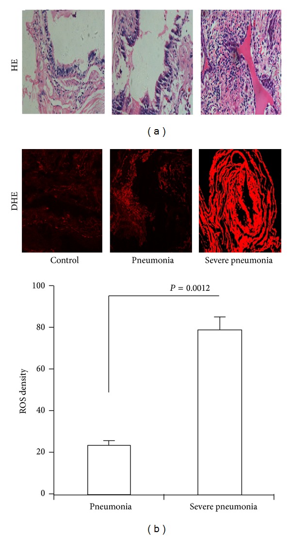
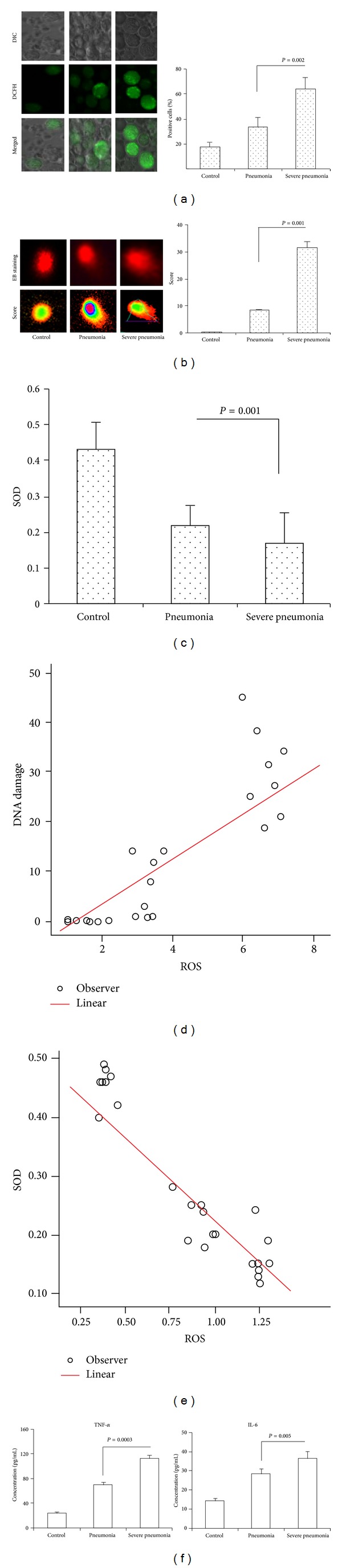
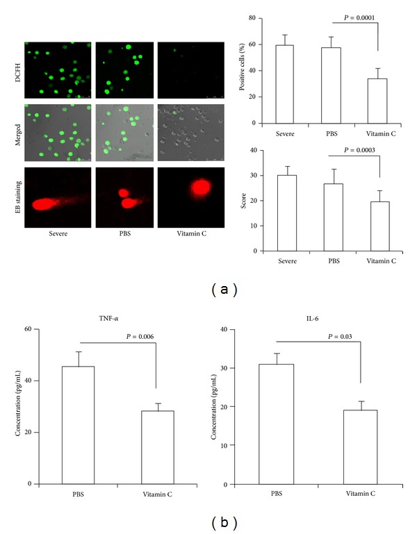
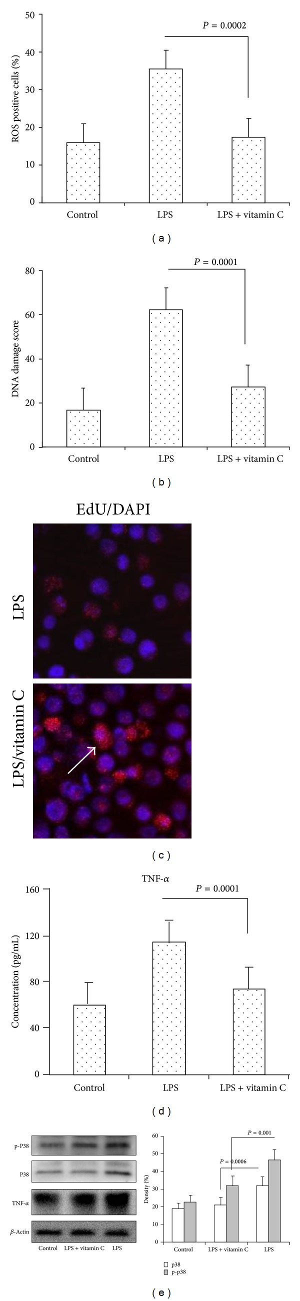
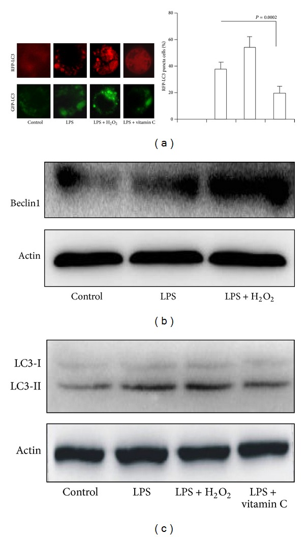
Oxidative stress is an important part of host innate immune response to foreign pathogens. However, the impact of vitamin C on oxidative stress and inflammation remains unclear in community-acquired pneumonia (CAP). We aimed to determine the effect of vitamin C on oxidative stress and inflammation. CAP patients were enrolled. Reactive oxygen species (ROS), DNA damage, superoxide dismutases (SOD) activity, tumor necrosis factor-alpha (TNF-α), and IL-6 were analyzed in CAP patients and LPS-stimulated macrophages cells. MH-S cells were transfected with RFP-LC3 plasmids. Autophagy was measured in LPS-stimulated macrophages cells. Severe CAP patients showed significantly increased ROS, DNA damage, TNF-α, and IL-6. SOD was significantly decreased in severe CAP. Vitamin C significantly decreased ROS, DNA damage, TNF-α, and IL-6. Vitamin C inhibited LPS-induced ROS, DNA damage, TNF-α, IL-6, and p38 in macrophages cells. Vitamin C inhibited autophagy in LPS-induced macrophages cells. These findings indicated that severe CAP exhibited significantly increased oxidative stress, DNA damage, and proinflammatory mediator. Vitamin C mitigated oxidative stress and proinflammatory mediator suggesting a possible mechanism for vitamin C in severe CAP.


Figures

Figure 1
Figure 1
Oxidative stress in severe CAP. (a) HE staining of lung tissue from no severe CAP and severe CAP patients. (b) ROS were detected by confocal microscope using DHE probe. ROS level was represented by fluorescence intensity (×200).
Figure 2
Figure 2
Oxidative stress, DNA damage, TNF-α, and IL-6 in severe CAP. (a) Intracellular ROS were detected by confocal microscope using DHE probe. ROS level was represented by fluorescence intensity (×400). (b) DNA damage was detected by comet assay using confocal microscope (×400). DNA damage score was analyzed with comet assay IV software. (c) SOD activity in serum was measured using the SOD assay kit WST. (d) Correlations between ROS and DNA damage were also evaluated. (e) Correlations between ROS and SOD were also evaluated. Spearman's correlation with a two-tailed test was used for statistical analyses. (f) Standard ELISA was performed to determine the levels of TNF-α and IL-6.
Figure 3
Figure 3
The effect of vitamin C on ROS, DNA damage, TNF-α, and IL-6 in vitro. Monocytes from severe CAP were treated with vitamin C for 12 h in vitro. (a) Intracellular ROS were detected by confocal microscope using DHE probe (×400). DNA damage was detected by comet assay using confocal microscope (×400). (b) Whole blood cells from severe CAP were treated with vitamin C for 12 h in vitro. Standard ELISA was performed to determine the levels of TNF-α and IL-6.
Figure 4
Figure 4
The effect of vitamin C on oxidative stress and DNA damage in LPS-induced MH-S cells. ROS and DNA damage in MH-S cells were measured in the presence or absence of vitamin C and treated with LPS for 12 hours. (a) ROS were detected by confocal microscope using DCFH-DA probe. ROS level was represented by fluorescence intensity. (b) DNA damage was measured by comet assay. DNA damage score was analyzed with comet assay IV software. (c) MH-S cells viability was detected by confocal microscope using Edu staining (×200). Arrow showed viability of cells. (d) TNF-α was measured by ELISA. (e) TNF-α, P38, and p-P38 were determined by western blotting. β-actin was used as the loading control.
Figure 5
Figure 5
The effect of vitamin C on LPS-induced autophagy in macrophages. (a) MH-S cells were transfected with RFP-LC3 or GFP-LC3 plasmids for 24 hours. MH-S cells were cultured in the presence or absence of vitamin C and treated with LPS for 12 hours. (b) Western blotting of Beclin1; (c) western blotting of LC3. β-actin was probed as a loading control in (b) and (c).

References

    1. Romero PV, Rodríguez B, Martínez S, Cañizares R, Sepúlveda D, Manresa F. Analysis of oxidative stress in exhaled breath condensate from patients with severe pulmonary infections. Archivos de Bronconeumologia. 2006;42(3):113–119. - PubMed
    1. Jensen PØ, Lykkesfeldt J, Bjarnsholt T, Hougen HP, Høiby N, Ciofu O. Poor antioxidant status exacerbates oxidative stress and inflammatory response to Pseudomonas aeruginosa lung infection in guinea pigs. Basic & Clinical Pharmacology & Toxicology. 2012;110(4):353–358. - PubMed
    1. Hosakote YM, Komaravelli N, Mautemps N, Liu T, Garofalo RP, Casola A. Antioxidant mimetics modulate oxidative stress and cellular signaling in airway epithelial cells infected with respiratory syncytial virus. The American Journal of Physiology—Lung Cellular and Molecular Physiology. 2012;303(11):L991–L1000. - PMC - PubMed
    1. Zhang W, Wei H, Frei B. Genetic deficiency of NADPH oxidase does not diminish, but rather enhances, LPS-induced acute inflammatory responses in vivo. Free Radical Biology and Medicine. 2009;46(6):791–798. - PMC - PubMed
    1. Cook-Mills JM, Marchese ME, Abdala-Valencia H. Vascular cell adhesion molecule-1 expression and signaling during disease: regulation by reactive oxygen species and antioxidants. Antioxidants & Redox Signaling. 2011;15(6):1607–1638. - PMC - PubMed

Publication types

MeSH terms

LinkOut - more resources